- Related Topics:
- eyeball
- tear duct and glands
- optic nerve
- eyelid
- visual pigment
Some basic facts of vision
So far, attention has been directed to what are essentially the preliminaries to vision. It is now time to examine some of the elementary facts of vision and to relate them to the structure of the retina and, later, to chemically identifiable events.
Measurement of the threshold
An important means of measuring a sensation is to determine the threshold stimulus—i.e., the minimum energy required to evoke the sensation. In the case of vision, this would be the minimum number of quanta of light entering the eye in unit time. If it is found that the threshold has altered because of a change of some sort, then this change can be said to have altered the subject’s sensitivity to light, and a numerical value can be assigned to the sensitivity by use of the reciprocal of the threshold energy.
Practically, a subject may be placed in the dark in front of a white screen, and the screen may be illuminated by flashes of light. For any given intensity of illumination of the screen, it is not difficult to calculate the flow of light energy entering the eye. One may begin with a low intensity of flash and increase this successively until the subject reports that he or she can see the flash. In fact, at this threshold level, the individual will not see every flash presented, even though the intensity of the light is kept constant. For this reason, a certain frequency of seeing—e.g., four times out of six—must be selected as the arbitrary point at which to fix the threshold.
When measurements of this sort are carried out, it is found that the threshold falls progressively as the subject is maintained in the dark room. This is not due to dilation of the pupil, because the same phenomenon occurs if the subject is made to look through an artificial pupil of fixed diameter. The eye, after about 30 minutes in the dark, may become about 10,000 times more sensitive to light. Vision under these conditions is, moreover, characteristically different from what it is under ordinary daylight conditions. Thus, in order to obtain best vision, the eye must look away from the screen so that the image of the screen does not fall on the fovea. If the screen is continuously illuminated at around this threshold level, it will be found to disappear if its image is brought onto the fovea, and it will become immediately visible on looking away. The same phenomenon may be demonstrated on a moonless night if the gaze is fixed on a dim star: it disappears on fixation and reappears on looking away. This feature of vision under these near-threshold, or scotopic, conditions suggests that the cones are effectively blind to weak light stimuli, since they are the only photoreceptors in the fovea. This is the basis of the duplicity theory of vision, which postulates that when the light stimulus is weak and the eye has been dark-adapted, it is the rods that are utilized because, under these conditions, their threshold is much lower than that of the cones. When the subject first enters the dark, the rods are the less sensitive type of photoreceptor, and the threshold stimulus is the light energy required to stimulate the cones. During the first five or more minutes, the threshold of the cones decreases; i.e., they become more sensitive. The rods then increase their sensitivity to the point that they are the more sensitive, and it is they that now determine the sensitivity of the whole eye, the threshold stimuli obtained after 10 minutes in the dark, for example, being too weak to activate the cones.
Scotopic sensitivity curve
When different wavelengths of light are employed for measuring the threshold, it is found, for example, that the eye is much more sensitive to blue-green light than to orange. The interesting feature of this kind of study is that the subject reports only that the light is light; he or she distinguishes no colour. If the intensity of a given wavelength of light is increased step by step above the threshold, a point comes when the subject states that it is coloured. The difference between the threshold for light appreciation and this, the chromatic threshold, is called the photochromatic interval. This suggests that the rods give only achromatic, or colourless, vision, and that it is the cones that permit wavelength discrimination. The photochromatic interval for long wavelengths (red light) is about zero, which means that the intensity required to reach the sensation of light is the same as that to reach the sensation of colour. This is because the rods are so insensitive to red light. If the dark-adaptation curve is plotted for a red stimulus, it is found that it follows the cone path, like that for foveal vision at all wavelengths.
Loss of dark adaptation
If, when the subject has become completely dark-adapted, one eye is held shut and the other exposed to a bright light for a little while, it is found that, whereas the dark-adapted eye retains its high sensitivity, that of the light-exposed eye has decreased greatly; it requires another period of dark adaptation for the two eyes to become equally sensitive.
These simple experiments pose several problems, the answers to which throw a great deal of light on the whole mechanism of vision. Why, for example, does it require time for both rods and cones to reach their maximum sensitivity in the dark? Again, why is visual acuity so low under scotopic conditions compared with that in daylight, although sensitivity to light is so high? Finally, why do the rods not serve to discriminate different wavelengths?
Bleaching of rhodopsin
It may be assumed that a photoreceptor is sensitive to light because it contains a substance that absorbs light and converts this vibrational type of energy into some other form that is eventually transmuted into electrical changes, which may be transmitted from the photoreceptor to the bipolar cell with which it is immediately connected. When the retina of a dark-adapted animal is removed and submitted to extraction procedures, a pigment, originally called visual purple but now called rhodopsin, may be obtained. If the eye is exposed to a bright light for some time before extraction, little or no rhodopsin is obtained. When retinas from animals that had been progressively dark-adapted were studied, a gradual increase in the amount of rhodopsin that could be extracted was observed. Thus, rhodopsin, on absorption of light energy, is changed to some other compound, but new rhodopsin is formed, or rhodopsin is regenerated, during dark adaptation. The obvious inference is that rhodopsin is the visual pigment of the rods and that, when it is exposed to relatively intense light, it becomes useless for vision. When the eye is allowed to remain in the dark, the rhodopsin regenerates and thus becomes available for vision. There is now conclusive proof that rhodopsin is, indeed, the visual pigment for the rods. It is obtained from retinas that have only rods and no cones—e.g., the retinas of the rat or guinea pig—and it is not obtained from the pure cone retina of the chicken.
When the absorption spectrum is measured, it is found that its maximum absorption occurs at the point of maximum sensitivity of the dark-adapted eye. Similar measurements may be carried out on animals, but the threshold sensitivity must be determined by some objective means—e.g., the response of the pupil, or, better still, the electrical changes occurring in the retina in response to light stimuli. Thus, the electroretinogram (ERG) is the record of changes in potential between an electrode placed on the surface of the cornea and an electrode placed on another part of the body, caused by illumination of the eye.
The high sensitivity of the rods by comparison with the cones may be a reflection of the greater concentration in them of pigment that would permit them to catch light more efficiently, or it may depend on other factors—e.g., the efficiency of transformation of the light energy into electrical energy. The pigments responsible for cone vision are not easily extracted or identified, and the problem will be considered in the material on colour vision. An important factor, so far as sensitivity is concerned, is the actual organization of the photoreceptors and neurons in the retina.
Synaptic organization of the retina
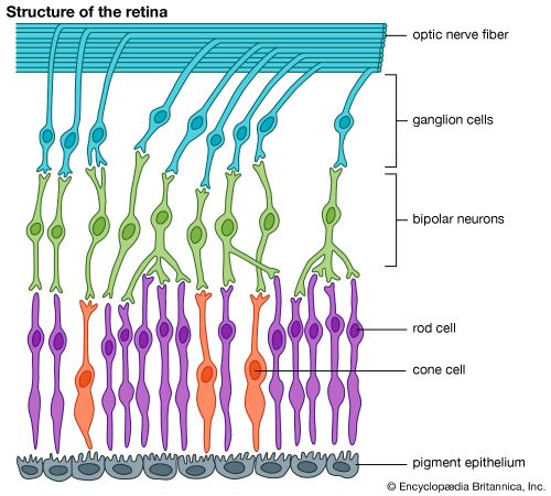
The basic structure of the retina has been indicated earlier. As in other parts of the nervous system, the messages initiated in one element are transmitted, or relayed, to others. The regions of transmission from one cell to another are areas of intimate contact known as synapses. An impulse conveyed from one cell to another travels from the first cell body along a projection called an axon, to a synapse, where the impulse is received by a projection, called a dendrite, of the second cell. The impulse is then conveyed to the second cell body, to be transmitted further, along the second cell’s axon.
It will be recalled that the functioning cells of the retina are the photoreceptor cells, the rods and cones; the ganglion cells, the axons of which form the optic nerve; and cells that act in a variety of ways as intermediaries between the photoreceptors and the ganglion cells. These intermediaries are named bipolar cells, horizontal cells, and amacrine cells.

