JP2006506118A - Lumen lining - Google Patents
Lumen lining Download PDFInfo
- Publication number
- JP2006506118A JP2006506118A JP2004551127A JP2004551127A JP2006506118A JP 2006506118 A JP2006506118 A JP 2006506118A JP 2004551127 A JP2004551127 A JP 2004551127A JP 2004551127 A JP2004551127 A JP 2004551127A JP 2006506118 A JP2006506118 A JP 2006506118A
- Authority
- JP
- Japan
- Prior art keywords
- cavity
- lining
- stent
- sphincter
- diameter
- Prior art date
- Legal status (The legal status is an assumption and is not a legal conclusion. Google has not performed a legal analysis and makes no representation as to the accuracy of the status listed.)
- Granted
Links
- 210000005070 sphincter Anatomy 0.000 claims abstract description 45
- 230000002572 peristaltic effect Effects 0.000 claims abstract description 18
- 210000001367 artery Anatomy 0.000 claims abstract description 15
- 210000000056 organ Anatomy 0.000 claims abstract description 12
- 238000010009 beating Methods 0.000 claims abstract description 8
- 238000003780 insertion Methods 0.000 claims abstract description 6
- 230000037431 insertion Effects 0.000 claims abstract description 6
- 238000000034 method Methods 0.000 claims description 14
- 239000000463 material Substances 0.000 claims description 9
- 230000002792 vascular Effects 0.000 claims 1
- 229910045601 alloy Inorganic materials 0.000 description 6
- 239000000956 alloy Substances 0.000 description 6
- 229910001285 shape-memory alloy Inorganic materials 0.000 description 6
- 230000005540 biological transmission Effects 0.000 description 5
- 210000000709 aorta Anatomy 0.000 description 4
- 210000003238 esophagus Anatomy 0.000 description 4
- 229920000642 polymer Polymers 0.000 description 4
- 210000001519 tissue Anatomy 0.000 description 4
- 206010002329 Aneurysm Diseases 0.000 description 3
- 208000001750 Endoleak Diseases 0.000 description 3
- 229910000639 Spring steel Inorganic materials 0.000 description 3
- 229920006125 amorphous polymer Polymers 0.000 description 3
- 229910001566 austenite Inorganic materials 0.000 description 3
- 230000007423 decrease Effects 0.000 description 3
- 239000012530 fluid Substances 0.000 description 3
- 229910000734 martensite Inorganic materials 0.000 description 3
- 206010064396 Stent-graft endoleak Diseases 0.000 description 2
- 208000002223 abdominal aortic aneurysm Diseases 0.000 description 2
- 210000004204 blood vessel Anatomy 0.000 description 2
- 239000013013 elastic material Substances 0.000 description 2
- 230000002452 interceptive effect Effects 0.000 description 2
- 239000012528 membrane Substances 0.000 description 2
- 210000004798 organs belonging to the digestive system Anatomy 0.000 description 2
- 210000002307 prostate Anatomy 0.000 description 2
- 230000002269 spontaneous effect Effects 0.000 description 2
- 229910000831 Steel Inorganic materials 0.000 description 1
- 230000003044 adaptive effect Effects 0.000 description 1
- 210000000013 bile duct Anatomy 0.000 description 1
- 239000008280 blood Substances 0.000 description 1
- 210000004369 blood Anatomy 0.000 description 1
- 210000001953 common bile duct Anatomy 0.000 description 1
- 230000008602 contraction Effects 0.000 description 1
- 238000001816 cooling Methods 0.000 description 1
- 230000010339 dilation Effects 0.000 description 1
- 210000001198 duodenum Anatomy 0.000 description 1
- 230000004064 dysfunction Effects 0.000 description 1
- 230000000694 effects Effects 0.000 description 1
- 230000007717 exclusion Effects 0.000 description 1
- 230000006870 function Effects 0.000 description 1
- 230000002496 gastric effect Effects 0.000 description 1
- 210000003736 gastrointestinal content Anatomy 0.000 description 1
- 239000011521 glass Substances 0.000 description 1
- 230000009477 glass transition Effects 0.000 description 1
- 238000010438 heat treatment Methods 0.000 description 1
- 238000002513 implantation Methods 0.000 description 1
- 210000000936 intestine Anatomy 0.000 description 1
- 230000001788 irregular Effects 0.000 description 1
- 230000006386 memory function Effects 0.000 description 1
- 230000003387 muscular Effects 0.000 description 1
- 229910001000 nickel titanium Inorganic materials 0.000 description 1
- HLXZNVUGXRDIFK-UHFFFAOYSA-N nickel titanium Chemical compound [Ti].[Ti].[Ti].[Ti].[Ti].[Ti].[Ti].[Ti].[Ti].[Ti].[Ti].[Ni].[Ni].[Ni].[Ni].[Ni].[Ni].[Ni].[Ni].[Ni].[Ni].[Ni].[Ni].[Ni].[Ni] HLXZNVUGXRDIFK-UHFFFAOYSA-N 0.000 description 1
- RVTZCBVAJQQJTK-UHFFFAOYSA-N oxygen(2-);zirconium(4+) Chemical compound [O-2].[O-2].[Zr+4] RVTZCBVAJQQJTK-UHFFFAOYSA-N 0.000 description 1
- 230000008855 peristalsis Effects 0.000 description 1
- 230000000704 physical effect Effects 0.000 description 1
- 210000001187 pylorus Anatomy 0.000 description 1
- 238000011084 recovery Methods 0.000 description 1
- 238000003303 reheating Methods 0.000 description 1
- 239000012858 resilient material Substances 0.000 description 1
- 229920000431 shape-memory polymer Polymers 0.000 description 1
- 239000010959 steel Substances 0.000 description 1
- 210000002784 stomach Anatomy 0.000 description 1
- 230000007704 transition Effects 0.000 description 1
- 238000011144 upstream manufacturing Methods 0.000 description 1
- 210000000626 ureter Anatomy 0.000 description 1
- 210000003708 urethra Anatomy 0.000 description 1
- 210000002700 urine Anatomy 0.000 description 1
Images
Classifications
-
- A—HUMAN NECESSITIES
- A61—MEDICAL OR VETERINARY SCIENCE; HYGIENE
- A61F—FILTERS IMPLANTABLE INTO BLOOD VESSELS; PROSTHESES; DEVICES PROVIDING PATENCY TO, OR PREVENTING COLLAPSING OF, TUBULAR STRUCTURES OF THE BODY, e.g. STENTS; ORTHOPAEDIC, NURSING OR CONTRACEPTIVE DEVICES; FOMENTATION; TREATMENT OR PROTECTION OF EYES OR EARS; BANDAGES, DRESSINGS OR ABSORBENT PADS; FIRST-AID KITS
- A61F2/00—Filters implantable into blood vessels; Prostheses, i.e. artificial substitutes or replacements for parts of the body; Appliances for connecting them with the body; Devices providing patency to, or preventing collapsing of, tubular structures of the body, e.g. stents
- A61F2/82—Devices providing patency to, or preventing collapsing of, tubular structures of the body, e.g. stents
-
- A—HUMAN NECESSITIES
- A61—MEDICAL OR VETERINARY SCIENCE; HYGIENE
- A61F—FILTERS IMPLANTABLE INTO BLOOD VESSELS; PROSTHESES; DEVICES PROVIDING PATENCY TO, OR PREVENTING COLLAPSING OF, TUBULAR STRUCTURES OF THE BODY, e.g. STENTS; ORTHOPAEDIC, NURSING OR CONTRACEPTIVE DEVICES; FOMENTATION; TREATMENT OR PROTECTION OF EYES OR EARS; BANDAGES, DRESSINGS OR ABSORBENT PADS; FIRST-AID KITS
- A61F2/00—Filters implantable into blood vessels; Prostheses, i.e. artificial substitutes or replacements for parts of the body; Appliances for connecting them with the body; Devices providing patency to, or preventing collapsing of, tubular structures of the body, e.g. stents
- A61F2/02—Prostheses implantable into the body
- A61F2/04—Hollow or tubular parts of organs, e.g. bladders, tracheae, bronchi or bile ducts
- A61F2/06—Blood vessels
- A61F2/07—Stent-grafts
- A61F2002/072—Encapsulated stents, e.g. wire or whole stent embedded in lining
Landscapes
- Health & Medical Sciences (AREA)
- Engineering & Computer Science (AREA)
- Biomedical Technology (AREA)
- General Health & Medical Sciences (AREA)
- Veterinary Medicine (AREA)
- Transplantation (AREA)
- Heart & Thoracic Surgery (AREA)
- Vascular Medicine (AREA)
- Life Sciences & Earth Sciences (AREA)
- Animal Behavior & Ethology (AREA)
- Cardiology (AREA)
- Public Health (AREA)
- Oral & Maxillofacial Surgery (AREA)
- Prostheses (AREA)
- Media Introduction/Drainage Providing Device (AREA)
- Consolidation Of Soil By Introduction Of Solidifying Substances Into Soil (AREA)
- Glass Compositions (AREA)
- Radiation-Therapy Devices (AREA)
- Chemical Or Physical Treatment Of Fibers (AREA)
- Undergarments, Swaddling Clothes, Handkerchiefs Or Underwear Materials (AREA)
- Other Investigation Or Analysis Of Materials By Electrical Means (AREA)
Abstract
拍動する動脈、蠕動器官、又は括約筋近くの腔のように、変動する内径を有する体腔中への挿入用の筒形状のライニング。このライニングは、腔の内径が変動するのに従って、ライニングを腔の形状に連続して一致させ得るように、腔によってライニングに加えられる径方向の力よりも小さい径方向の抵抗を有する。A cylindrical lining for insertion into a body cavity having a variable inner diameter, such as a beating artery, a peristaltic organ, or a cavity near the sphincter. This lining has a radial resistance that is less than the radial force applied to the lining by the cavity so that the lining can be continuously matched to the shape of the cavity as the inner diameter of the cavity varies.
Description
本発明は、体腔中に挿入するための医療装置に関する。 The present invention relates to a medical device for insertion into a body cavity.
ステントは、腔の開存性を維持するために、体腔中に挿入されて拡大される管腔(endoluminal)用の装置である。例えば、動脈、尿道、又は胃腸器官の開存性を維持するためにステントを使用することは知られている。 A stent is an endoluminal device that is inserted and expanded into a body cavity to maintain the patency of the cavity. For example, it is known to use stents to maintain patency of arteries, urethra, or gastrointestinal organs.
ステントは、本質的に、2つの形態で存在し得る筒形状の装置である。小径の形態では、ステントは、体内に挿入され、処理される腔へと送られる。腔中に正確に位置されると、ステントは、管腔の内壁へと径方向外方の力を加えて大径にさせることで配置される。このステントは、腔壁によってステントに加えられる径方向内方の全ての力に耐え得るように構成されている。この結果、ステントの直径は、腔に配置された後は変わらない。 A stent is essentially a tubular device that can exist in two forms. In the small diameter form, the stent is inserted into the body and delivered to the cavity to be processed. When correctly positioned in the lumen, the stent is placed by applying a radially outward force to the inner wall of the lumen to increase its diameter. The stent is configured to withstand all radially inward forces applied to the stent by the cavity wall. As a result, the diameter of the stent does not change after being placed in the cavity.
ステントは、例えば、大径の形態の時には、非緊張の弾性材料から形成されることができる。従って、このステントは、小径の形態にされるように機械的に構成されている。このステントは、制限スリーブ中へと挿入されることによって、小径の形態に構成されることができる。体内に位置させた後に、この制限スリーブは取り除かれる。ステントの弾性性質によって、ステントは、大径の形態へと自然に変形する。 The stent can be formed from a non-tensioned elastic material, for example, when in the large diameter form. Accordingly, the stent is mechanically configured to have a small diameter configuration. The stent can be configured in a small diameter configuration by being inserted into a limiting sleeve. After being placed in the body, the restriction sleeve is removed. Due to the elastic nature of the stent, the stent naturally deforms into a larger diameter configuration.
緊張されるとプラスチックになる材料でステントを形成することも知られている。このステントは、非緊張の小径の形態に形成されている。バルーンが、ステントの腔中に挿入される。そして、ステントは体内に位置され、バルーンは膨張される。これは、ステント材料をプラスチック変形させることによってステントを大径の形態に拡大させる。 It is also known to form stents from materials that become plastic when tensioned. The stent is formed in a non-tensioned small diameter form. A balloon is inserted into the lumen of the stent. The stent is then placed in the body and the balloon is inflated. This expands the stent to a larger diameter configuration by plastic deformation of the stent material.
ニチロール(商標登録)のように、形状記憶合金からステントを形成することがさらに知られている。形状記憶合金は、2つの形態:超弾性の形態(オーステナイト形態)と柔軟な形態(マルテンサイト形態)で存在することができる。オーステナイト形態の合金は、大径の形態のステントに形成されることができる。従って、合金は、冷却又は緊張されて、マルテンサイト形態にされる。このマルテンサイト形態で、合金は、処理される腔へと送られるように小径の形態に変形される。位置された後に、合金は、加熱されることによってオーステナイト形態にされる。このオーステナイトの形態で、ステントは、合金の形状記憶性質により、大径の形態を取り戻す。 It is further known to form stents from shape memory alloys, such as Nichirol. Shape memory alloys can exist in two forms: a superelastic form (austenite form) and a flexible form (martensite form). An austenitic form of the alloy can be formed into a large diameter form of the stent. Thus, the alloy is cooled or strained into the martensite form. In this martensite form, the alloy is transformed into a smaller diameter form for delivery to the cavity being processed. After being positioned, the alloy is brought into the austenite form by heating. In this austenite form, the stent regains its large diameter form due to the shape memory nature of the alloy.
生物適合、又は生物分解可能な弾性形状記憶ポリマーからステントを形成することはさらに知られている。これらポリマーの形状記憶機能は、これら材料で形成されたステントを、小さな開口部を通して挿入可能にし、そして、温度を上げることによって内腔を拡大可能である。ポリマーの形状記憶の効果は、非晶質ポリマーによって示される物理的性質であり、非晶質ポリマーのガラス転移温度は、室温よりもわずかに高く、非晶質ポリマーのガラスからゴムへの転移は特に急である。この場合、捻転エネルギーが、冷却に従う機械的な変形によって(例えば、ストレッチングによって)ポリマー中にストアされ得る。形状記憶の回復は、伸ばされたポリマー鎖を均一なコイル構造に戻し得るように、冷却された温度よりも高く材料を再加熱することで示される。 It is further known to form stents from biocompatible or biodegradable elastic shape memory polymers. The shape memory function of these polymers allows stents made of these materials to be inserted through small openings and expand the lumen by raising the temperature. The effect of polymer shape memory is a physical property exhibited by amorphous polymers, where the glass transition temperature of amorphous polymers is slightly higher than room temperature, and the transition of amorphous polymers from glass to rubber is Especially steep. In this case, the torsion energy can be stored in the polymer by mechanical deformation following cooling (eg by stretching). Shape memory recovery is indicated by reheating the material above the cooled temperature so that the stretched polymer chains can be returned to a uniform coil structure.
内径が時間の経過と共に動的に変動する多くの体腔がある。このような体腔は、例えば、拍動する動脈、蠕動する消化器官、又は括約筋近くの腔の部分を含む。 There are many body cavities whose inner diameter varies dynamically over time. Such body cavities include, for example, beating arteries, peristaltic digestive organs, or portions of cavities near the sphincter.
括約筋は、体腔の一部を囲む筋肉組織体である。この括約筋の構造は、括約筋の片側から他側へと、腔中への材料の通過を防止するように腔を塞ぐ。例えば、胃食道の括約筋は、食道と胃との間の結合部に位置されている。この括約筋が閉じられると、食道への胃の内容物の逆流は防止される。自発的な尿道口は、前立腺の下方に位置されており、膀胱からの尿流量をコントロールしている。 A sphincter is a muscular tissue that surrounds a portion of a body cavity. This sphincter structure blocks the cavity to prevent passage of material into the cavity from one side of the sphincter to the other. For example, the gastroesophageal sphincter is located at the junction between the esophagus and the stomach. When this sphincter is closed, backflow of stomach contents into the esophagus is prevented. A spontaneous urethral orifice is located below the prostate and controls urine flow from the bladder.
内径が時間の経過と共に動的に変化する体腔中へとステントを位置させることがしばしば望ましい。例えば、括約筋近くの体腔の領域にステントを位置させることが望ましいかもしれない。かくして、自発的な尿道括約筋近くの尿道前立腺部にステントを位置させることが望ましいかもしれない。また、胃食道括約筋近くの十二指腸にステントを位置させるか、幽門括約筋近くの胆管にステントを位置させるか、又はオッディ括約筋近くの総胆管にステントを位置させることが望ましい。ステントによって括約筋近くの腔の内壁に与えられる径方向外方の力は、括約筋が収縮したときに、腔を完全に塞ぐことを防止することができる。 It is often desirable to position the stent into a body cavity whose inner diameter changes dynamically over time. For example, it may be desirable to position the stent in a region of the body cavity near the sphincter. Thus, it may be desirable to position the stent in the urethral prostate near the spontaneous urethral sphincter. It is also desirable to place the stent in the duodenum near the gastroesophageal sphincter, the stent in the bile duct near the pyloric sphincter, or the stent in the common bile duct near the oddy sphincter. The radially outward force exerted by the stent on the inner wall of the cavity near the sphincter can prevent the cavity from being completely occluded when the sphincter contracts.
拍動する血管は、体腔の他の例であり、この体腔の内径は、ステントを位置させることがしばしば望ましいように、時間の経過と共に動的に変化する。血管移植装置(Endovascular grafts)は、ステント、又は移植材料と一体的なステントのような足場(scaffolding)から構成されている。収縮期と弛緩期との間に、大動脈の直径への変更に動的に調整される不正確な移植片のサイズ、及び移植片の無機能が、血管壁と移植片(エンドリーク(endo-leaks))との間の動脈瘤に流路を生じさせ得る。これは、動脈瘤の拡大並びに破裂を招き得る。エンドリークは、ステントによる動脈循環から動脈瘤の不完全な除外によって引き起こされる。エンドリークは、一般的な合併症であり、腹部大動脈瘤(AAA)の治療のためにステント移植を受けている45%もの患者に起きている。 A beating blood vessel is another example of a body cavity, and the inner diameter of the body cavity changes dynamically over time, as it is often desirable to position the stent. Endovascular grafts are composed of scaffolds such as stents or stents integral with the graft material. Inaccurate graft size, dynamically adjusted to changes in the diameter of the aorta during systole and relaxation, and dysfunction of the graft can lead to vessel wall and graft (endo- leaks)) can create a flow path in the aneurysm. This can lead to enlargement and rupture of the aneurysm. Endoleak is caused by incomplete exclusion of the aneurysm from the arterial circulation by the stent. Endoleak is a common complication and has occurred in as many as 45% of patients undergoing stent implantation for the treatment of abdominal aortic aneurysms (AAA).
蠕動器官の腔は、体腔の更なる他の例であり、体腔の内径は、ステントが位置されることがしばしば望まれるように、時間の経過と共に動的に変化する。しかし、蠕動器官でのステントの存在は、蠕動波の伝達に干渉し得る。このような場合、蠕動波は、ステントを横切ることができず、かくして、ステントに対する下流への蠕動を阻止する。 The peristaltic cavity is yet another example of a body cavity, where the inner diameter of the body cavity changes dynamically over time, as it is often desirable for the stent to be located. However, the presence of a stent in a peristaltic organ can interfere with the transmission of peristaltic waves. In such cases, the peristaltic wave cannot traverse the stent, thus preventing downstream peristalsis with respect to the stent.
本発明は、内径が時間の経過と共に変化する体腔への挿入のための管腔用装置を提供する。上述したように、このような体腔は、拍動する動脈、蠕動器官、又は括約筋近くの腔を含んでいる。この明細書では“ライニング”として称されている装置は、ほぼ円筒形状であり、腔に位置されたとき腔壁の内側を覆う。本発明に従えば、このライニングは、弾性であり、ほぼ円筒形状であり、ライニングを腔の断面形状に一致させ得るような断面エリアを有している。かくして、例えば、このライニングは、用途の必要に応じて、円形、三角形、又は不規則な断面形状を有することができる。このライニングは、配置される体腔の最大内径よりもわずかに大きい非緊張の直径を有している。ライニングの直径は、腔が収縮したときに、径方向内方の力が装置に加えられると小さくなる。以下に詳細に説明されているように、ライニングは、直径が小さくなったときに崩壊されないように構成されている。ライニングの弾性の特徴により、径方向の力が取り除かれると、ライニングは、非緊張の直径に戻る。従って、本発明の装置が体腔中に挿入されると、この装置は、腔の内径が時間の経過と共に動的に変動するのに従って、腔の形状に連続して一致する。ライニングの表面は、例えば、可撓性の材料でライニングの弾性部材を埋め、又は可撓性の円筒シースで弾性部材を覆うことにより連続的であることができる。 The present invention provides a luminal device for insertion into a body cavity where the inner diameter changes over time. As described above, such body cavities include pulsating arteries, peristaltic organs, or cavities near the sphincter. The device referred to herein as a “lining” is generally cylindrical in shape and covers the inside of the cavity wall when positioned in the cavity. In accordance with the present invention, the lining is elastic, generally cylindrical, and has a cross-sectional area that allows the lining to conform to the cross-sectional shape of the cavity. Thus, for example, the lining can have a circular, triangular, or irregular cross-sectional shape, depending on the needs of the application. This lining has an unstrained diameter that is slightly larger than the maximum inner diameter of the body cavity in which it is placed. The diameter of the lining decreases when a radially inward force is applied to the device when the cavity is contracted. As described in detail below, the lining is configured so that it does not collapse when the diameter decreases. Due to the elastic nature of the lining, the lining returns to the non-tensioned diameter when the radial force is removed. Thus, when the device of the present invention is inserted into a body cavity, it continuously conforms to the shape of the cavity as the inner diameter of the cavity dynamically changes over time. The surface of the lining can be continuous, for example by filling the elastic member of the lining with a flexible material or covering the elastic member with a flexible cylindrical sheath.
本発明のライニングは、ステントと結合して使用されることができる。例えば、ステントに隣接する本発明のライニングの使用が、ステントの両端部周辺の腔壁に別に存在する急な圧力勾配を排除する。急な圧力勾配の領域は、ステントの両端部で腔を部分的若しくは完全に塞いでいる、ステントの両端部で内方に延びた組織を誘発するように知られている。 The lining of the present invention can be used in conjunction with a stent. For example, the use of the lining of the present invention adjacent to a stent eliminates a steep pressure gradient that exists separately in the cavity walls around the ends of the stent. Regions of steep pressure gradients are known to induce inwardly extending tissue at both ends of the stent that partially or completely occludes the cavity at both ends of the stent.
本発明のライニングは、括約筋近くの腔に位置されることができる。ライニングに隣接して、ステントは腔に位置されることができ、この結果、ライニングは、括約筋に近い一側とステントに近い他側とに位置される。ライニングとステントとは、単一ユニットとして組立てられるか、腔中に挿入される前に結合され、一体的な単一ユニットして一緒に挿入される別々の2つのユニットとして形成されることができる。若しくは、ライニングとステントとは、別々に挿入されることができる。ステントは、ライニングによって括約筋から分離されている、所定領域の腔の開存性を維持している。括約筋が収縮すると、括約筋近くの腔の内径は小さくなり、ライニングの直径も腔壁の形状に一致しながら小さくなる。かくして、ライニングは、括約筋の開閉の間に、括約筋近くの腔の形状に動的に一致する。 The lining of the present invention can be located in a cavity near the sphincter. Adjacent to the lining, the stent can be positioned in the cavity so that the lining is positioned on one side close to the sphincter and on the other side close to the stent. The lining and stent can be assembled as a single unit or combined before being inserted into the cavity and formed as two separate units that are inserted together as an integral single unit. . Alternatively, the lining and stent can be inserted separately. The stent maintains the patency of a predetermined area of the cavity that is separated from the sphincter by the lining. When the sphincter contracts, the inner diameter of the cavity near the sphincter becomes smaller, and the diameter of the lining also becomes smaller while matching the shape of the cavity wall. Thus, the lining dynamically conforms to the shape of the cavity near the sphincter during opening and closing of the sphincter.
括約筋近くの腔でステントと結合したライニングを使用することには、幾つかの利点がある。第1に、ステントを括約筋近くの腔に正確に位置させることができる。括約筋とステントを必要とする腔の領域との間の距離が測定され、本発明に従ったライニングは、この距離に等しい長さを有するようになっている。従って、ステントとライニングとは、上述したように位置される。また、括約筋近くのライニングの存在は、括約筋の変動を干渉することなく、括約筋近くの腔壁に支持を与える。 There are several advantages to using a lining combined with a stent in the space near the sphincter. First, the stent can be accurately positioned in the cavity near the sphincter. The distance between the sphincter and the area of the cavity requiring the stent is measured, and the lining according to the invention has a length equal to this distance. Thus, the stent and lining are positioned as described above. Also, the presence of the lining near the sphincter provides support to the cavity wall near the sphincter without interfering with sphincter variability.
ステント、又はステントを含む移植片が、大動脈のような拍動する動脈内に挿入されるとき、この移植片又はステントは、本発明の1つ以上のライニングと結合して使用される。ステント又は移植片は、本発明のライニングが片側又は両側に位置された動脈内に位置される。前記ライニングは、収縮期に生じる動脈の内径の増加の間に拡大され、弛緩期に生じる動脈の内径の減少の間に収縮する。血管壁の内径に動的に一致することによって、ステント又は移植片と血管壁との間の流体のフローが防止される。 When a stent, or a graft containing a stent, is inserted into a beating artery such as the aorta, the graft or stent is used in conjunction with one or more linings of the present invention. The stent or graft is located in an artery in which the lining of the invention is located on one or both sides. The lining expands during the increase in the inner diameter of the artery that occurs during systole and contracts during the decrease in the inner diameter of the artery that occurs during the relaxation period. By dynamically matching the inner diameter of the vessel wall, fluid flow between the stent or graft and the vessel wall is prevented.
本発明に従ったライニングは、尿管、腸、又は食道のような蠕動器官の腔中で、2つのステントと結合して使用されることができる。このライニングは、各端部にステントが位置された腔中に位置される。使用されているステントは、腔を抑えるほど強く、一方、蠕動波の伝達を阻止しないように十分に柔軟である。ライニングの弾性性質により、ライニングは、蠕動波の伝達を干渉しない。 The lining according to the invention can be used in conjunction with two stents in the cavity of a peristaltic organ such as the ureter, intestine or esophagus. This lining is located in the cavity where the stent is located at each end. The stents used are strong enough to constrain the cavity, while being sufficiently flexible so as not to block the transmission of peristaltic waves. Due to the elastic nature of the lining, the lining does not interfere with the transmission of peristaltic waves.
かくして、本発明の第1の態様では、本発明は、変動する内径を有する体腔中へ挿入されるための円筒形状のライニングであって、腔の内径が変動するのに従って、ライニングを腔の形状に連続して一致させ得るように、腔によって加えられる径方向の力より小さな径方向の抵抗を有するライニングを提供している。 Thus, in a first aspect of the present invention, the present invention provides a cylindrical lining for insertion into a body cavity having a varying inner diameter, wherein the lining is shaped into the cavity as the cavity inner diameter varies. A lining having a radial resistance that is less than the radial force applied by the cavity is provided.
本発明の第2の態様では、本発明は、腔の内径が変動するのに従って、ライニングを腔の形状に連続して一致させ得るように、腔によって加えられる径方向の力より小さい径方向の抵抗を有する円筒形状のライニングを腔中に挿入することを有する、体腔を処理するための方法を提供している。 In a second aspect of the present invention, the present invention provides a radial force that is less than the radial force applied by the cavity so that the lining can be continuously matched to the cavity shape as the cavity inner diameter varies. A method for treating a body cavity is provided that includes inserting a cylindrical lining with resistance into the cavity.
図1Aは、本発明の一実施形態に従ったライニング(lining)を示している。10で一般的に示されているこのライニングは、波型の螺旋形状に構成された弾性フィラメント15から形成されている。このフィラメントは、ばね鋼、ニチノール(登録商標)のような超弾性形状記憶合金、又は形状記憶合金で形成されることができる。このライニングは、非緊張の形態で図1Aに示されている。径方向内方の力が、ライニング10に加えられると、ライニング10は、図1Bに示されているように、ほぼ円筒形状を維持しながら収縮する。ライニング10が収縮するのに従って、ライニング10の壁のフィラメント15の面密度は増える。フィラメント15の弾性によって、径方向内方の力がライニングから取り除かれると、ライニングは、図1Bに示されている小径の形状から、図1Aに示されている非緊張の大径に戻る。
FIG. 1A illustrates a lining according to one embodiment of the present invention. This lining, generally indicated at 10, is formed from
前記ライニング10は、このライニングが中に配置される体腔の最大径よりもわずかに大きい、非緊張の大径を有するようなディメンションである。径方向内方に加えられる力に対するライニングの抵抗は、ライニングが構成されている幾何学構造が組み込まれているフィラメント15のゲージと範囲とのようなファクターによって決定されている。かくして、ライニング10のために、所望の抵抗が、これらファクターの値の適切な選択によって与えられ得る。実際には、ライニング10の弾性抵抗は、腔が収縮したときに、ライニングが中に配置される腔壁によってライニングに加えられる径方向内方の力よりも小さいように決定されている。
The lining 10 is dimensioned to have an unstrained large diameter that is slightly larger than the maximum diameter of the body cavity in which the lining is placed. The resistance of the lining to the force applied radially inward is determined by factors such as the gauge and extent of the
本発明のライニングを体腔中に配置するために、このライニングは、制限スリーブ中へとこのライニングを挿入することによって、小径に維持されることができる。従って、ライニングとスリーブとは、カテーテルによって配置場所へと送られる。ライニングの正確な位置付けの後に、前記制限スリーブは取り除かれ、ライニングの直径は、ライニングが中に配置された腔の内径と腔の形状とに一致するように大きくなる。腔の内径が、時間の経過と共に動的に変動するのに従って、ライニングは、この腔の内径と形状とに連続して一致する。 In order to place the lining of the present invention in a body cavity, the lining can be maintained at a small diameter by inserting the lining into a restriction sleeve. Thus, the lining and sleeve are delivered to the placement location by the catheter. After precise positioning of the lining, the limiting sleeve is removed and the lining diameter is increased to match the inner diameter of the cavity in which the lining is placed and the shape of the cavity. As the inner diameter of the cavity dynamically changes over time, the lining continuously matches the inner diameter and shape of the cavity.
図2Aは、ステント103と一体的な本発明のライニング110を示している。このステント103は、当業者に知られたステントであり得る。この好ましい実施形態では、ライニング110は、フィラメント105から構成され、ステント103は、フィラメント106から構成されている。これらフィラメント105,106は、図1を参照して上述されているように、ばね鋼、又は形状記憶合金のような弾性材料から形成さている。フィラメント105,106は、波型の螺旋形状に構成されている。フィラメント105とこれの幾何学構造とは、図1のライニングを参照して上述されたように、ライニングが時間の経過と共に動的に変動するのに従って、ライニングを腔の内径に一致可能にさせる径方向の抵抗を、ライニング110に与えるように選択されている。フィラメント106とこれの幾何学構造とは、ステントが、腔壁によってステントに働く径方向の力に耐えることを可能にする径方向の抵抗を、ステント103に与えるように選択されている。図2Aに示されている実施形態では、ライニング110とステント103との波型の螺旋形状は、同じ幾何学構造を有している。しかし、フィラメント105は、フィラメント106よりも小さなゲージを有している。例えば、フィラメント106は、直径が0.3mmの超弾性のニチノールワイヤーで形成されることができ、一方、エンドセグメント110は、0.2mmの直径を有した同じ合金のワイヤーで形成されることができる。
FIG. 2A shows the
図2Bは、ライニング120が、ステント123と一体的である本発明の他の実施形態を示している。このライニング120は、フィラメント125から構成され、ステント123は、フィラメント126から構成されている。これらフィラメント125,126は、図1を参照して上述されたように、ばね鋼、又は形状記憶合金のような弾性の材料から形成されている。また、フィラメント125,126は、波型の螺旋形状に構成されている。図2Aに示された実施形態のように、フィラメント125とこれの幾何学構造とは、図1のライニングを参照して上述されたように、ライニングが時間の経過と共に動的に変動するのに従って、ライニングを腔の内径に一致可能にさせる抵抗を、ライニング120に与えるように選択されている。フィラメント126とこれの幾何学構造とは、ステントが、腔壁によってステントに働く径方向の力に耐えることを可能にする径方向の抵抗を、ステント123に与えるように選択されている。図2Bに示された実施形態では、これらフィラメント125,126は、同じゲージを有している。しかし、フィラメント126は、フィラメント125の幾何学構造よりも、さらに複雑な幾何学構造に構成されている。
FIG. 2B illustrates another embodiment of the invention in which the
図3は、本発明に従った図2Aの2つのライニング130a,130bを有している装置を示している。ライニング130bは、ステント133と一体的である。これらライニング130a,130bは、つなぎ網(tether)135によって互いに繋がれている。以下の図4Dに示されているように、図3に示された装置は、括約筋の両側のライニング130a,130bと一緒に、括約筋近くの腔中に位置される。
FIG. 3 shows a device having the two
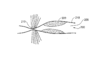
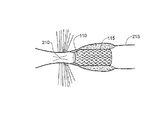
図4Aは、括約筋210近くに閉塞組織(obstructing tissue)220を有した中空器官205の体腔200を示しており、図4Bは、腔200中への配置の後の装置100を示している。ステント103は、腔の開存性を維持するように、器官の壁面に径方向で適合する。ライニング110は、括約筋に最も近い。図4Bでは、括約筋210は、閉じられており、ライニング110は、腔を閉じるという括約筋の機能を干渉することなく、最大限可能な開存性の支持を与えるように、括約筋近くの腔の最小径面に一致している。図4Cは、ライニング110が、括約筋に最も近い腔の大径に従って拡大された開存性の括約筋210を示している。
FIG. 4A shows the
図4Dは、括約筋210近くの腔に位置された後の、図3の装置を示している。ライニング130a,130bは、括約筋210の両側にそれぞれ位置されている。つなぎ網135は、括約筋210を貫通している。ライニング130aは、括約筋の動作を干渉せず、一方、ライニング130bとステント133とが括約筋210から離れるのを防止する固定装置(anchor)として機能している。
FIG. 4D shows the device of FIG. 3 after being positioned in the cavity near the
図5Aは、本発明の他の実施形態に従ったライニングを有する装置300を示している。この装置300は、大動脈のような脈管に配置されるようになっている。また、装置300は、ステント320と一体的で、このステントの両側にそれぞれ位置されている、本発明に従った2つのライニング310a,310bを有している。これら2つのライニング310a,310bは、腔の最大径よりもわずかに大きい非緊張の直径を有している。装置300では、フィラメントの部分間のスペースは、生体適応可能な重膜305で充填されている。図5Bと図5Cとは、大動脈、又は消化器のように、脈壁225を有する腔に配置された後の装置300を示している。図5Bでは、小径を有する腔が示されており、一方、図5Cでは、大径を有する腔が示されている。例えば、腔が動脈である場合、図5Bと図5Cは、弛緩期と収縮期とにそれぞれ対応しているだろう。図5Bと図5Cとに示されているように、ライニング310a,310bは、腔壁が拍動しても、腔壁225と接触したままである。収縮期間に、上流のライニング310aは、動脈壁自体にぴったりと適合するように、動脈の拡張と共に拡大される。膜305の存在は、血液のような流体が、ステント320と腔壁225との間に流れることを防止している。
FIG. 5A shows an
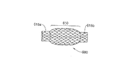
図6は、2つのステント615a,615bと一体的で本発明に従ったライニング610を有する装置600を示している。これら2つのステント615は、ライニング610の両側に位置するように、装置600の両端にそれぞれ位置されている。この装置610は、食道のような蠕動器官内に使用されることができる。ステント615は、蠕動波(peristaltic waves)の伝達を阻止しないように十分に短く設計されている。ライニング610は、蠕動波の伝達を阻止しないように十分柔軟であり、一方、腔に蠕動波を教えないように十分強く設計されている。
FIG. 6 shows a device 600 that is integral with two
図7Aは、ステント710と一体的なライニング705を有する装置700を示している。この装置700は、ライニングを、前に挿入したステントに加えるために使用されることができる。図7Bに示されているように、ステント710は、体腔720中の前に挿入されたステント715の腔内を拡大可能なようなディメンションである。ライニング705は、腔720の最大径よりもわずかに大きい非緊張の直径を有するように構成されている。上述されたように、ライニング705をステント715近くに位置付けることによって、ステントの両端周辺の腔720の壁面に以前存在した急な圧力勾配(sharp pressure gradient)は除去される。スチールの圧力勾配の領域は、ステントの両端で腔を部分的、又は完全に塞ぎ、かつステントの両端で内方に延びた組織を誘導するように知られている。ライニング705は、流体がステント715と腔壁720との間に流入するのを防止するように、可撓性の材料725によって囲まれることができる。
FIG. 7A shows a device 700 having a lining 705 integral with the
Claims (21)
Applications Claiming Priority (3)
| Application Number | Priority Date | Filing Date | Title |
|---|---|---|---|
| US10/292,753 | 2002-11-13 | ||
| US10/292,753 US8282678B2 (en) | 2002-11-13 | 2002-11-13 | Endoluminal lining |
| PCT/IL2003/000942 WO2004043296A1 (en) | 2002-11-13 | 2003-11-11 | Endoluminal lining |
Publications (2)
| Publication Number | Publication Date |
|---|---|
| JP2006506118A true JP2006506118A (en) | 2006-02-23 |
| JP5138868B2 JP5138868B2 (en) | 2013-02-06 |
Family
ID=32229520
Family Applications (1)
| Application Number | Title | Priority Date | Filing Date |
|---|---|---|---|
| JP2004551127A Expired - Fee Related JP5138868B2 (en) | 2002-11-13 | 2003-11-11 | Lumen lining |
Country Status (9)
| Country | Link |
|---|---|
| US (1) | US8282678B2 (en) |
| EP (1) | EP1562516B1 (en) |
| JP (1) | JP5138868B2 (en) |
| AT (1) | ATE438356T1 (en) |
| AU (1) | AU2003282343A1 (en) |
| CA (1) | CA2505417C (en) |
| DE (1) | DE60328707D1 (en) |
| ES (1) | ES2332268T3 (en) |
| WO (1) | WO2004043296A1 (en) |
Cited By (3)
| Publication number | Priority date | Publication date | Assignee | Title |
|---|---|---|---|---|
| JP2010188117A (en) * | 2009-02-19 | 2010-09-02 | Taewoong Medical Co Ltd | Biodegradable stent for preventing reflux of food |
| JP4669480B2 (en) * | 2003-12-09 | 2011-04-13 | ジーアイ・ダイナミックス・インコーポレーテッド | Intestinal sleeve |
| JP2020121054A (en) * | 2019-01-31 | 2020-08-13 | センチュリーメディカル株式会社 | Stent |
Families Citing this family (104)
| Publication number | Priority date | Publication date | Assignee | Title |
|---|---|---|---|---|
| US7837669B2 (en) | 2002-11-01 | 2010-11-23 | Valentx, Inc. | Devices and methods for endolumenal gastrointestinal bypass |
| US7794447B2 (en) | 2002-11-01 | 2010-09-14 | Valentx, Inc. | Gastrointestinal sleeve device and methods for treatment of morbid obesity |
| US9060844B2 (en) | 2002-11-01 | 2015-06-23 | Valentx, Inc. | Apparatus and methods for treatment of morbid obesity |
| US8070743B2 (en) | 2002-11-01 | 2011-12-06 | Valentx, Inc. | Devices and methods for attaching an endolumenal gastrointestinal implant |
| US7766973B2 (en) | 2005-01-19 | 2010-08-03 | Gi Dynamics, Inc. | Eversion resistant sleeves |
| US7678068B2 (en) | 2002-12-02 | 2010-03-16 | Gi Dynamics, Inc. | Atraumatic delivery devices |
| US7695446B2 (en) | 2002-12-02 | 2010-04-13 | Gi Dynamics, Inc. | Methods of treatment using a bariatric sleeve |
| US7122058B2 (en) | 2002-12-02 | 2006-10-17 | Gi Dynamics, Inc. | Anti-obesity devices |
| US7608114B2 (en) | 2002-12-02 | 2009-10-27 | Gi Dynamics, Inc. | Bariatric sleeve |
| US7025791B2 (en) | 2002-12-02 | 2006-04-11 | Gi Dynamics, Inc. | Bariatric sleeve |
| US9333102B2 (en) * | 2003-02-24 | 2016-05-10 | Allium Medical Solutions Ltd. | Stent |
| US20040220682A1 (en) * | 2003-03-28 | 2004-11-04 | Gi Dynamics, Inc. | Anti-obesity devices |
| EP1610719B1 (en) * | 2003-03-28 | 2010-01-13 | GI Dynamics, Inc. | Sleeve for delayed introduction of enzymes into the intestine |
| US8057420B2 (en) | 2003-12-09 | 2011-11-15 | Gi Dynamics, Inc. | Gastrointestinal implant with drawstring |
| US20050131515A1 (en) * | 2003-12-16 | 2005-06-16 | Cully Edward H. | Removable stent-graft |
| US20050228473A1 (en) * | 2004-04-05 | 2005-10-13 | David Brown | Device and method for delivering a treatment to an artery |
| US7758633B2 (en) * | 2004-04-12 | 2010-07-20 | Boston Scientific Scimed, Inc. | Varied diameter vascular graft |
| EP3195829A1 (en) * | 2004-09-17 | 2017-07-26 | GI Dynamics, Inc. | Gastrointestinal achor |
| JP2006141881A (en) * | 2004-11-24 | 2006-06-08 | Tohoku Univ | Peristaltic motion conveying apparatus |
| US7328063B2 (en) * | 2004-11-30 | 2008-02-05 | Cardiac Pacemakers, Inc. | Method and apparatus for arrhythmia classification using atrial signal mapping |
| US7771382B2 (en) * | 2005-01-19 | 2010-08-10 | Gi Dynamics, Inc. | Resistive anti-obesity devices |
| US7976488B2 (en) | 2005-06-08 | 2011-07-12 | Gi Dynamics, Inc. | Gastrointestinal anchor compliance |
| US8518100B2 (en) * | 2005-12-19 | 2013-08-27 | Advanced Cardiovascular Systems, Inc. | Drug eluting stent for the treatment of dialysis graft stenoses |
| AU2006327539A1 (en) | 2005-12-23 | 2007-06-28 | Vysera Biomedical Limited | A medical device suitable for treating reflux from a stomach to an oesophagus |
| US7881797B2 (en) | 2006-04-25 | 2011-02-01 | Valentx, Inc. | Methods and devices for gastrointestinal stimulation |
| US7819836B2 (en) | 2006-06-23 | 2010-10-26 | Gi Dynamics, Inc. | Resistive anti-obesity devices |
| EP2080242A4 (en) * | 2006-09-25 | 2013-10-30 | Valentx Inc | Toposcopic access and delivery devices |
| US20080082154A1 (en) * | 2006-09-28 | 2008-04-03 | Cook Incorporated | Stent Graft Delivery System for Accurate Deployment |
| US8388679B2 (en) | 2007-01-19 | 2013-03-05 | Maquet Cardiovascular Llc | Single continuous piece prosthetic tubular aortic conduit and method for manufacturing the same |
| US8801647B2 (en) | 2007-02-22 | 2014-08-12 | Gi Dynamics, Inc. | Use of a gastrointestinal sleeve to treat bariatric surgery fistulas and leaks |
| WO2008154450A1 (en) * | 2007-06-08 | 2008-12-18 | Valentx, Inc. | Methods and devices for intragastric support of functional or prosthetic gastrointestinal devices |
| US20090012544A1 (en) * | 2007-06-08 | 2009-01-08 | Valen Tx, Inc. | Gastrointestinal bypass sleeve as an adjunct to bariatric surgery |
| WO2009036244A1 (en) * | 2007-09-12 | 2009-03-19 | Endometabolic Solutions, Inc. | Devices and methods for treatment of obesity |
| US8585713B2 (en) | 2007-10-17 | 2013-11-19 | Covidien Lp | Expandable tip assembly for thrombus management |
| US11337714B2 (en) | 2007-10-17 | 2022-05-24 | Covidien Lp | Restoring blood flow and clot removal during acute ischemic stroke |
| US10123803B2 (en) | 2007-10-17 | 2018-11-13 | Covidien Lp | Methods of managing neurovascular obstructions |
| US8088140B2 (en) | 2008-05-19 | 2012-01-03 | Mindframe, Inc. | Blood flow restorative and embolus removal methods |
| WO2009153770A1 (en) | 2008-06-20 | 2009-12-23 | Vysera Biomedical Limited | Esophageal valve |
| US9402707B2 (en) | 2008-07-22 | 2016-08-02 | Neuravi Limited | Clot capture systems and associated methods |
| US20100049307A1 (en) * | 2008-08-25 | 2010-02-25 | Aga Medical Corporation | Stent graft having extended landing area and method for using the same |
| EP2512574B1 (en) | 2009-12-18 | 2017-09-27 | Coloplast A/S | A urological device |
| AU2015218421B2 (en) * | 2009-12-31 | 2017-08-03 | Covidien Lp | Blood flow restoration and thrombus management |
| US9579193B2 (en) * | 2010-09-23 | 2017-02-28 | Transmural Systems Llc | Methods and systems for delivering prostheses using rail techniques |
| EP2629684B1 (en) | 2010-10-22 | 2018-07-25 | Neuravi Limited | Clot engagement and removal system |
| US8992410B2 (en) | 2010-11-03 | 2015-03-31 | Vysera Biomedical Limited | Urological device |
| US8696741B2 (en) | 2010-12-23 | 2014-04-15 | Maquet Cardiovascular Llc | Woven prosthesis and method for manufacturing the same |
| US9839540B2 (en) | 2011-01-14 | 2017-12-12 | W. L. Gore & Associates, Inc. | Stent |
| US11259824B2 (en) | 2011-03-09 | 2022-03-01 | Neuravi Limited | Clot retrieval device for removing occlusive clot from a blood vessel |
| US12076037B2 (en) | 2011-03-09 | 2024-09-03 | Neuravi Limited | Systems and methods to restore perfusion to a vessel |
| EP3871617A1 (en) | 2011-03-09 | 2021-09-01 | Neuravi Limited | A clot retrieval device for removing occlusive clot from a blood vessel |
| CA2858301C (en) * | 2011-12-19 | 2021-01-12 | Vysera Biomedical Limited | A luminal prosthesis and a gastrointestinal implant device |
| JP6179949B2 (en) * | 2012-01-30 | 2017-08-16 | 川澄化学工業株式会社 | Biliary stent |
| US9681975B2 (en) | 2012-05-31 | 2017-06-20 | Valentx, Inc. | Devices and methods for gastrointestinal bypass |
| US9451960B2 (en) | 2012-05-31 | 2016-09-27 | Valentx, Inc. | Devices and methods for gastrointestinal bypass |
| US9050168B2 (en) | 2012-05-31 | 2015-06-09 | Valentx, Inc. | Devices and methods for gastrointestinal bypass |
| BR112015000384A2 (en) | 2012-07-13 | 2017-06-27 | Gi Dynamics Inc | gastrointestinal implant device, treatment method and method of removing gastrointestinal implant |
| US9931193B2 (en) | 2012-11-13 | 2018-04-03 | W. L. Gore & Associates, Inc. | Elastic stent graft |
| US9642635B2 (en) | 2013-03-13 | 2017-05-09 | Neuravi Limited | Clot removal device |
| US9757264B2 (en) | 2013-03-13 | 2017-09-12 | Valentx, Inc. | Devices and methods for gastrointestinal bypass |
| US9433429B2 (en) | 2013-03-14 | 2016-09-06 | Neuravi Limited | Clot retrieval devices |
| EP3536253B1 (en) | 2013-03-14 | 2024-04-10 | Neuravi Limited | Devices for removal of acute blockages from blood vessels |
| CN109157304B (en) | 2013-03-14 | 2021-12-31 | 尼尔拉维有限公司 | A clot retrieval device for removing a clogged clot from a blood vessel |
| US10842918B2 (en) | 2013-12-05 | 2020-11-24 | W.L. Gore & Associates, Inc. | Length extensible implantable device and methods for making such devices |
| US10285720B2 (en) | 2014-03-11 | 2019-05-14 | Neuravi Limited | Clot retrieval system for removing occlusive clot from a blood vessel |
| WO2015138763A1 (en) * | 2014-03-14 | 2015-09-17 | The Board Of Trustees Of The Leland Stanford Junior University | Indwelling body lumen expander |
| WO2015189354A1 (en) | 2014-06-13 | 2015-12-17 | Neuravi Limited | Devices for removal of acute blockages from blood vessels |
| US10792056B2 (en) | 2014-06-13 | 2020-10-06 | Neuravi Limited | Devices and methods for removal of acute blockages from blood vessels |
| CN106659563B (en) | 2014-06-26 | 2019-03-08 | 波士顿科学国际有限公司 | Medical device and method for preventing that bile reflux occurs after bariatric surgery |
| US10265086B2 (en) | 2014-06-30 | 2019-04-23 | Neuravi Limited | System for removing a clot from a blood vessel |
| US11253278B2 (en) | 2014-11-26 | 2022-02-22 | Neuravi Limited | Clot retrieval system for removing occlusive clot from a blood vessel |
| JP2017535352A (en) | 2014-11-26 | 2017-11-30 | ニューラヴィ・リミテッド | Clot collection device for removing obstructive clots from blood vessels |
| US10617435B2 (en) | 2014-11-26 | 2020-04-14 | Neuravi Limited | Clot retrieval device for removing clot from a blood vessel |
| US20160354193A1 (en) * | 2015-06-05 | 2016-12-08 | Bruce Gordon McNAB, SR. | Prostatic urethral stent |
| JP7086935B2 (en) | 2016-08-17 | 2022-06-20 | ニューラヴィ・リミテッド | Thrombus recovery system for removing thromboangiitis obliterans from blood vessels |
| ES2972736T3 (en) | 2016-09-06 | 2024-06-14 | Neuravi Ltd | Clot retrieval device to remove clots from a blood vessel |
| EP3551140A4 (en) | 2016-12-09 | 2020-07-08 | Zenflow, Inc. | Systems, devices, and methods for the accurate deployment of an implant in the prostatic urethra |
| US10842498B2 (en) | 2018-09-13 | 2020-11-24 | Neuravi Limited | Systems and methods of restoring perfusion to a vessel |
| US11406416B2 (en) | 2018-10-02 | 2022-08-09 | Neuravi Limited | Joint assembly for vasculature obstruction capture device |
| JP7483409B2 (en) | 2019-03-04 | 2024-05-15 | ニューラヴィ・リミテッド | Actuated Clot Retrieval Catheter |
| US11529495B2 (en) | 2019-09-11 | 2022-12-20 | Neuravi Limited | Expandable mouth catheter |
| US11712231B2 (en) | 2019-10-29 | 2023-08-01 | Neuravi Limited | Proximal locking assembly design for dual stent mechanical thrombectomy device |
| EP4061292A4 (en) | 2019-11-19 | 2023-12-27 | Zenflow, Inc. | Systems, devices, and methods for the accurate deployment and imaging of an implant in the prostatic urethra |
| US11779364B2 (en) | 2019-11-27 | 2023-10-10 | Neuravi Limited | Actuated expandable mouth thrombectomy catheter |
| US11839725B2 (en) | 2019-11-27 | 2023-12-12 | Neuravi Limited | Clot retrieval device with outer sheath and inner catheter |
| US11517340B2 (en) | 2019-12-03 | 2022-12-06 | Neuravi Limited | Stentriever devices for removing an occlusive clot from a vessel and methods thereof |
| US11633198B2 (en) | 2020-03-05 | 2023-04-25 | Neuravi Limited | Catheter proximal joint |
| US11944327B2 (en) | 2020-03-05 | 2024-04-02 | Neuravi Limited | Expandable mouth aspirating clot retrieval catheter |
| US11883043B2 (en) | 2020-03-31 | 2024-01-30 | DePuy Synthes Products, Inc. | Catheter funnel extension |
| US11759217B2 (en) | 2020-04-07 | 2023-09-19 | Neuravi Limited | Catheter tubular support |
| US11730501B2 (en) | 2020-04-17 | 2023-08-22 | Neuravi Limited | Floating clot retrieval device for removing clots from a blood vessel |
| US11717308B2 (en) | 2020-04-17 | 2023-08-08 | Neuravi Limited | Clot retrieval device for removing heterogeneous clots from a blood vessel |
| US11871946B2 (en) | 2020-04-17 | 2024-01-16 | Neuravi Limited | Clot retrieval device for removing clot from a blood vessel |
| US11737771B2 (en) | 2020-06-18 | 2023-08-29 | Neuravi Limited | Dual channel thrombectomy device |
| US11937836B2 (en) | 2020-06-22 | 2024-03-26 | Neuravi Limited | Clot retrieval system with expandable clot engaging framework |
| US11395669B2 (en) | 2020-06-23 | 2022-07-26 | Neuravi Limited | Clot retrieval device with flexible collapsible frame |
| US11439418B2 (en) | 2020-06-23 | 2022-09-13 | Neuravi Limited | Clot retrieval device for removing clot from a blood vessel |
| US11864781B2 (en) | 2020-09-23 | 2024-01-09 | Neuravi Limited | Rotating frame thrombectomy device |
| US11937837B2 (en) | 2020-12-29 | 2024-03-26 | Neuravi Limited | Fibrin rich / soft clot mechanical thrombectomy device |
| US12029442B2 (en) | 2021-01-14 | 2024-07-09 | Neuravi Limited | Systems and methods for a dual elongated member clot retrieval apparatus |
| US11872354B2 (en) | 2021-02-24 | 2024-01-16 | Neuravi Limited | Flexible catheter shaft frame with seam |
| US12064130B2 (en) | 2021-03-18 | 2024-08-20 | Neuravi Limited | Vascular obstruction retrieval device having sliding cages pinch mechanism |
| US11974764B2 (en) | 2021-06-04 | 2024-05-07 | Neuravi Limited | Self-orienting rotating stentriever pinching cells |
| US11937839B2 (en) | 2021-09-28 | 2024-03-26 | Neuravi Limited | Catheter with electrically actuated expandable mouth |
| US12011186B2 (en) | 2021-10-28 | 2024-06-18 | Neuravi Limited | Bevel tip expandable mouth catheter with reinforcing ring |
Citations (21)
| Publication number | Priority date | Publication date | Assignee | Title |
|---|---|---|---|---|
| JPH01145076A (en) * | 1987-10-19 | 1989-06-07 | Medtronic Inc | Stent radially expansible in blood vessel and transplantation method |
| JPH05507215A (en) * | 1990-06-28 | 1993-10-21 | シュナイダー・(ユーエスエイ)・インコーポレーテッド | Fixation device in body cavity |
| JPH067454A (en) * | 1992-03-25 | 1994-01-18 | Cook Inc | Stent for vessel |
| US5383892A (en) * | 1991-11-08 | 1995-01-24 | Meadox France | Stent for transluminal implantation |
| JPH07136282A (en) * | 1993-06-24 | 1995-05-30 | Synthelabo Sa | Tube introducing prosthesis |
| JPH08502428A (en) * | 1992-10-13 | 1996-03-19 | ボストン・サイエンティフィック・コーポレイション | Stent for body lumen showing peristaltic motion |
| JPH08509394A (en) * | 1993-02-19 | 1996-10-08 | デボネ,マリア | Prostheses for treatment of natural lumens or ducts such as prostheses in the urethra |
| JPH08509899A (en) * | 1994-04-01 | 1996-10-22 | プログラフト メディカル,インコーポレイテッド | Self-expandable stents and stent-grafts and methods of use thereof |
| JPH08336597A (en) * | 1994-11-15 | 1996-12-24 | Advanced Cardeovascular Syst Inc | Intratubal stent for installation of transplantation piece |
| JPH0999058A (en) * | 1995-07-10 | 1997-04-15 | Devonec Marian | Separatable catheter |
| FR2745172A1 (en) * | 1996-02-26 | 1997-08-29 | Braun Celsa Sa | Variable=diameter prosthetic implant, especially for blood vessel |
| JPH09285548A (en) * | 1996-04-10 | 1997-11-04 | Advanced Cardeovascular Syst Inc | Stent having its structural strength changed along longitudinal direction |
| US5741333A (en) * | 1995-04-12 | 1998-04-21 | Corvita Corporation | Self-expanding stent for a medical device to be introduced into a cavity of a body |
| JPH10506297A (en) * | 1994-07-13 | 1998-06-23 | デヴォネ,マリアン | Therapeutic device for selective cell reduction treatment of a disorder in a natural lumen or passage in a human or animal body |
| WO1998043695A1 (en) * | 1997-03-31 | 1998-10-08 | Kabushikikaisha Igaki Iryo Sekkei | Stent for vessels |
| JPH11347133A (en) * | 1998-05-28 | 1999-12-21 | Medtronic Ave Inc | Endoluminal supporting assembly with end cap |
| JP2000312721A (en) * | 1999-04-08 | 2000-11-14 | Cordis Corp | Wall thickness variable stent |
| JP2002500533A (en) * | 1997-05-22 | 2002-01-08 | ボストン サイエンティフィック リミテッド | Stent with variable expansion force |
| JP2002531169A (en) * | 1998-08-31 | 2002-09-24 | ウイルソンークック メディカル インク. | Anti-reflux esophageal prosthesis |
| JP2002532136A (en) * | 1998-12-18 | 2002-10-02 | クック インコーポレイティド | Cannula tent |
| WO2003017882A2 (en) * | 2001-08-27 | 2003-03-06 | Synecor, Llc | Satiation devices and methods |
Family Cites Families (19)
| Publication number | Priority date | Publication date | Assignee | Title |
|---|---|---|---|---|
| US5123917A (en) * | 1990-04-27 | 1992-06-23 | Lee Peter Y | Expandable intraluminal vascular graft |
| US5662713A (en) * | 1991-10-09 | 1997-09-02 | Boston Scientific Corporation | Medical stents for body lumens exhibiting peristaltic motion |
| US5876445A (en) * | 1991-10-09 | 1999-03-02 | Boston Scientific Corporation | Medical stents for body lumens exhibiting peristaltic motion |
| US5683448A (en) * | 1992-02-21 | 1997-11-04 | Boston Scientific Technology, Inc. | Intraluminal stent and graft |
| US5282823A (en) * | 1992-03-19 | 1994-02-01 | Medtronic, Inc. | Intravascular radially expandable stent |
| US6576008B2 (en) * | 1993-02-19 | 2003-06-10 | Scimed Life Systems, Inc. | Methods and device for inserting and withdrawing a two piece stent across a constricting anatomic structure |
| SE505436C2 (en) * | 1993-04-27 | 1997-08-25 | Ams Medinvent Sa | prostatic stent |
| US5549663A (en) * | 1994-03-09 | 1996-08-27 | Cordis Corporation | Endoprosthesis having graft member and exposed welded end junctions, method and procedure |
| CH688174A5 (en) * | 1995-03-28 | 1997-06-13 | Norman Godin | Prosthesis to oppose the gastric reflux into the esophagus. |
| US5830179A (en) * | 1996-04-09 | 1998-11-03 | Endocare, Inc. | Urological stent therapy system and method |
| WO1998020810A1 (en) * | 1996-11-12 | 1998-05-22 | Medtronic, Inc. | Flexible, radially expansible luminal prostheses |
| US5938697A (en) * | 1998-03-04 | 1999-08-17 | Scimed Life Systems, Inc. | Stent having variable properties |
| WO1999056663A2 (en) | 1998-05-05 | 1999-11-11 | Scimed Life Systems, Inc. | Stent with smooth ends |
| US6042597A (en) * | 1998-10-23 | 2000-03-28 | Scimed Life Systems, Inc. | Helical stent design |
| US6273911B1 (en) * | 1999-04-22 | 2001-08-14 | Advanced Cardiovascular Systems, Inc. | Variable strength stent |
| US7169175B2 (en) | 2000-05-22 | 2007-01-30 | Orbusneich Medical, Inc. | Self-expanding stent |
| GB0311221D0 (en) | 2003-05-15 | 2003-06-18 | Orthogem Ltd | Biomaterial |
| US7857834B2 (en) | 2004-06-14 | 2010-12-28 | Zimmer Spine, Inc. | Spinal implant fixation assembly |
| JP2008509899A (en) | 2004-08-12 | 2008-04-03 | グラクソスミスクライン・イストラジヴァッキ・センタル・ザグレブ・ドルズバ・ゼー・オメイェノ・オドゴヴォルノスティオ | Use of cell-specific conjugates for the treatment of inflammatory diseases of the gastrointestinal tract |
-
2002
- 2002-11-13 US US10/292,753 patent/US8282678B2/en not_active Expired - Lifetime
-
2003
- 2003-11-11 AT AT03773957T patent/ATE438356T1/en not_active IP Right Cessation
- 2003-11-11 WO PCT/IL2003/000942 patent/WO2004043296A1/en active Application Filing
- 2003-11-11 CA CA2505417A patent/CA2505417C/en not_active Expired - Fee Related
- 2003-11-11 DE DE60328707T patent/DE60328707D1/en not_active Expired - Lifetime
- 2003-11-11 AU AU2003282343A patent/AU2003282343A1/en not_active Abandoned
- 2003-11-11 JP JP2004551127A patent/JP5138868B2/en not_active Expired - Fee Related
- 2003-11-11 EP EP03773957A patent/EP1562516B1/en not_active Expired - Lifetime
- 2003-11-11 ES ES03773957T patent/ES2332268T3/en not_active Expired - Lifetime
Patent Citations (21)
| Publication number | Priority date | Publication date | Assignee | Title |
|---|---|---|---|---|
| JPH01145076A (en) * | 1987-10-19 | 1989-06-07 | Medtronic Inc | Stent radially expansible in blood vessel and transplantation method |
| JPH05507215A (en) * | 1990-06-28 | 1993-10-21 | シュナイダー・(ユーエスエイ)・インコーポレーテッド | Fixation device in body cavity |
| US5383892A (en) * | 1991-11-08 | 1995-01-24 | Meadox France | Stent for transluminal implantation |
| JPH067454A (en) * | 1992-03-25 | 1994-01-18 | Cook Inc | Stent for vessel |
| JPH08502428A (en) * | 1992-10-13 | 1996-03-19 | ボストン・サイエンティフィック・コーポレイション | Stent for body lumen showing peristaltic motion |
| JPH08509394A (en) * | 1993-02-19 | 1996-10-08 | デボネ,マリア | Prostheses for treatment of natural lumens or ducts such as prostheses in the urethra |
| JPH07136282A (en) * | 1993-06-24 | 1995-05-30 | Synthelabo Sa | Tube introducing prosthesis |
| JPH08509899A (en) * | 1994-04-01 | 1996-10-22 | プログラフト メディカル,インコーポレイテッド | Self-expandable stents and stent-grafts and methods of use thereof |
| JPH10506297A (en) * | 1994-07-13 | 1998-06-23 | デヴォネ,マリアン | Therapeutic device for selective cell reduction treatment of a disorder in a natural lumen or passage in a human or animal body |
| JPH08336597A (en) * | 1994-11-15 | 1996-12-24 | Advanced Cardeovascular Syst Inc | Intratubal stent for installation of transplantation piece |
| US5741333A (en) * | 1995-04-12 | 1998-04-21 | Corvita Corporation | Self-expanding stent for a medical device to be introduced into a cavity of a body |
| JPH0999058A (en) * | 1995-07-10 | 1997-04-15 | Devonec Marian | Separatable catheter |
| FR2745172A1 (en) * | 1996-02-26 | 1997-08-29 | Braun Celsa Sa | Variable=diameter prosthetic implant, especially for blood vessel |
| JPH09285548A (en) * | 1996-04-10 | 1997-11-04 | Advanced Cardeovascular Syst Inc | Stent having its structural strength changed along longitudinal direction |
| WO1998043695A1 (en) * | 1997-03-31 | 1998-10-08 | Kabushikikaisha Igaki Iryo Sekkei | Stent for vessels |
| JP2002500533A (en) * | 1997-05-22 | 2002-01-08 | ボストン サイエンティフィック リミテッド | Stent with variable expansion force |
| JPH11347133A (en) * | 1998-05-28 | 1999-12-21 | Medtronic Ave Inc | Endoluminal supporting assembly with end cap |
| JP2002531169A (en) * | 1998-08-31 | 2002-09-24 | ウイルソンークック メディカル インク. | Anti-reflux esophageal prosthesis |
| JP2002532136A (en) * | 1998-12-18 | 2002-10-02 | クック インコーポレイティド | Cannula tent |
| JP2000312721A (en) * | 1999-04-08 | 2000-11-14 | Cordis Corp | Wall thickness variable stent |
| WO2003017882A2 (en) * | 2001-08-27 | 2003-03-06 | Synecor, Llc | Satiation devices and methods |
Cited By (3)
| Publication number | Priority date | Publication date | Assignee | Title |
|---|---|---|---|---|
| JP4669480B2 (en) * | 2003-12-09 | 2011-04-13 | ジーアイ・ダイナミックス・インコーポレーテッド | Intestinal sleeve |
| JP2010188117A (en) * | 2009-02-19 | 2010-09-02 | Taewoong Medical Co Ltd | Biodegradable stent for preventing reflux of food |
| JP2020121054A (en) * | 2019-01-31 | 2020-08-13 | センチュリーメディカル株式会社 | Stent |
Also Published As
| Publication number | Publication date |
|---|---|
| DE60328707D1 (en) | 2009-09-17 |
| JP5138868B2 (en) | 2013-02-06 |
| WO2004043296A1 (en) | 2004-05-27 |
| US20040093065A1 (en) | 2004-05-13 |
| ATE438356T1 (en) | 2009-08-15 |
| EP1562516B1 (en) | 2009-08-05 |
| CA2505417C (en) | 2012-05-01 |
| ES2332268T3 (en) | 2010-02-01 |
| EP1562516A1 (en) | 2005-08-17 |
| AU2003282343A1 (en) | 2004-06-03 |
| CA2505417A1 (en) | 2004-05-27 |
| US8282678B2 (en) | 2012-10-09 |
Similar Documents
| Publication | Publication Date | Title |
|---|---|---|
| JP5138868B2 (en) | Lumen lining | |
| US11564818B2 (en) | Vascular implant | |
| US11241304B2 (en) | Method for fluid flow through body passages | |
| JP5719327B2 (en) | Helical stent | |
| US5876445A (en) | Medical stents for body lumens exhibiting peristaltic motion | |
| US6270524B1 (en) | Flexible, radially expansible luminal prostheses | |
| US7318835B2 (en) | Endoluminal prosthesis having expandable graft sections | |
| USRE39335E1 (en) | Surgical graft/stent system | |
| US6505654B1 (en) | Medical stents for body lumens exhibiting peristaltic motion | |
| WO2003034948A1 (en) | Prostheses for curved lumens | |
| JP2003325672A (en) | Device for expansion | |
| IL168532A (en) | Device for insertion into a body lumen | |
| AU2002348080B2 (en) | Prostheses for curved lumens | |
| JP2004089382A (en) | Stent graft |
Legal Events
| Date | Code | Title | Description |
|---|---|---|---|
| A621 | Written request for application examination |
Free format text: JAPANESE INTERMEDIATE CODE: A621 Effective date: 20061013 |
|
| A131 | Notification of reasons for refusal |
Free format text: JAPANESE INTERMEDIATE CODE: A131 Effective date: 20090825 |
|
| A601 | Written request for extension of time |
Free format text: JAPANESE INTERMEDIATE CODE: A601 Effective date: 20091125 |
|
| A602 | Written permission of extension of time |
Free format text: JAPANESE INTERMEDIATE CODE: A602 Effective date: 20091202 |
|
| A521 | Request for written amendment filed |
Free format text: JAPANESE INTERMEDIATE CODE: A523 Effective date: 20091225 |
|
| A131 | Notification of reasons for refusal |
Free format text: JAPANESE INTERMEDIATE CODE: A131 Effective date: 20100330 |
|
| A521 | Request for written amendment filed |
Free format text: JAPANESE INTERMEDIATE CODE: A523 Effective date: 20100611 |
|
| A131 | Notification of reasons for refusal |
Free format text: JAPANESE INTERMEDIATE CODE: A131 Effective date: 20110104 |
|
| A601 | Written request for extension of time |
Free format text: JAPANESE INTERMEDIATE CODE: A601 Effective date: 20110404 |
|
| A602 | Written permission of extension of time |
Free format text: JAPANESE INTERMEDIATE CODE: A602 Effective date: 20110411 |
|
| A601 | Written request for extension of time |
Free format text: JAPANESE INTERMEDIATE CODE: A601 Effective date: 20110506 |
|
| A602 | Written permission of extension of time |
Free format text: JAPANESE INTERMEDIATE CODE: A602 Effective date: 20110513 |
|
| A711 | Notification of change in applicant |
Free format text: JAPANESE INTERMEDIATE CODE: A711 Effective date: 20110526 |
|
| A601 | Written request for extension of time |
Free format text: JAPANESE INTERMEDIATE CODE: A601 Effective date: 20110606 |
|
| A602 | Written permission of extension of time |
Free format text: JAPANESE INTERMEDIATE CODE: A602 Effective date: 20110628 |
|
| A521 | Request for written amendment filed |
Free format text: JAPANESE INTERMEDIATE CODE: A523 Effective date: 20110704 |
|
| A02 | Decision of refusal |
Free format text: JAPANESE INTERMEDIATE CODE: A02 Effective date: 20120131 |
|
| A01 | Written decision to grant a patent or to grant a registration (utility model) |
Free format text: JAPANESE INTERMEDIATE CODE: A01 |
|
| A61 | First payment of annual fees (during grant procedure) |
Free format text: JAPANESE INTERMEDIATE CODE: A61 Effective date: 20121115 |
|
| R150 | Certificate of patent or registration of utility model |
Ref document number: 5138868 Country of ref document: JP Free format text: JAPANESE INTERMEDIATE CODE: R150 Free format text: JAPANESE INTERMEDIATE CODE: R150 |
|
| FPAY | Renewal fee payment (event date is renewal date of database) |
Free format text: PAYMENT UNTIL: 20151122 Year of fee payment: 3 |
|
| R250 | Receipt of annual fees |
Free format text: JAPANESE INTERMEDIATE CODE: R250 |
|
| R250 | Receipt of annual fees |
Free format text: JAPANESE INTERMEDIATE CODE: R250 |
|
| R250 | Receipt of annual fees |
Free format text: JAPANESE INTERMEDIATE CODE: R250 |
|
| S303 | Written request for registration of pledge or change of pledge |
Free format text: JAPANESE INTERMEDIATE CODE: R316303 |
|
| S531 | Written request for registration of change of domicile |
Free format text: JAPANESE INTERMEDIATE CODE: R313531 |
|
| R350 | Written notification of registration of transfer |
Free format text: JAPANESE INTERMEDIATE CODE: R350 |
|
| R250 | Receipt of annual fees |
Free format text: JAPANESE INTERMEDIATE CODE: R250 |
|
| R250 | Receipt of annual fees |
Free format text: JAPANESE INTERMEDIATE CODE: R250 |
|
| S111 | Request for change of ownership or part of ownership |
Free format text: JAPANESE INTERMEDIATE CODE: R313113 |
|
| R350 | Written notification of registration of transfer |
Free format text: JAPANESE INTERMEDIATE CODE: R350 |
|
| R250 | Receipt of annual fees |
Free format text: JAPANESE INTERMEDIATE CODE: R250 |
|
| R250 | Receipt of annual fees |
Free format text: JAPANESE INTERMEDIATE CODE: R250 |
|
| LAPS | Cancellation because of no payment of annual fees |


