4BSU

Structure of the ectodomain of LGR5 in complex with R-spondin-1 (Fu1Fu2) in C2 crystal form


Experimental Data Snapshot

  • Method: X-RAY DIFFRACTION
  • Resolution: 3.20 Å
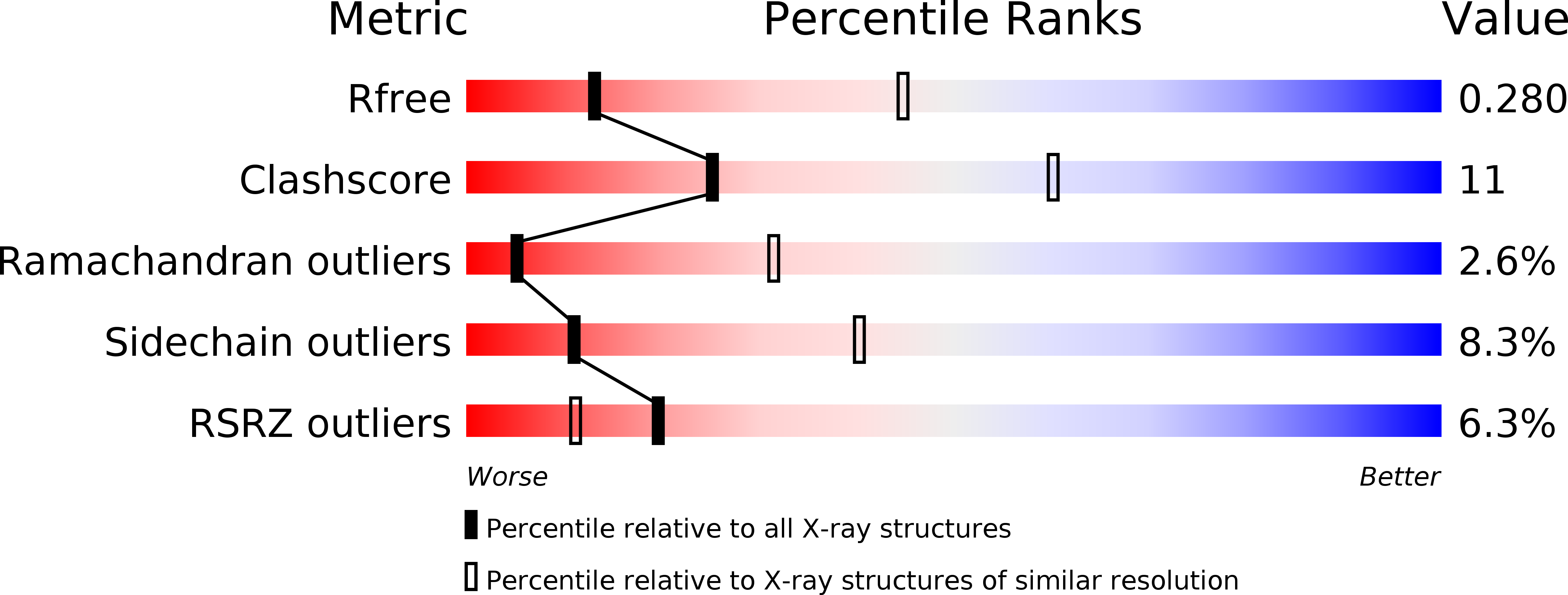
  • R-Value Free: 0.287 
  • R-Value Work: 0.240 
  • R-Value Observed: 0.242 

wwPDB Validation   3D Report Full Report


Ligand Structure Quality Assessment 


This is version 2.1 of the entry. See complete history


Literature

Structure of Stem Cell Growth Factor R-Spondin 1 in Complex with the Ectodomain of its Receptor Lgr5.

Peng, W.C.De Lau, W.Forneris, F.Granneman, J.C.M.Huch, M.Clevers, H.Gros, P.

(2013) Cell Rep 3: 1885

  • DOI: https://doi.org/10.1016/j.celrep.2013.06.009
  • Primary Citation of Related Structures:  
    4BSO, 4BSP, 4BSR, 4BSS, 4BST, 4BSU

  • PubMed Abstract: 

    Leucine-rich repeat-containing G protein-coupled receptors 4-6 (LGR4-LGR6) are receptors for R-spondins, potent Wnt agonists that exert profound trophic effects on Wnt-driven stem cells compartments. We present crystal structures of a signaling-competent fragment of R-spondin 1 (Rspo1) at a resolution of 2.0 Å and its complex with the LGR5 ectodomain at a resolution of 3.2 Å. Ecto-LGR5 binds Rspo1 at its concave leucine-rich-repeat (LRR) surface, forming a dimeric 2:2 complex. Fully conserved residues on LGR4-LGR6 explain promiscuous binding of R-spondins. A phenylalanine clamp formed by Rspo1 Phe106 and Phe110 pinches Ala190 of LGR5 and is critical for binding. Mutations related to congenital anonychia reduce signaling, but not binding of Rspo1 to LGR5. Furthermore, antibody binding to the extended loop of the C-terminal LRR cap of LGR5 activates signaling in a ligand-independent manner. Thus, our data reveal binding of R-spondins to conserved sites on LGR4-LGR6 and, in analogy to FSHR and related receptors, suggest a direct signaling role for LGR4-LGR6 in addition to its formation of Wnt receptor and coreceptor complexes.


  • Organizational Affiliation

    Crystal and Structural Chemistry, Bijvoet Center for Biomolecular Research, Department of Chemistry, Faculty of Science, Utrecht University, Padualaan 8, 3584 CH Utrecht, The Netherlands.


Macromolecules
Find similar proteins by:  (by identity cutoff)  |  3D Structure
Entity ID: 1
MoleculeChains Sequence LengthOrganismDetailsImage
LEUCINE-RICH REPEAT-CONTAINING G-PROTEIN COUPLED RECEPTOR 5
A, B, E, F
539Homo sapiensMutation(s): 0 
UniProt & NIH Common Fund Data Resources
Find proteins for O75473 (Homo sapiens)
Explore O75473 
Go to UniProtKB:  O75473
PHAROS:  O75473
GTEx:  ENSG00000139292 
Entity Groups  
Sequence Clusters30% Identity50% Identity70% Identity90% Identity95% Identity100% Identity
UniProt GroupO75473
Glycosylation
Glycosylation Sites: 2Go to GlyGen: O75473-1
Sequence Annotations
Expand
  • Reference Sequence
Find similar proteins by:  (by identity cutoff)  |  3D Structure
Entity ID: 2
MoleculeChains Sequence LengthOrganismDetailsImage
R-SPONDIN-1
C, D, G, H
126Homo sapiensMutation(s): 0 
UniProt & NIH Common Fund Data Resources
Find proteins for Q2MKA7 (Homo sapiens)
Explore Q2MKA7 
Go to UniProtKB:  Q2MKA7
PHAROS:  Q2MKA7
GTEx:  ENSG00000169218 
Entity Groups  
Sequence Clusters30% Identity50% Identity70% Identity90% Identity95% Identity100% Identity
UniProt GroupQ2MKA7
Sequence Annotations
Expand
  • Reference Sequence
Oligosaccharides

Help

Entity ID: 3
MoleculeChains Length2D Diagram Glycosylation3D Interactions
2-acetamido-2-deoxy-beta-D-glucopyranose-(1-4)-2-acetamido-2-deoxy-beta-D-glucopyranose
I
2N-Glycosylation
Glycosylation Resources
GlyTouCan:  G42666HT
GlyCosmos:  G42666HT
GlyGen:  G42666HT
Entity ID: 4
MoleculeChains Length2D Diagram Glycosylation3D Interactions
beta-D-mannopyranose-(1-4)-2-acetamido-2-deoxy-beta-D-glucopyranose-(1-4)-2-acetamido-2-deoxy-beta-D-glucopyranose
J, K, L
3N-Glycosylation
Glycosylation Resources
GlyTouCan:  G15407YE
GlyCosmos:  G15407YE
GlyGen:  G15407YE
Experimental Data & Validation

Experimental Data

  • Method: X-RAY DIFFRACTION
  • Resolution: 3.20 Å
  • R-Value Free: 0.287 
  • R-Value Work: 0.240 
  • R-Value Observed: 0.242 
  • Space Group: C 1 2 1
Unit Cell:
Length ( Å )Angle ( ˚ )
a = 495.13α = 90
b = 62.15β = 91.19
c = 104.869γ = 90
Software Package:
Software NamePurpose
PHENIXrefinement

Structure Validation

View Full Validation Report


Ligand Structure Quality Assessment 


Entry History 

Deposition Data

Revision History  (Full details and data files)

  • Version 1.0: 2013-06-26
    Type: Initial release
  • Version 1.1: 2013-07-24
    Changes: Database references
  • Version 2.0: 2020-07-29
    Type: Remediation
    Reason: Carbohydrate remediation
    Changes: Advisory, Atomic model, Data collection, Derived calculations, Other, Structure summary
  • Version 2.1: 2024-11-06
    Changes: Advisory, Data collection, Database references, Structure summary