pediagenosis: Cell
Article Update
Loading...
Showing posts with label Cell. Show all posts

Friday, September 29, 2023

The Nucleus

The Nucleus


The Nucleus

The nucleus of the cell appears as a rounded or elongated structure situated near the center of the cell (see Fig. 4.1). All eukaryotic cells have at least one nucleus (prokaryotic cells, such as bacteria, lack a nucleus and nuclear membrane). Some cells contain more than one nucleus; osteoclasts (a type of bone cell) typically contain 12 nuclei or more. The platelet-producing cell, the megakaryocyte, has only one nucleus but usually contains 16 times the normal chromatin amount.
The nucleus can be regarded as the control center for the cell. It contains the deoxyribonucleic acid (DNA) that is essential to the cell because its genes encode the information necessary for the synthesis of proteins that the cell must produce to stay alive. These proteins include structural proteins and enzymes used to synthesize other substances, including carbohydrates and lipids. Genes also represent the individual units of inheritance that transmit information from one generation to another. The nucleus also is the site for the synthesis of the three types of ribonucleic acid (messenger RNA [mRNA], ribosomal RNA [rRNA], and transfer RNA [tRNA]) that move to the cytoplasm and carry out the actual synthesis of proteins. mRNA copies and carries the DNA instructions for protein synthesis to the cytoplasm; rRNA is the site of protein synthesis; and tRNA transports amino acids to the site of proteins synthesis for incorporation into the protein being synthesized.
The Nucleus

Chromatin is the term denoting the complex structure of DNA and DNA-associated proteins dispersed in the nuclear matrix. Depending on its transcriptional activity, chromatin may be condensed as an inactive form of chromatin called heterochromatin or extended as a more active form called euchromatin. Because heterochromatic regions of the nucleus stain more intensely than regions consisting of euchromatin, nuclear staining can be a guide to cell activity. Evidence suggests the importance that alteration in the chromatin, along with DNA hypermethylation, in neoplastic progression. It seems both of these processes work symbiotically not separately in their role regarding cancer.

Tuesday, September 26, 2023

Blood

Blood


Blood

Blood

The primary function of blood is to deliver O2 and energy to the tissues, and remove CO2 and waste products. It is also important for the defence and immune systems, regulation of temperature, and transport of hormones and signalling molecules between tissues. Blood consists of plasma (Chapter 2) and blood cells. Red blood cells contain haemoglobin and transport respiratory gases (Chapter 28), whereas white cells form part of the defence system (Chapter 10). In adults, all blood cells are produced in the red bone marrow. Normal values for cell counts, haemoglobin and proportion of blood volume due to red cells (haematocrit or packed cell volume; estimated by centrifuging a blood sample) are shown in Figure 8a. Platelets are discussed in Chapter 9.
Biological Electricity

Biological Electricity


Biological Electricity

Biological Electricity

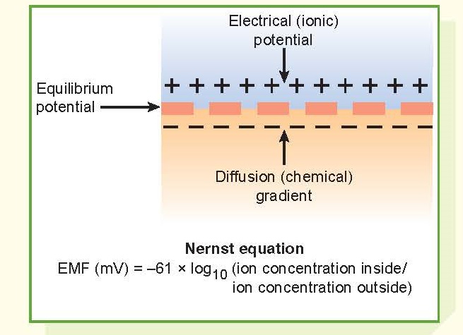
Electrical events in biological tissues are caused by the movement of ions across the membrane. A potential difference exists across the membranes of all cells (membrane potential, Em), but only excitable tissues can generate action potentials (transient depolarization of a cell as a result of ion channel activity). Action potentials transmit information in nerve cells (Chapter 6) and trigger contractions in muscle cells (Chapter 12). Cell membranes are electrically polarized so that the inside is negative relative to the outside. In excitable tissues, resting Em  is usually between –60 and –90 mV.

Sunday, March 26, 2023

Genetic Imprinting

Genetic Imprinting


Genetic Imprinting

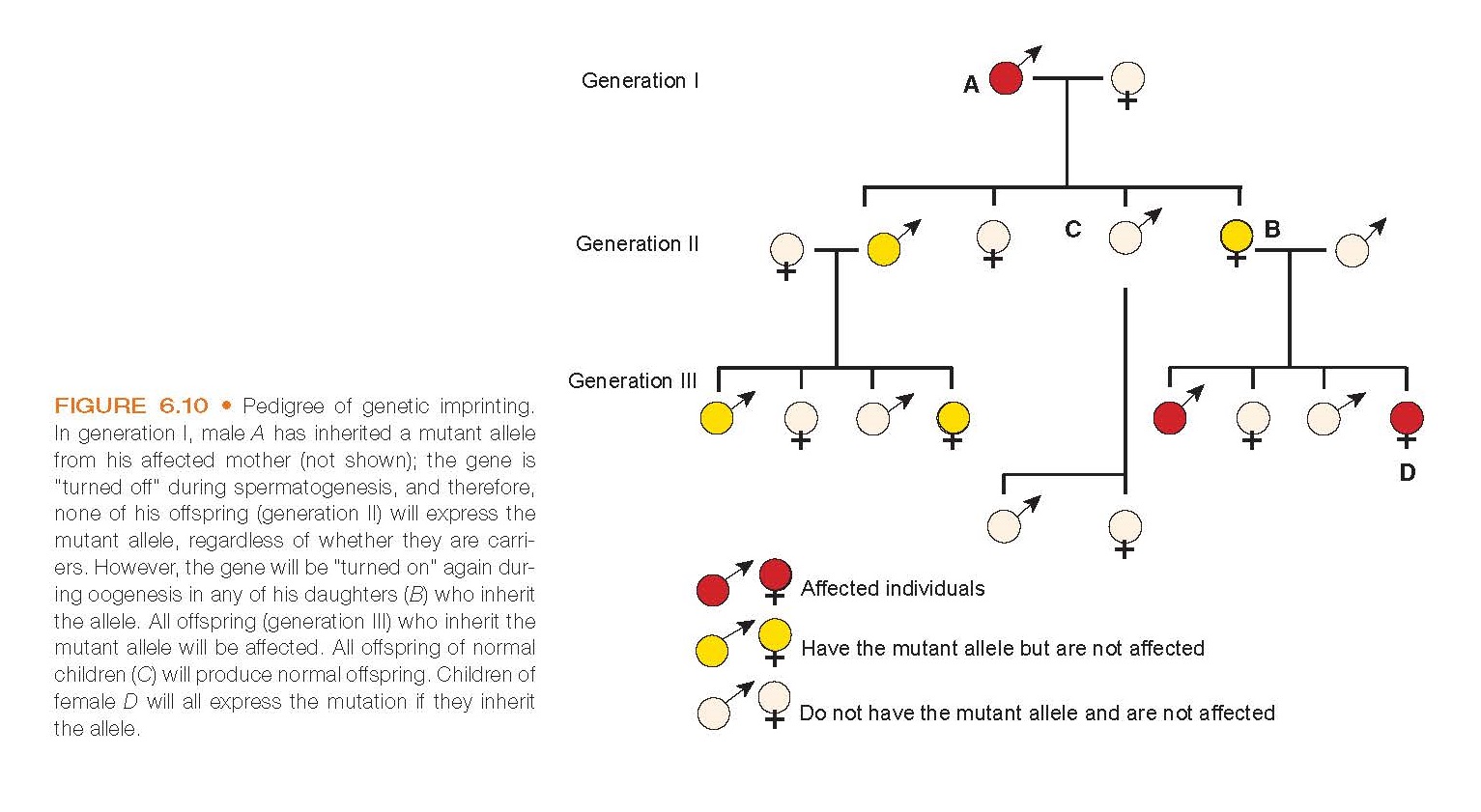
Pedigree of genetic imprinting. In generation I, male A has inherited a mutant allele from his affected mother (not shown); the gene is “turned off” during spermatogenesis, and therefore, none of his offspring (generation II) will express the mutant allele, regardless of whether they are carriers. However, the gene will be “turned on” again during oogenesis in any of his daughters (B) who inherit the allele. All offspring (generation III) who inherit the mutant allele will be affected. All offspring of normal children (C) will produce normal offspring. Children of female D will all express the mutation if they inherit the allele.

Besides autosomal and sex-linked genes and mitochondrial inheritance, it was found that certain genes exhibit a “parent of origin” type of transmission in which the parental genomes do not always contribute equally in the development of a person (Fig. 6.10). The transmission of this phenomenon is called genetic imprinting. Although rare, it is estimated  that  approximately  100  genes  exhibit  genetic imprinting. Evidence suggests a genetic conflict occurs in the developing embryo: the male genome attempts to establish larger offspring, whereas the female prefers smaller off-spring to conserve her energy for the current and subsequent pregnancies.

Thursday, March 23, 2023

Membrane Potentials

Membrane Potentials


Membrane Potentials

Diffusion Of Current-Carrying Ions

Electrochemical potentials are present across the membranes of virtually all cells in the body. Some cells, such as nerve and muscle cells, are capable of generating rapidly changing electrical impulses, and these impulses are used to transmit signals along their membranes. In other cells, such as glandular cells, membrane potentials are used to signal the release of hormones or activate other functions of the cell. Generation of membrane potentials relies on (1) diffusion of current-carrying ions, (2) development of an electrochemical equilibrium, (3) establishment of a RMP, and (4) triggering of action potentials.
Movement of Substances across the Cell Membrane

Movement of Substances across the Cell Membrane


Movement of Substances across the Cell Membrane


Movement through the cell membrane occurs in essentially two ways: passively, without an expenditure of energy, or actively, using energy-consuming processes. The cell membrane can also engulf a particle, forming a membrane-coated vesicle; this membrane-coated vesicle is moved into the cell by endocytosis or out of the cell by exocytosis.

Tuesday, March 21, 2023

From Genes to Proteins

From Genes to Proteins


From Genes to Proteins

The DNA helix and transcription of messenger RNA (mRNA). The DNA helix unwinds and a new mRNA strand is built on the template strand of the DNA

Although DNA determines the type of biochemical product needed by the cell and directs its synthesis, it is RNA through the process of translation, which is responsible for the actual assembly of the products.

Monday, March 20, 2023

DnA structure and Function

DnA structure and Function


DnA structure and Function.


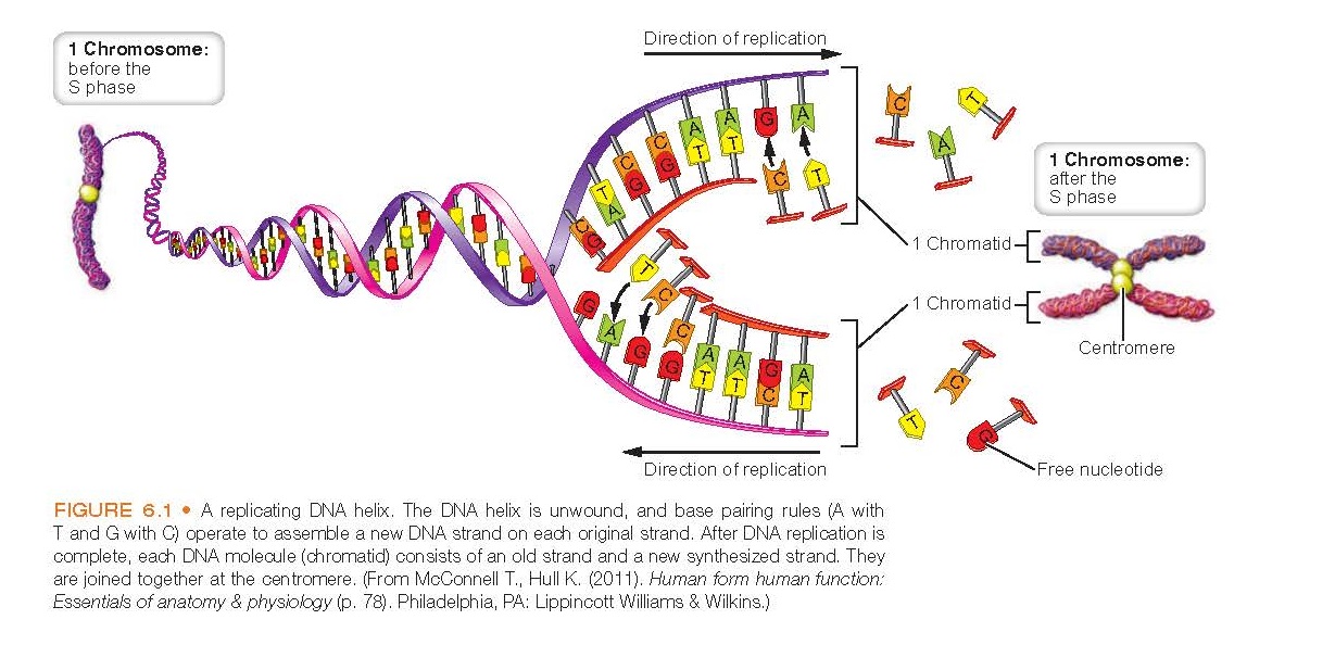
replicating DNA helix. The DNA helix is unwound, and base pairing rules (A with T and G with C) operate to assemble a new DNA strand on each original strand


The DNA molecule that stores the genetic information in the nucleus is a long, double-stranded, helical structure. DNA is composed of nucleotides, which consist of phosphoric acid, a five-carbon sugar called deoxyribose, and one of four nitrogenous bases (Fig. 6.1). These nitrogenous bases carry the genetic information and are divided into two groups: the pyrimidine bases, thymine (T) and cytosine (C), which have one nitrogen ring, and the purine bases, adenine (A) and guanine (G), which have two. The backbone of DNA consists of alternating groups of sugar and phosphoric acid, with the paired bases projecting inward from the sides of the sugar molecule.
Mechanisms of Cell Injury

Mechanisms of Cell Injury


Mechanisms of Cell Injury.


Mechanisms of cell injury. The injurious agents tend to cause hypoxia/ischemia (see middle arrow that illustrates the manifestations that trigger anaerobic metabolism to develop and cellular injury). Also on the left aspect of the figure, the free radical formation causes oxidation of cell structures leading to decreased ATP, and on the right side, the increased intracellular calcium damages many aspects of the cell that also causes ATP depletion. These three paths illustrate how injurious agents cause cell injury and death.


The mechanisms by which injurious agents cause cell injury and death are complex. Some agents, such as heat, produce direct cell injury. Other factors, such as genetic derangements, produce their effects indirectly through metabolic disturbances and altered immune responses. There seem to be at least three major mechanisms whereby most injurious agents exert their effects:
Cell Metabolism

Cell Metabolism


Cell Metabolism.

Glycolytic Pathway


Cell metabolism is the process that converts dietary fuels from carbohydrates, proteins, and fats into ATP, which provides for the energy needs of the cell. ATP is formed through three major pathways: (1) the glycolytic pathway, (2) the citric acid cycle, and (3) the electron transport chain. In fuel metabolism, which is an oxidation–reduction reaction, the fuel donates electrons and is oxidized, and the coenzymes NAD+ and FAD accept electrons and are reduced.

Sunday, February 19, 2023

Reversible Cell Injury and Cell Death

Reversible Cell Injury and Cell Death


Reversible Cell Injury and Cell Death.

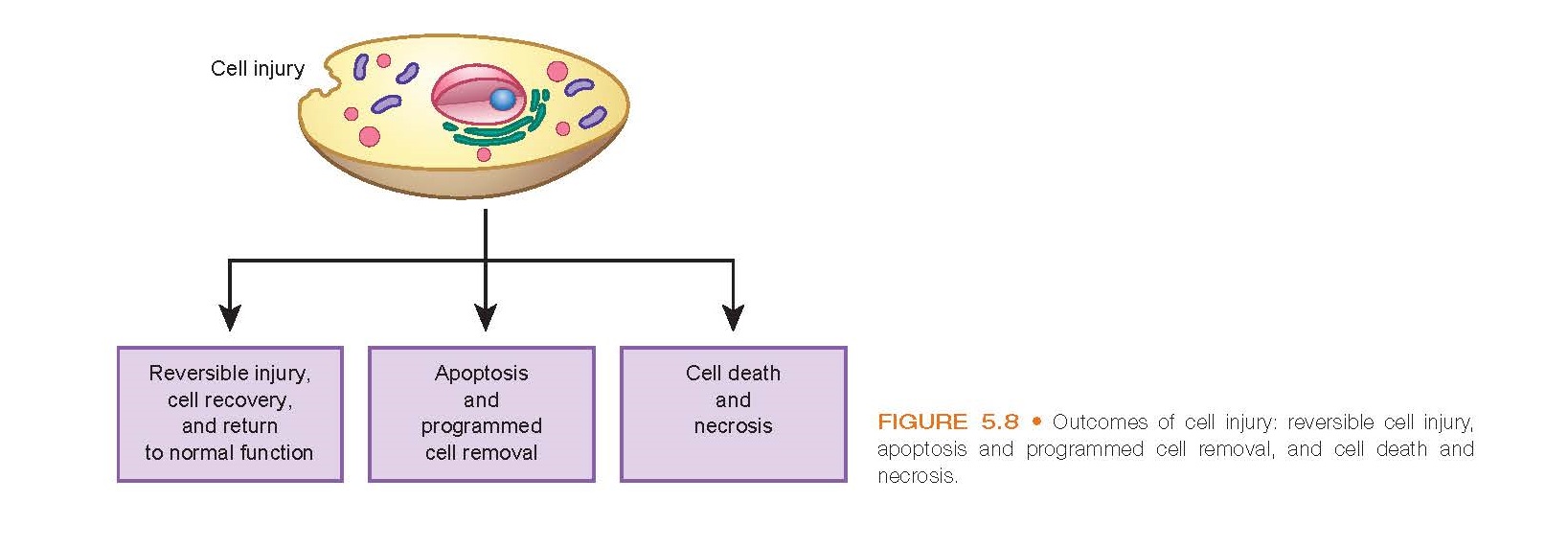
Outcomes of cell injury: reversible cell injury, apoptosis and programmed cell removal, and cell death and necrosis.


The mechanisms of cell injury can produce sublethal and reversible cellular damage or lead to irreversible injury with cell destruction or death (Fig.  5.8). Celldestruction and removal can involve one of two mechanisms:
       Apoptosis, which is designed to remove injured or worn-out cells
       Cell death or necrosis, which occurs in irreversibly damaged cells
Pathologic Calcifications

Pathologic Calcifications


Pathologic Calcifications.

Calcific aortic stenosis. Large deposits of calcium salts are evident in the cusps and free margins of the thickened aortic valve as viewed from above.

Pathologic calcification involves the abnormal tissue deposition of calcium salts, together with smaller amounts of iron, magnesium, and other minerals. It is known as dystrophic calcification when it occurs in dead or dying tissue and as meta- static calcification when it occurs in normal tissue.

Tuesday, February 14, 2023

Embryonic Origin of Tissue Types

Embryonic Origin of Tissue Types


Embryonic Origin of Tissue Types

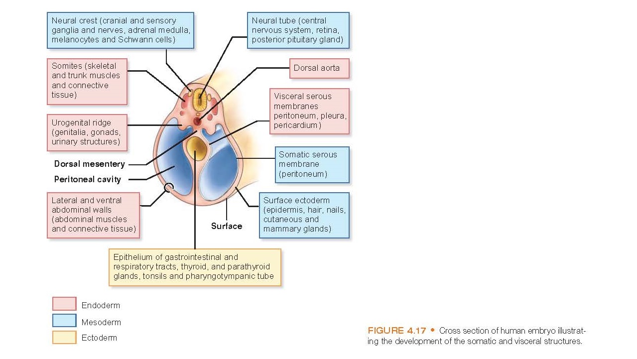
Cross section of human embryo illustrating the development of the somatic and visceral structures.


All of the approximately 200 different types of body cells can be classified into four basic or primary tissue types: epithelial, connective, muscle, and nervous (Table 4.1). These basic tissue types are often described by their embryonic origin. The embryo is essentially a three-layered tubular structure (Fig. 4.17). The outer layer of the tube is called the ectoderm; the middle layer, the mesoderm; and the inner layer, the endoderm. All of the adult body tissues originate from these three cellular layers. Epithelium has its origin in all three embryonic layers, connective tissue and muscle develop mainly from the mesoderm, and nervous tissue develops from the ectoderm.

Saturday, February 11, 2023

Cell Receptors

Cell Receptors

Cell Receptors


Activation of a G-protein–linked receptor and production of cyclic adenosine monophosphate (cAMP).


Signaling systems consist of receptors that reside either in the cell membrane (surface receptors) or within the cells (intracellular receptors). Receptors are activated by a variety of extracellular signals or first messengers, including neurotransmitters, protein hormones and growth factors, steroids, and other chemical messengers. Some lipid-soluble chemical messengers move through the membrane and bind to cytoplasmic or nuclear receptors to exert their physiologic effects. Signaling systems also include transducers and effectors that are involved in conversion of the signal into a physiologic response. The pathway may include additional intracellular mechanisms, called second messengers. Many molecules involved in signal transduction are proteins. A unique property of proteins that allows them to function in this way is their ability to change their shape or conformation, thereby changing their function and consequently the functions of the cell. Proteins often accomplish these conformational changes through enzymes called protein kinases that catalyze the phosphorylation of amino acids in the protein structure.
Protoplasm

Protoplasm


Protoplasm

Although diverse in their organization, all eukaryotic cells have in common structures that perform unique functions. Eukaryote cells are larger and have more specific parts in compartments divided by membranes called organelles. The prokaryote cells do not have compartments and they do not possess a demarcated nucleus as the eukaryotes do. When seen under a microscope, three major components of the cell become evident the nucleus, the cytoplasm, and the cell membrane in the eukaryote cells (Fig. 4.1).

Friday, February 10, 2023

Epithelial Tissue

Epithelial Tissue


Epithelial Tissue.

Typical arrangement of epithelial cells in relation to underlying tissues and blood supply. Epithelial tissue has no blood supply of its own but relies on the blood vessels in the underlying connective tissue for nutrition (N) and elimination of wastes (W).

Epithelial tissue covers the body’s outer surface and lines the internal closed cavities (including blood vessels) and body tubes that communicate with the exterior (gastrointestinal, respiratory, and genitourinary tracts). Epithelium also forms the secretory portion of glands and their ducts.
The Cell Plasma Membrane

The Cell Plasma Membrane


The Cell (Plasma) Membrane.

The Cell Plasma Membrane

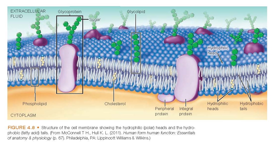
The cell is enclosed in a thin membrane that separates the intracellular contents from the extracellular environment. To differentiate it from the other cell membranes, such as the mitochondrial or nuclear membranes, the cell membrane is often called the plasma membrane. In many respects, the plasma membrane is one of the most important parts of the cell. It acts as a semipermeable structure that separates the intracellular and extracellular environments. It provides receptors for hormones and other biologically active substances, participates in the electrical events that occur in nerve and muscle cells, and aids in the regulation of cell growth and proliferation.
Cell Communication

Cell Communication


Cell Communication.

Cell Communication

Cells in multicellular organisms need to communicate with one another to coordinate their function and control their growth. The human body has several means of transmitting information between cells. These mechanisms include direct communication between adjacent cells through gap junctions, autocrine and paracrine signaling, and endocrine or synaptic signaling. Autocrine signaling occurs when a cell releases a chemical into the extracellular fluid that affects its own activity (Fig. 4.9). 

Tuesday, February 7, 2023

Single-Gene Disorders

Single-Gene Disorders


Single-Gene Disorders.


Single-gene disorders are caused by a defective or mutant allele at a single gene locus and follow mendelian patterns of inheritance. Single-gene disorders are primarily disorders of the pediatric age group. Less than 10% manifest after puberty and only 1% after the reproductive years.

Sunday, February 5, 2023

Cell Metabolism and Energy Sources

Cell Metabolism and Energy Sources


Cell Metabolism and Energy Sources.

ATP is the major source of cellular energy. (A) Each molecule of ATP contains two high-energy bonds, each containing about 12 kcal of potential energy. (B) The high-energy ATP bonds are in constant flux. They are generated by substrate (glucose, amino acid, and fat) metabolism and are consumed as the energy is expended.


Energy is the ability to do work. Cells use oxygen to transform the breakdown products of the foods we eat into the energy needed for muscle contraction; the transport of ions and other molecules across cell membranes; and the synthesis of enzymes, hormones, and other macromolecules. Energy metabolism refers to the processes by which fats, proteins, and carbohydrates from the foods we eat are converted into energy or complex energy sources in the cell. Catabolism and anabolism are the two phases of metabolism. Catabolism consists of breaking down stored nutrients and body tissues to produce energy. Anabolism is a constructive process in which more complex molecules are formed from simpler ones.

Anatomy Physiology

[AnatomyPhysiology][recentbylabel2]

Featured

[Featured][recentbylabel2]
Notification
This is just an example, you can fill it later with your own note.
Done