Quick filters:
Imaging Stock Photos and Images
RFHNF3FB–Vector isometric low poly MRI room icon
 Isometric vector of a radiologist doctor and nurse performing patient brain body scan on MRI machine Stock Vectorhttps://www.alamy.com/image-license-details/?v=1https://www.alamy.com/isometric-vector-of-a-radiologist-doctor-and-nurse-performing-patient-brain-body-scan-on-mri-machine-image243002648.html
RFT39MA0–Isometric vector of a radiologist doctor and nurse performing patient brain body scan on MRI machine
RFKBPBFT–MRI machine icon, Magnetic Resonance Imaging vector symbol
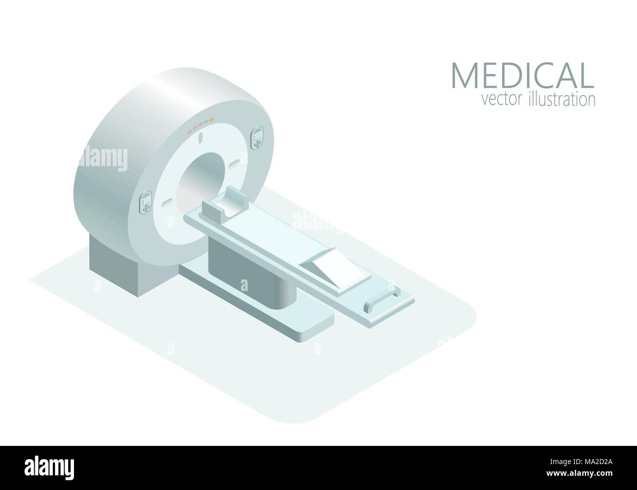 MRI computed tomography concept magnetic resonance imaging scanner vector illustration isometric flat 3d gray monochrome color Stock Vectorhttps://www.alamy.com/image-license-details/?v=1https://www.alamy.com/mri-computed-tomography-concept-magnetic-resonance-imaging-scanner-vector-illustration-isometric-flat-3d-gray-monochrome-color-image178304402.html
RFMA2D2A–MRI computed tomography concept magnetic resonance imaging scanner vector illustration isometric flat 3d gray monochrome color
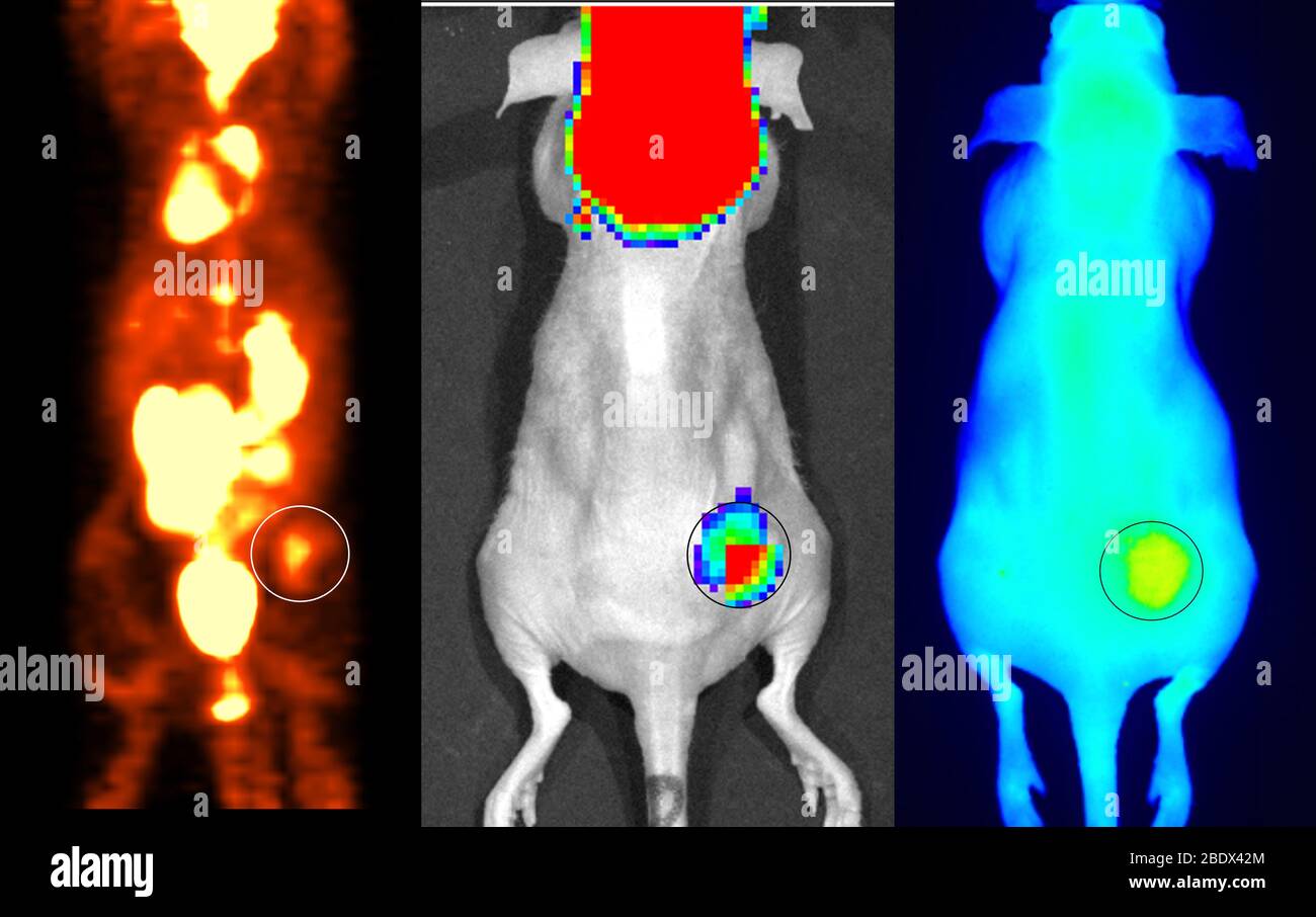 Multimodal Imaging with Nanoparticles Stock Photohttps://www.alamy.com/image-license-details/?v=1https://www.alamy.com/multimodal-imaging-with-nanoparticles-image352771852.html
RM2BDX42M–Multimodal Imaging with Nanoparticles
 A laboratory technician using a photomicroscope which is a combination microscope and digital camera for imaging samples during examination. Stock Photohttps://www.alamy.com/image-license-details/?v=1https://www.alamy.com/stock-photo-a-laboratory-technician-using-a-photomicroscope-which-is-a-combination-49040647.html
RMCRNYRK–A laboratory technician using a photomicroscope which is a combination microscope and digital camera for imaging samples during examination.
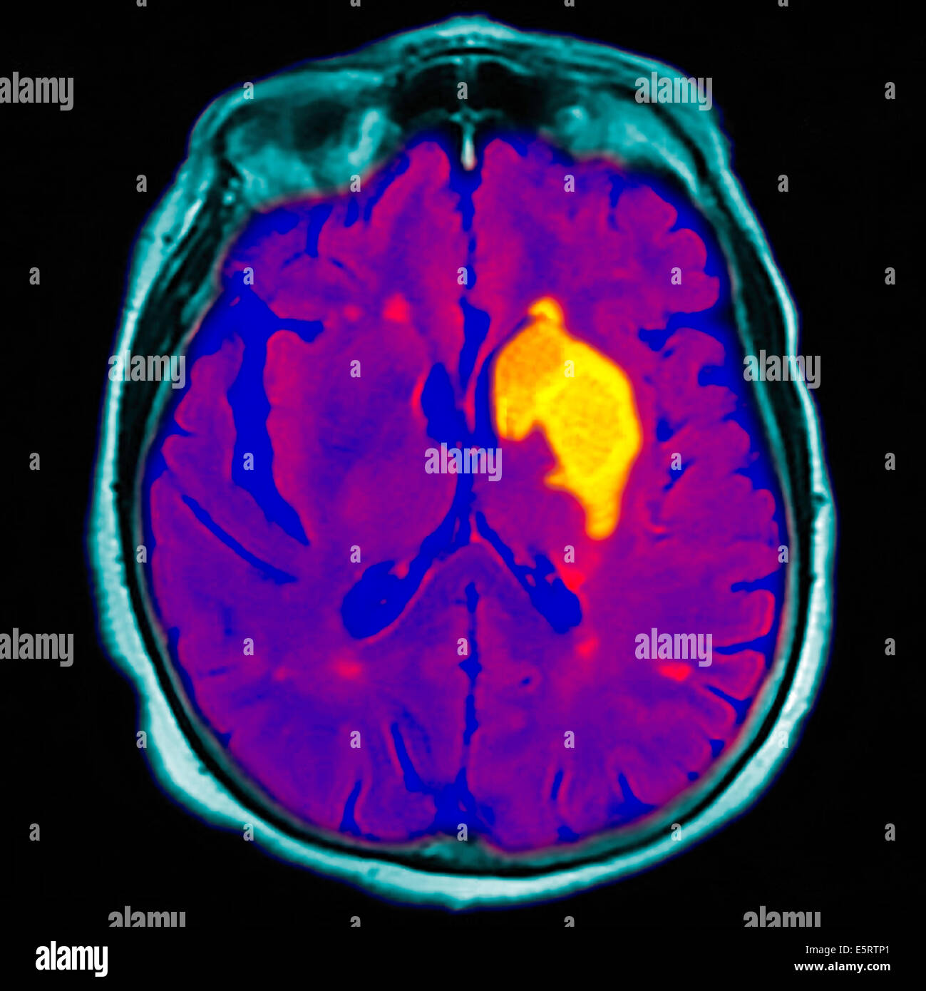 Coloured magnetic resonance imaging (MRI) scan and CT of an axial section through the brain of patient, showing the damage Stock Photohttps://www.alamy.com/image-license-details/?v=1https://www.alamy.com/stock-photo-coloured-magnetic-resonance-imaging-mri-scan-and-ct-of-an-axial-section-72439081.html
RME5RTP1–Coloured magnetic resonance imaging (MRI) scan and CT of an axial section through the brain of patient, showing the damage
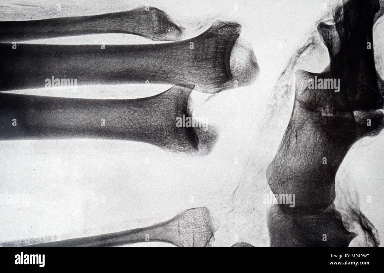 X-ray imaging of the leg of an Egyptian mummy. Dated 19th century Stock Photohttps://www.alamy.com/image-license-details/?v=1https://www.alamy.com/x-ray-imaging-of-the-leg-of-an-egyptian-mummy-dated-19th-century-image186347332.html
RMMR4RWT–X-ray imaging of the leg of an Egyptian mummy. Dated 19th century
 Entrance hall at Photokina digital imaging trade show in Cologne Germany Stock Photohttps://www.alamy.com/image-license-details/?v=1https://www.alamy.com/stock-photo-entrance-hall-at-photokina-digital-imaging-trade-show-in-cologne-germany-31656915.html
RMBRE2M3–Entrance hall at Photokina digital imaging trade show in Cologne Germany
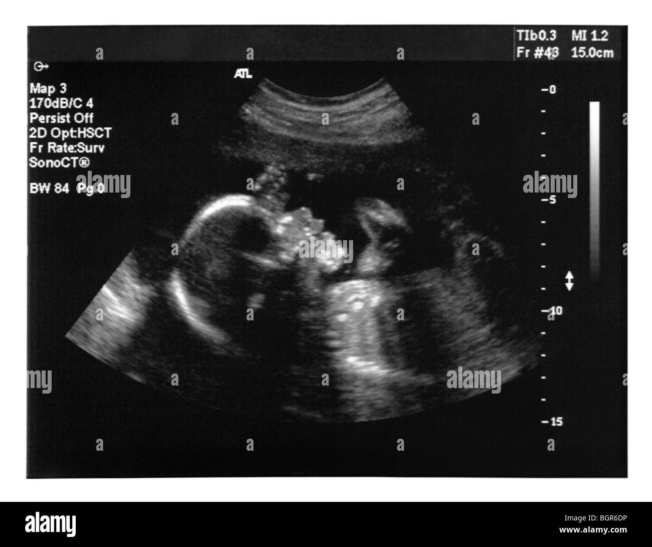 License available at MaximImages.com - Ultrasound imaging of a baby. Five months old fetus. Stock Photohttps://www.alamy.com/image-license-details/?v=1https://www.alamy.com/stock-photo-license-available-at-maximimagescom-ultrasound-imaging-of-a-baby-five-27554850.html
RMBGR6DP–License available at MaximImages.com - Ultrasound imaging of a baby. Five months old fetus.
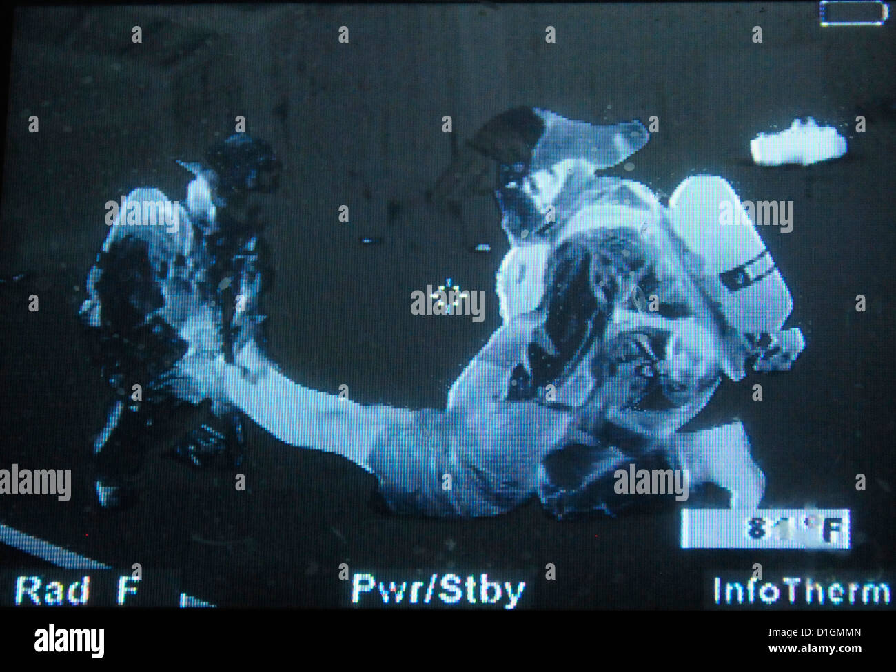 US Air Force fire fighters participate in a search and rescue training mission using thermo imaging August 5, 2012 in Battle Creek, MI. Stock Photohttps://www.alamy.com/image-license-details/?v=1https://www.alamy.com/stock-photo-us-air-force-fire-fighters-participate-in-a-search-and-rescue-training-52613253.html
RMD1GMMN–US Air Force fire fighters participate in a search and rescue training mission using thermo imaging August 5, 2012 in Battle Creek, MI.
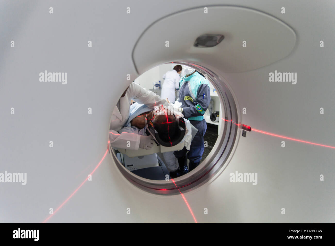 Patient in medical MRI scanner, magnetic resonance imaging, a medical imaging technique used in radiology to image the anatomy and the physiological processes of the body in both health and disease - MRI scanners use strong magnetic fields, radio waves, and field gradients to form images of the body. Stock Photohttps://www.alamy.com/image-license-details/?v=1https://www.alamy.com/stock-photo-patient-in-medical-mri-scanner-magnetic-resonance-imaging-a-medical-121956797.html
RMH2BH3W–Patient in medical MRI scanner, magnetic resonance imaging, a medical imaging technique used in radiology to image the anatomy and the physiological processes of the body in both health and disease - MRI scanners use strong magnetic fields, radio waves, and field gradients to form images of the body.
 CAPE CANAVERAL, FLORIDA, USA - 09 December 2021 - A SpaceX Falcon 9 rocket launches with NASA’s Imaging X-ray Polarimetry Explorer (IXPE) spacecraft o Stock Photohttps://www.alamy.com/image-license-details/?v=1https://www.alamy.com/cape-canaveral-florida-usa-09-december-2021-a-spacex-falcon-9-rocket-launches-with-nasas-imaging-x-ray-polarimetry-explorer-ixpe-spacecraft-o-image454084488.html
RM2HAN9B4–CAPE CANAVERAL, FLORIDA, USA - 09 December 2021 - A SpaceX Falcon 9 rocket launches with NASA’s Imaging X-ray Polarimetry Explorer (IXPE) spacecraft o
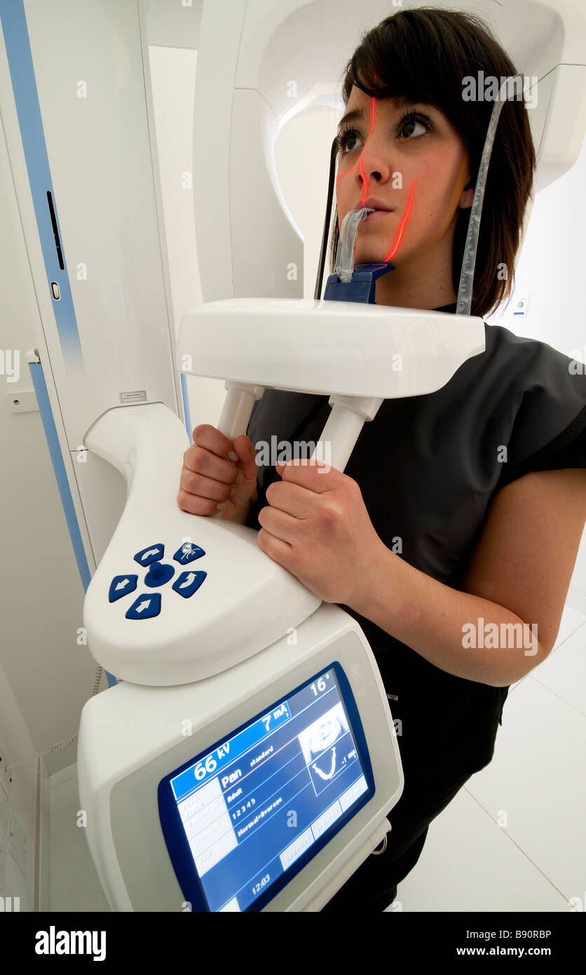 Computerized dental imaging technology Stock Photohttps://www.alamy.com/image-license-details/?v=1https://www.alamy.com/stock-photo-computerized-dental-imaging-technology-22760634.html
RMB90RBP–Computerized dental imaging technology
 : Magnetic Resonance Imaging ( MRI ) : cross-sectional images of a knee, , , Stock Photohttps://www.alamy.com/image-license-details/?v=1https://www.alamy.com/stock-photo-magnetic-resonance-imaging-mri-cross-sectional-images-of-a-knee-111744848.html
RMGDPBKC–: Magnetic Resonance Imaging ( MRI ) : cross-sectional images of a knee, , ,
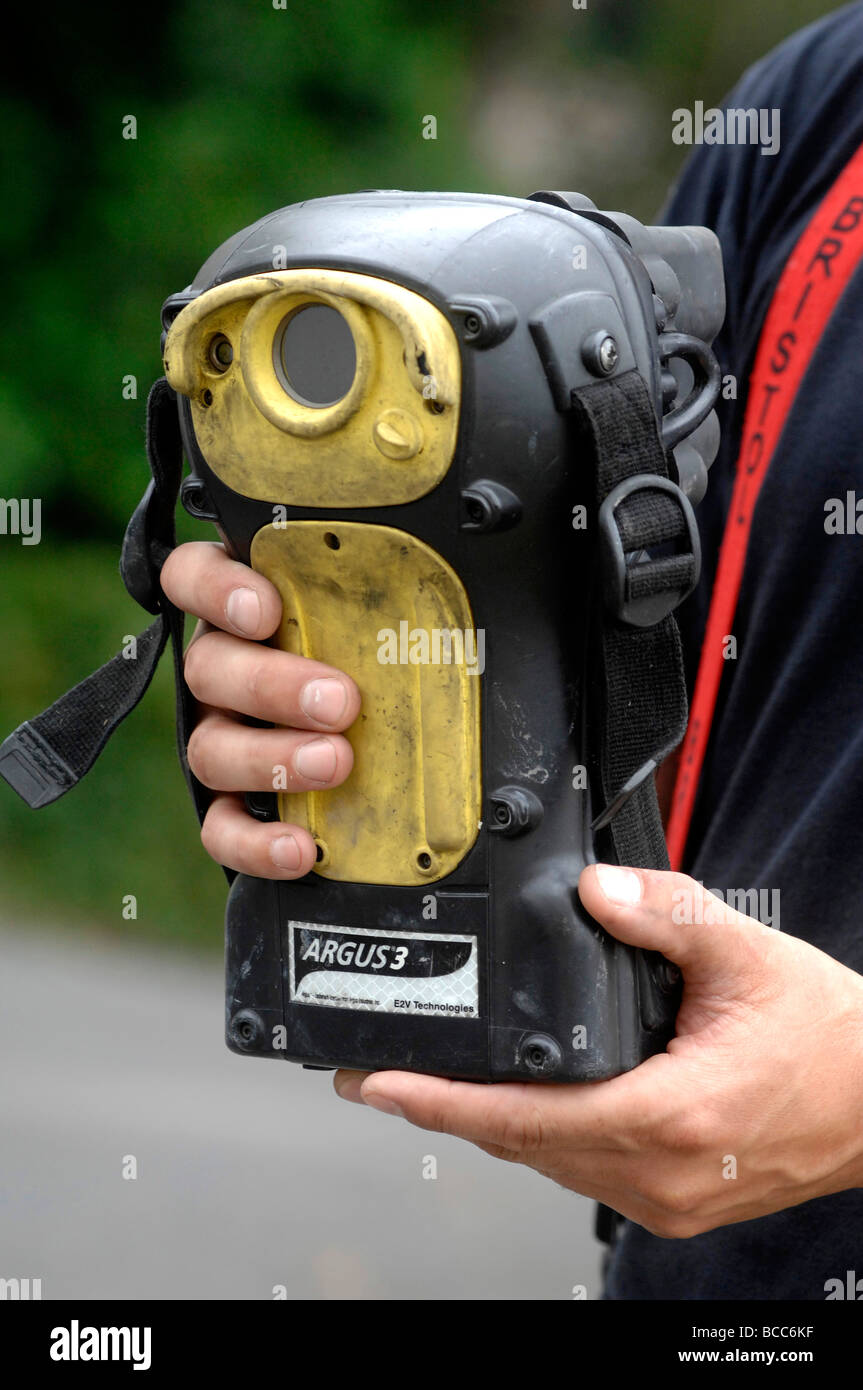 Fireman using a 'thermal imaging camera' , Britain UK Stock Photohttps://www.alamy.com/image-license-details/?v=1https://www.alamy.com/stock-photo-fireman-using-a-thermal-imaging-camera-britain-uk-24854915.html
RMBCC6KF–Fireman using a 'thermal imaging camera' , Britain UK
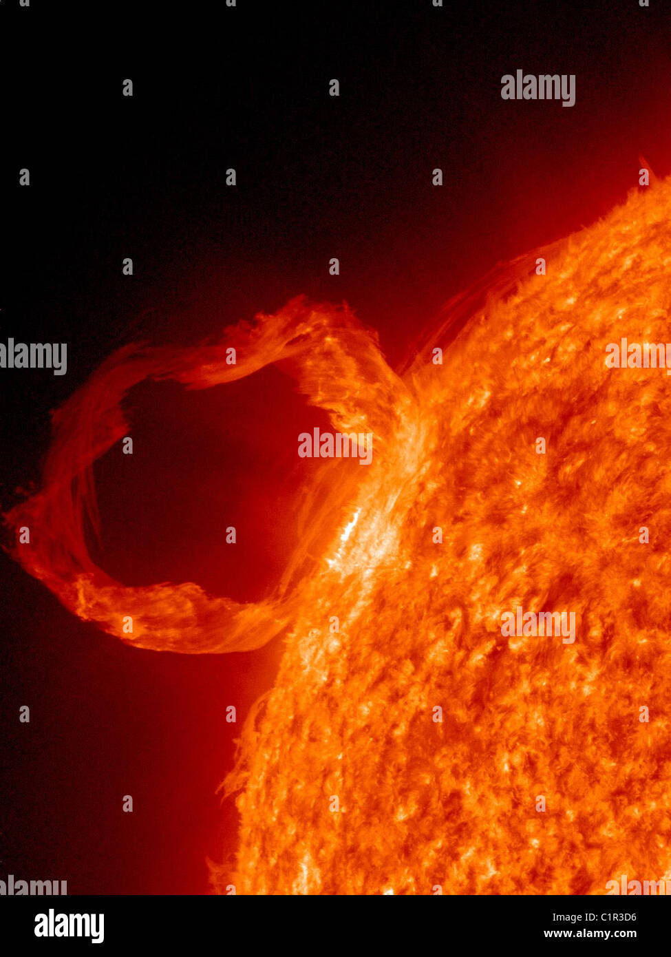 Solar Dynamics Observatory’s Atmospheric Imaging Assembly (AIA) shows in great detail a solar prominence eruption. Stock Photohttps://www.alamy.com/image-license-details/?v=1https://www.alamy.com/stock-photo-solar-dynamics-observatorys-atmospheric-imaging-assembly-aia-shows-35543010.html
RMC1R3D6–Solar Dynamics Observatory’s Atmospheric Imaging Assembly (AIA) shows in great detail a solar prominence eruption.
 s Brain Research Imaging Centre (CUBRIC) building during his visit to the Cardiff inWales, prior to delivering a speech. Stock Photohttps://www.alamy.com/image-license-details/?v=1https://www.alamy.com/stock-photo-s-brain-research-imaging-centre-cubric-building-during-his-visit-to-108573867.html
RMG8HY23–s Brain Research Imaging Centre (CUBRIC) building during his visit to the Cardiff inWales, prior to delivering a speech.
 Researcher operating at VERITAS (Very Energetic Radiation Imaging Telescope Array System), is a major ground-based gamma-ray observatory, Arizona. Stock Photohttps://www.alamy.com/image-license-details/?v=1https://www.alamy.com/researcher-operating-at-veritas-very-energetic-radiation-imaging-telescope-array-system-is-a-major-ground-based-gamma-ray-observatory-arizona-image470518112.html
RM2J9DXJ8–Researcher operating at VERITAS (Very Energetic Radiation Imaging Telescope Array System), is a major ground-based gamma-ray observatory, Arizona.
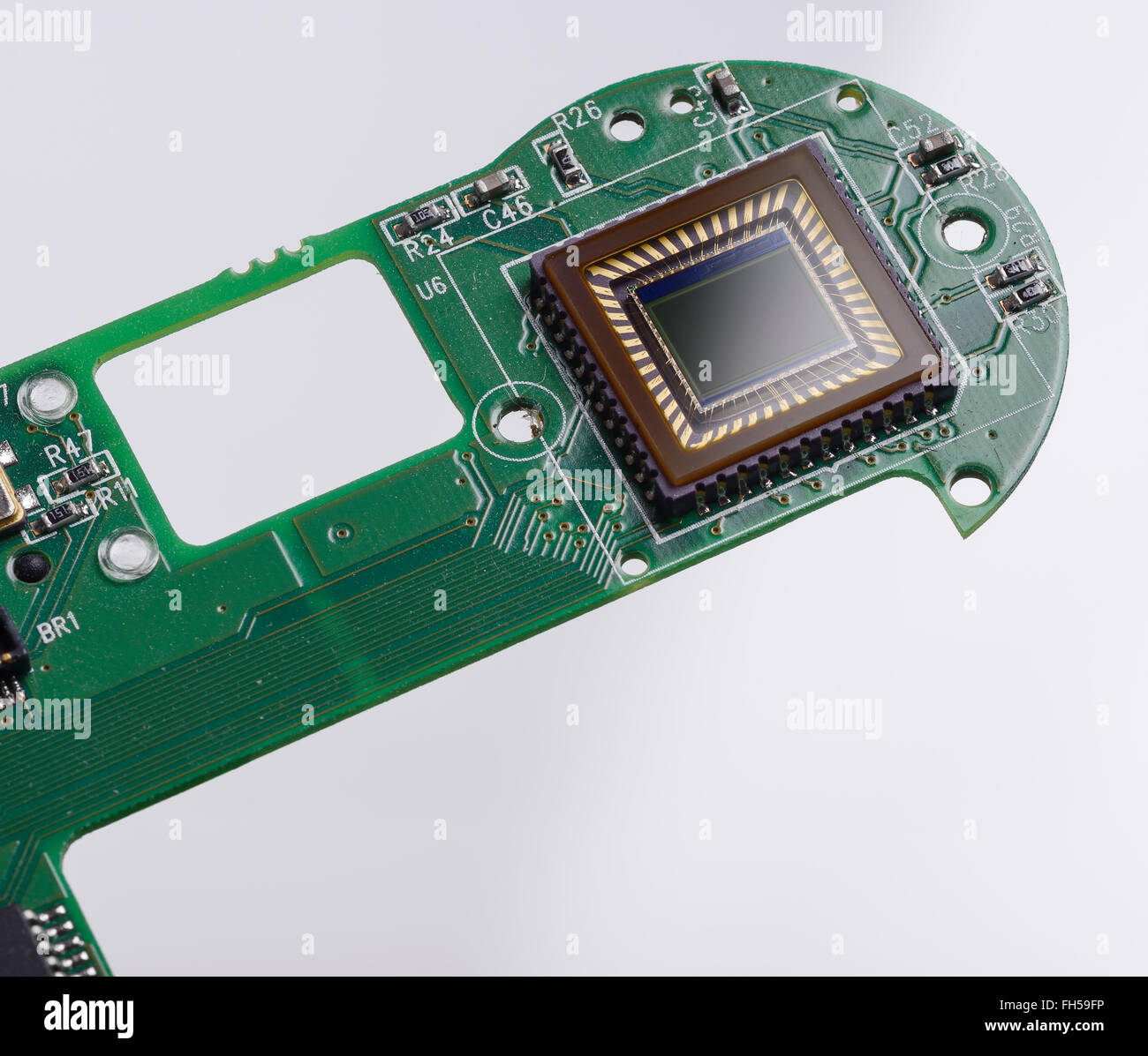 The imaging sensor and circuit board from a compact digital camera Stock Photohttps://www.alamy.com/image-license-details/?v=1https://www.alamy.com/stock-photo-the-imaging-sensor-and-circuit-board-from-a-compact-digital-camera-96618250.html
RMFH59FP–The imaging sensor and circuit board from a compact digital camera
 Isometric vector of a medical clinic hospital inpatient care with rooms, patients, doctors and nurses. Healthcare technology and imaging studies conce Stock Vectorhttps://www.alamy.com/image-license-details/?v=1https://www.alamy.com/isometric-vector-of-a-medical-clinic-hospital-inpatient-care-with-rooms-patients-doctors-and-nurses-healthcare-technology-and-imaging-studies-conce-image224314082.html
RFR0XAW6–Isometric vector of a medical clinic hospital inpatient care with rooms, patients, doctors and nurses. Healthcare technology and imaging studies conce
 children's Brain MRI magnetic resonance imaging or NMRI nuclear magnetic resonance imaging of Stock Photohttps://www.alamy.com/image-license-details/?v=1https://www.alamy.com/stock-photo-childrens-brain-mri-magnetic-resonance-imaging-or-nmri-nuclear-magnetic-31518175.html
RMBR7NN3–children's Brain MRI magnetic resonance imaging or NMRI nuclear magnetic resonance imaging of
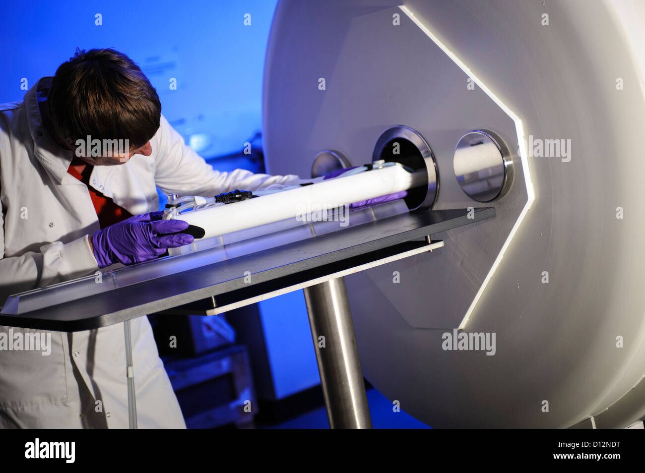 Scientist or technician loads a specimen on a tray into a small bore Magnetic Resonance Imaging (MRI) scanner Stock Photohttps://www.alamy.com/image-license-details/?v=1https://www.alamy.com/stock-photo-scientist-or-technician-loads-a-specimen-on-a-tray-into-a-small-bore-52306516.html
RMD12NDT–Scientist or technician loads a specimen on a tray into a small bore Magnetic Resonance Imaging (MRI) scanner
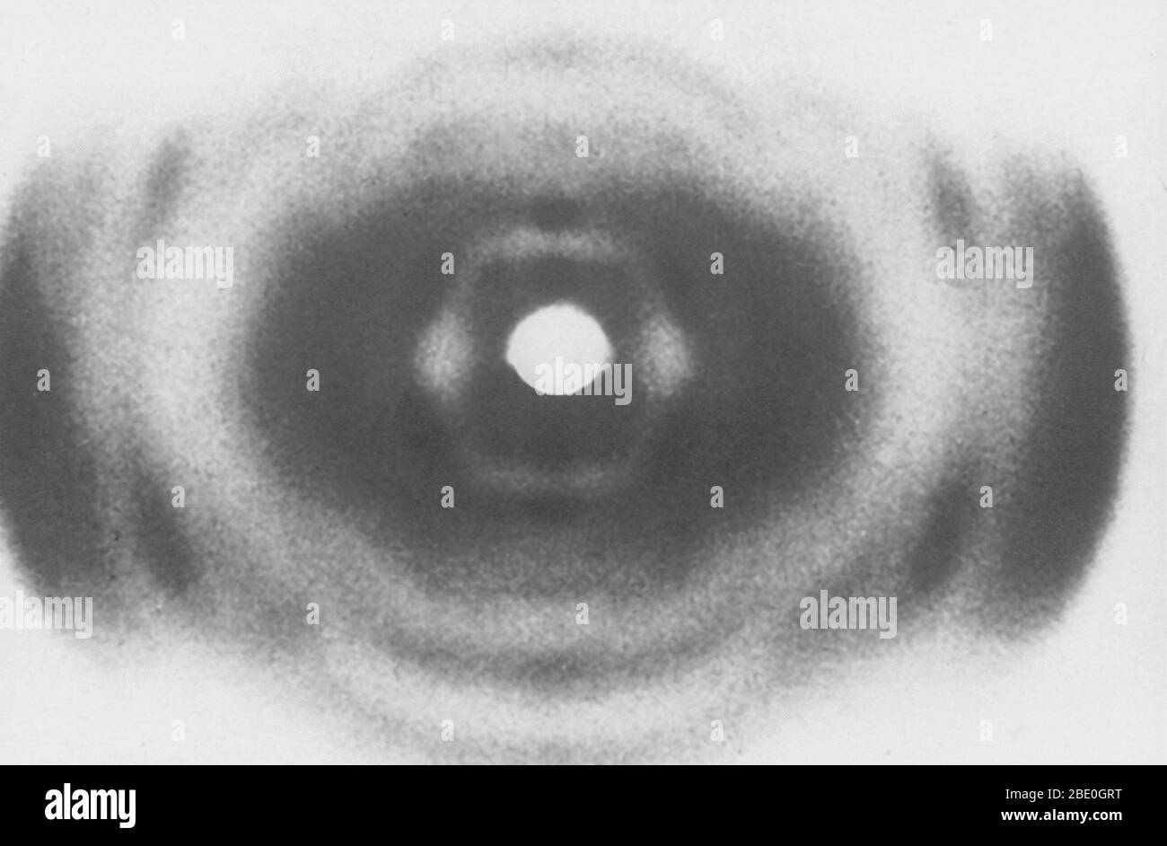 An X-ray of DNA, produced circa 1937, by William Thomas Astbury (1898-1961), a pioneer of DNA imaging. Stock Photohttps://www.alamy.com/image-license-details/?v=1https://www.alamy.com/an-x-ray-of-dna-produced-circa-1937-by-william-thomas-astbury-1898-1961-a-pioneer-of-dna-imaging-image352825756.html
RM2BE0GRT–An X-ray of DNA, produced circa 1937, by William Thomas Astbury (1898-1961), a pioneer of DNA imaging.
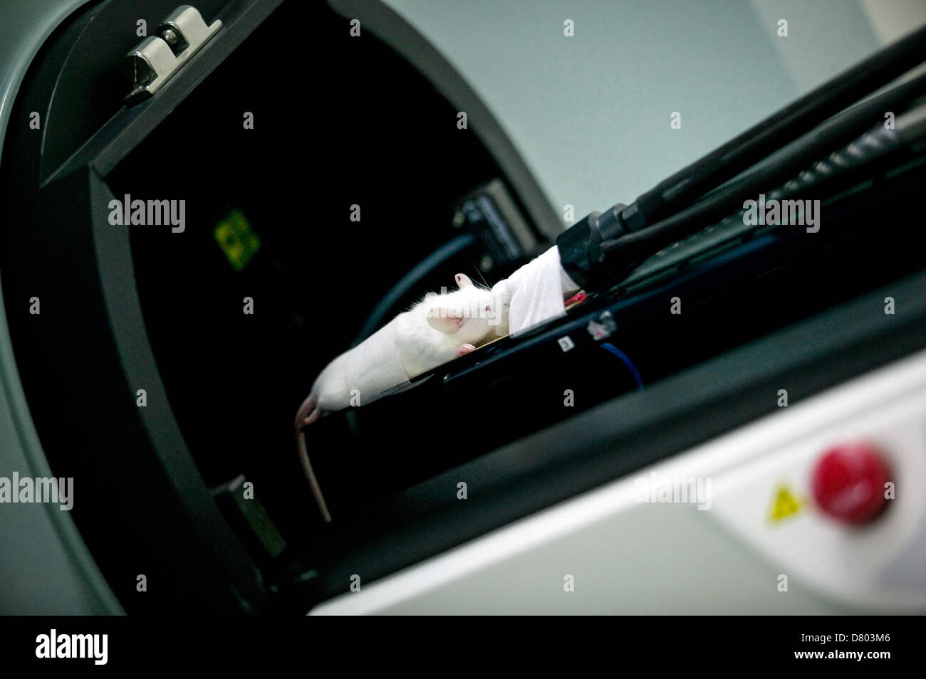 CT Imaging room machine Stock Photohttps://www.alamy.com/image-license-details/?v=1https://www.alamy.com/stock-photo-ct-imaging-room-machine-56551270.html
RMD803M6–CT Imaging room machine
 Coloured magnetic resonance imaging (MRI) scan and CT of an axial section through the brain of patient, showing the damage Stock Photohttps://www.alamy.com/image-license-details/?v=1https://www.alamy.com/stock-photo-coloured-magnetic-resonance-imaging-mri-scan-and-ct-of-an-axial-section-72439082.html
RME5RTP2–Coloured magnetic resonance imaging (MRI) scan and CT of an axial section through the brain of patient, showing the damage
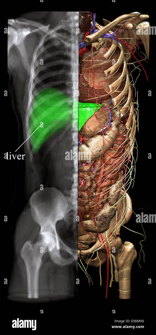 Different viewing modes such as X-ray imaging may be chosen from any direction and for any part of the image. The X-ray image may be interrogated as to which object contributes to the image. Stock Photohttps://www.alamy.com/image-license-details/?v=1https://www.alamy.com/stock-photo-different-viewing-modes-such-as-x-ray-imaging-may-be-chosen-from-any-84970648.html
RMEX6MX0–Different viewing modes such as X-ray imaging may be chosen from any direction and for any part of the image. The X-ray image may be interrogated as to which object contributes to the image.
 Samsung display stand at Photokina digital imaging trade show in Cologne Germany Stock Photohttps://www.alamy.com/image-license-details/?v=1https://www.alamy.com/stock-photo-samsung-display-stand-at-photokina-digital-imaging-trade-show-in-cologne-31657696.html
RMBRE3M0–Samsung display stand at Photokina digital imaging trade show in Cologne Germany
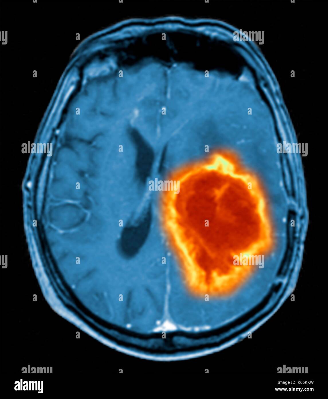 Brain tumour. Coloured Magnetic Resonance Imaging (MRI) scan of an axial section through the brain showing a metastatic tumour. At bottom left is the tumour (red-yellow) This tumour occurs within one cerebral hemisphere; the other hemisphere is at right. The eyeballs - not visible -are at top. Metastatic cancer is a secondary disease spread from cancer elsewhere in the body. Metastatic brain tumours are malignant. Typically they cause brain compression and nerve damage Stock Photohttps://www.alamy.com/image-license-details/?v=1https://www.alamy.com/brain-tumour-coloured-magnetic-resonance-imaging-mri-scan-of-an-axial-image158728413.html
RFK66KKW–Brain tumour. Coloured Magnetic Resonance Imaging (MRI) scan of an axial section through the brain showing a metastatic tumour. At bottom left is the tumour (red-yellow) This tumour occurs within one cerebral hemisphere; the other hemisphere is at right. The eyeballs - not visible -are at top. Metastatic cancer is a secondary disease spread from cancer elsewhere in the body. Metastatic brain tumours are malignant. Typically they cause brain compression and nerve damage
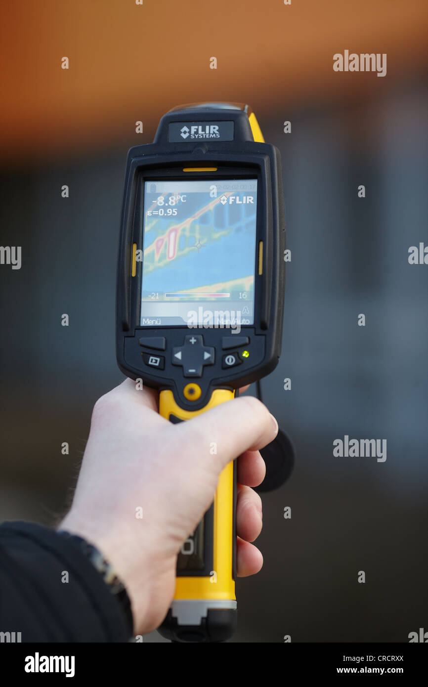 Thermal imaging camera, Muelheim-Kaerlich, Rhineland-Palatinate, Germany, Europe Stock Photohttps://www.alamy.com/image-license-details/?v=1https://www.alamy.com/stock-photo-thermal-imaging-camera-muelheim-kaerlich-rhineland-palatinate-germany-48840034.html
RMCRCRXX–Thermal imaging camera, Muelheim-Kaerlich, Rhineland-Palatinate, Germany, Europe
 Forensics expert using RUVIS Reflected Ultra-violet Imaging Systems to examine evidence. Nebraska State patrol Crime Lab. Stock Photohttps://www.alamy.com/image-license-details/?v=1https://www.alamy.com/stock-photo-forensics-expert-using-ruvis-reflected-ultra-violet-imaging-systems-29362224.html
RMBKNFPT–Forensics expert using RUVIS Reflected Ultra-violet Imaging Systems to examine evidence. Nebraska State patrol Crime Lab.
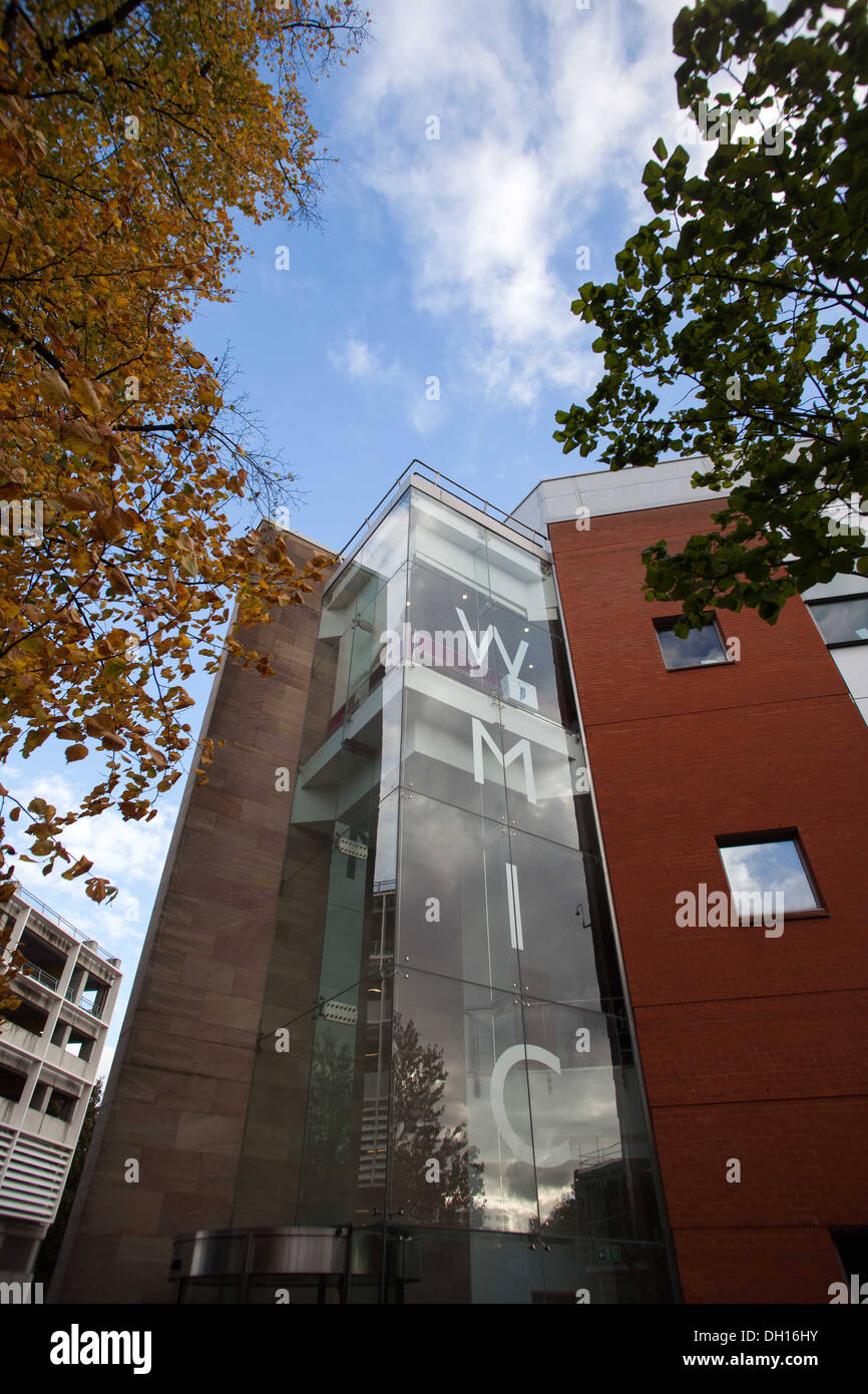 The Cancer Imaging Centre at The University of Manchester Wolfson Molecular Imaging Centre. Stock Photohttps://www.alamy.com/image-license-details/?v=1https://www.alamy.com/the-cancer-imaging-centre-at-the-university-of-manchester-wolfson-image62107415.html
RMDH16HY–The Cancer Imaging Centre at The University of Manchester Wolfson Molecular Imaging Centre.
 Los Angeles, USA - 1 February 2021: Pentax Ricoh Imaging website page. ricoh-imaging.co.jp logo on display screen, Illustrative Editorial Stock Photohttps://www.alamy.com/image-license-details/?v=1https://www.alamy.com/los-angeles-usa-1-february-2021-pentax-ricoh-imaging-website-page-ricoh-imagingcojp-logo-on-display-screen-illustrative-editorial-image401809940.html
RF2E9M0K0–Los Angeles, USA - 1 February 2021: Pentax Ricoh Imaging website page. ricoh-imaging.co.jp logo on display screen, Illustrative Editorial
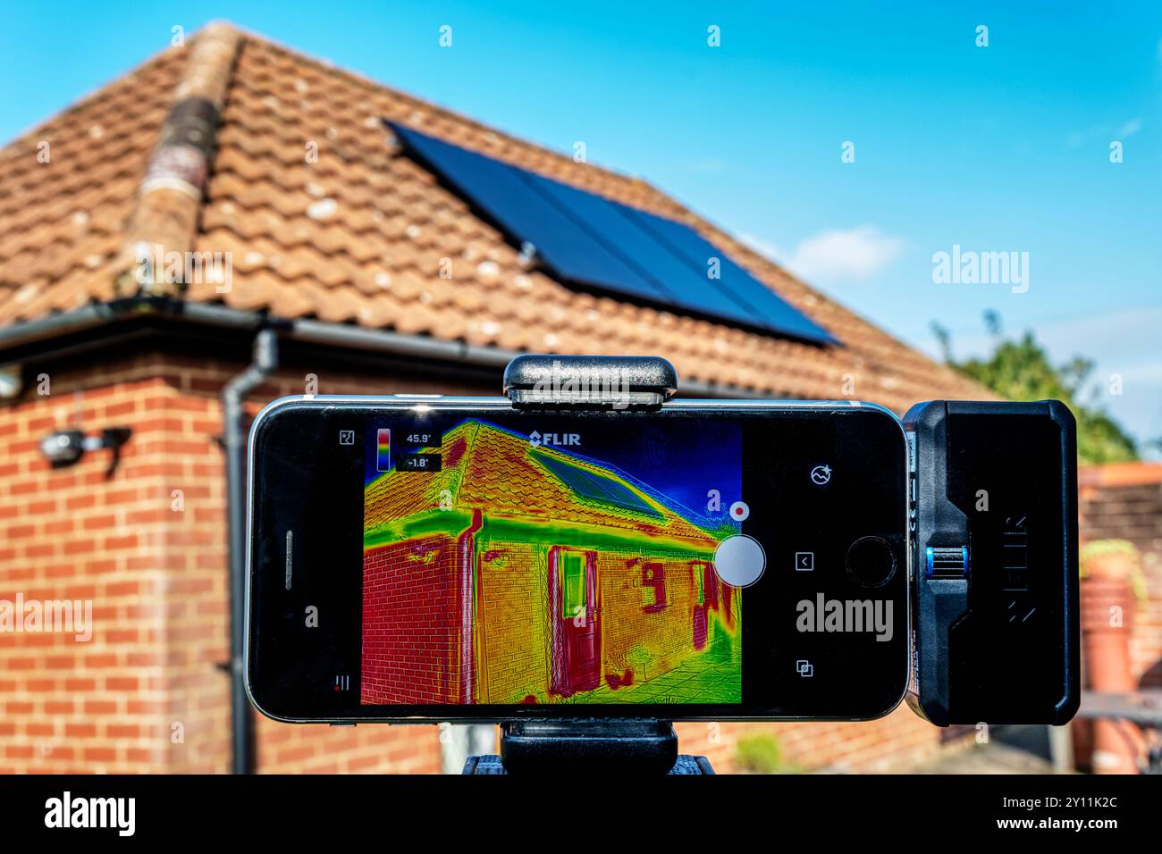 An infrared thermal imaging device being used to assess heat loss from a bungalow fitted with solar panels on the roof. Stock Photohttps://www.alamy.com/image-license-details/?v=1https://www.alamy.com/an-infrared-thermal-imaging-device-being-used-to-assess-heat-loss-from-a-bungalow-fitted-with-solar-panels-on-the-roof-image620224820.html
RM2Y11K2C–An infrared thermal imaging device being used to assess heat loss from a bungalow fitted with solar panels on the roof.
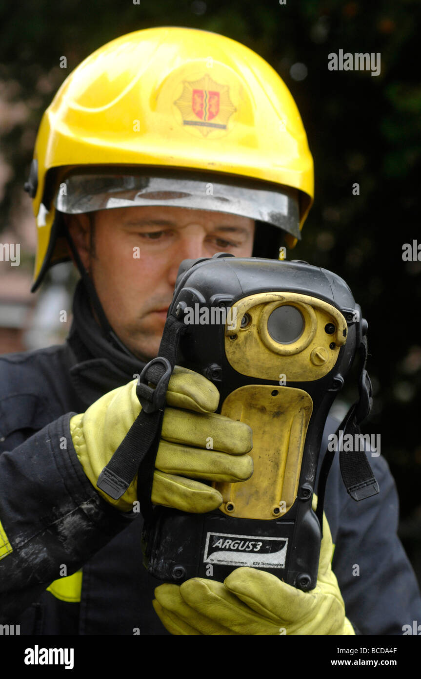 Fireman using a 'thermal imaging camera' , Britain UK Stock Photohttps://www.alamy.com/image-license-details/?v=1https://www.alamy.com/stock-photo-fireman-using-a-thermal-imaging-camera-britain-uk-24879583.html
RMBCDA4F–Fireman using a 'thermal imaging camera' , Britain UK
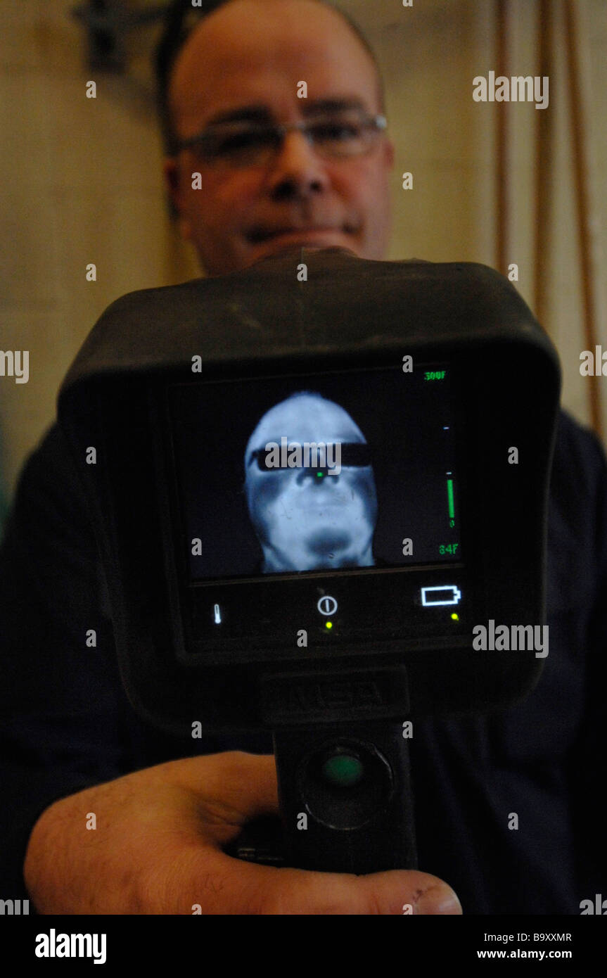 Fireman showing display on MSA thermal imaging Camera. Note glasses block out some of heat signature. Stock Photohttps://www.alamy.com/image-license-details/?v=1https://www.alamy.com/stock-photo-fireman-showing-display-on-msa-thermal-imaging-camera-note-glasses-23333991.html
RMB9XXMR–Fireman showing display on MSA thermal imaging Camera. Note glasses block out some of heat signature.
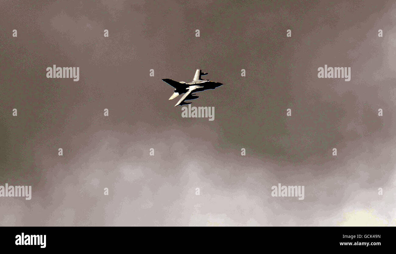 A RAF Tornado with imaging equipment flies over Rothbury as the search for Raoul Moat continues. Stock Photohttps://www.alamy.com/image-license-details/?v=1https://www.alamy.com/stock-photo-a-raf-tornado-with-imaging-equipment-flies-over-rothbury-as-the-search-111058577.html
RMGCK49N–A RAF Tornado with imaging equipment flies over Rothbury as the search for Raoul Moat continues.
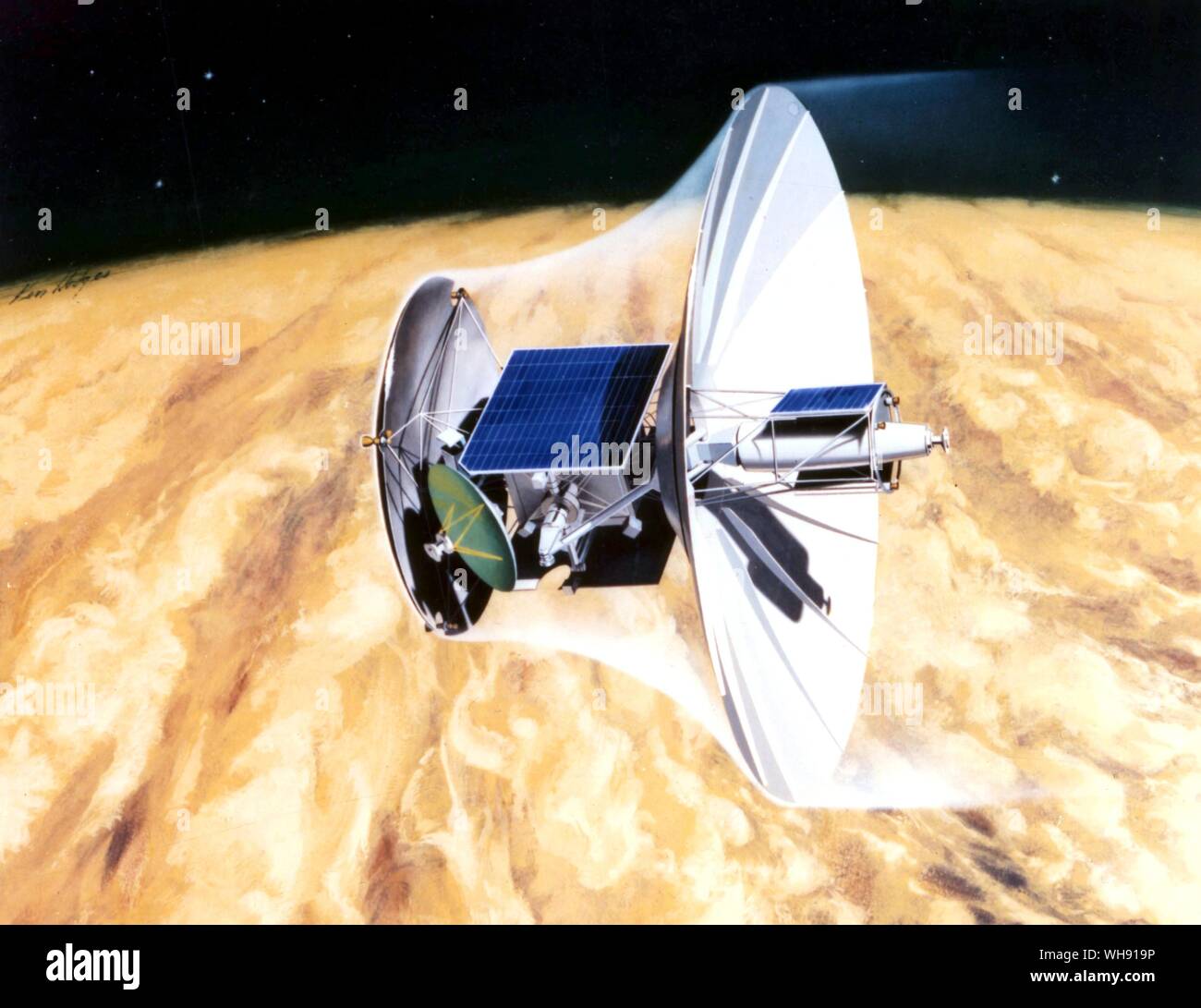 Space - artist impressions concept of Venus orbiting imaging radar. Stock Photohttps://www.alamy.com/image-license-details/?v=1https://www.alamy.com/space-artist-impressions-concept-of-venus-orbiting-imaging-radar-image268803298.html
RMWH919P–Space - artist impressions concept of Venus orbiting imaging radar.
 NASA International Space Station Expedition 51 prime crew member American astronaut Peggy Whitson changes out the Bone Densitometer Imaging Unit inside the Harmony Module June 19, 2017 in Earth orbit. (photo by NASA Photo via Planetpix) Stock Photohttps://www.alamy.com/image-license-details/?v=1https://www.alamy.com/stock-image-nasa-international-space-station-expedition-51-prime-crew-member-american-162691581.html
RMKCK6NH–NASA International Space Station Expedition 51 prime crew member American astronaut Peggy Whitson changes out the Bone Densitometer Imaging Unit inside the Harmony Module June 19, 2017 in Earth orbit. (photo by NASA Photo via Planetpix)
 : Magnetic Resonance Imaging ( MRI ) : cross-sectional images of a knee, , , Stock Photohttps://www.alamy.com/image-license-details/?v=1https://www.alamy.com/stock-photo-magnetic-resonance-imaging-mri-cross-sectional-images-of-a-knee-111744980.html
RMGDPBT4–: Magnetic Resonance Imaging ( MRI ) : cross-sectional images of a knee, , ,
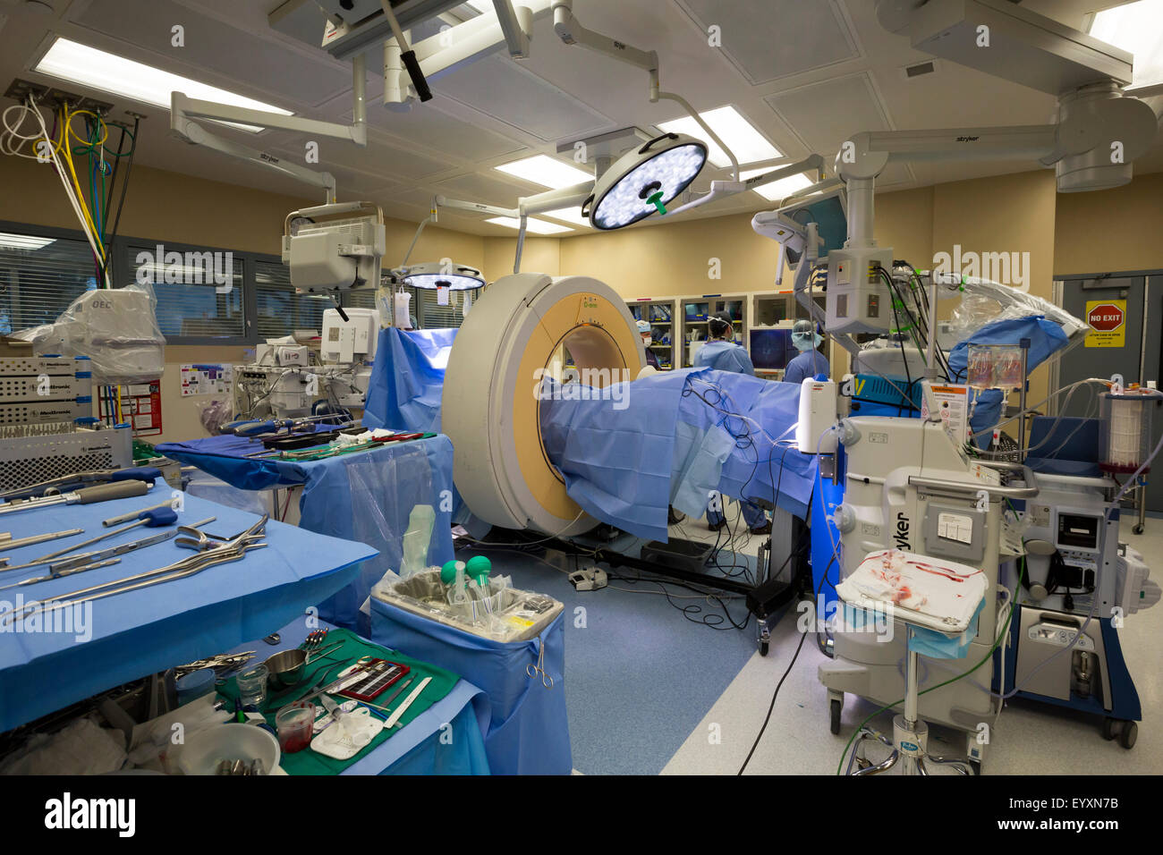 Englewood, Colorado - A CT scan machine, the Stealth O-arm spinal navigation, is used to obtain imaging before spine surgery. Stock Photohttps://www.alamy.com/image-license-details/?v=1https://www.alamy.com/stock-photo-englewood-colorado-a-ct-scan-machine-the-stealth-o-arm-spinal-navigation-86024607.html
RMEYXN7B–Englewood, Colorado - A CT scan machine, the Stealth O-arm spinal navigation, is used to obtain imaging before spine surgery.
 Soil mite, AU-17142 Queensland, Australia. Different imaging illumination examples. Stock Photohttps://www.alamy.com/image-license-details/?v=1https://www.alamy.com/soil-mite-au-17142-queensland-australia-different-imaging-illumination-examples-image329779658.html
RM2A4EN8X–Soil mite, AU-17142 Queensland, Australia. Different imaging illumination examples.
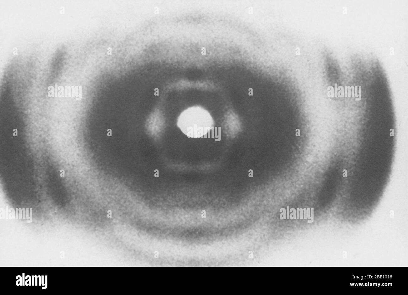 An X-ray of DNA, produced circa 1937, by William Thomas Astbury (1898-1961), a pioneer of DNA imaging. Stock Photohttps://www.alamy.com/image-license-details/?v=1https://www.alamy.com/an-x-ray-of-dna-produced-circa-1937-by-william-thomas-astbury-1898-1961-a-pioneer-of-dna-imaging-image352834532.html
RM2BE1018–An X-ray of DNA, produced circa 1937, by William Thomas Astbury (1898-1961), a pioneer of DNA imaging.
 Close up of the control panel to an ultrasound imaging system. Stock Photohttps://www.alamy.com/image-license-details/?v=1https://www.alamy.com/stock-photo-close-up-of-the-control-panel-to-an-ultrasound-imaging-system-49199736.html
RMCT16NC–Close up of the control panel to an ultrasound imaging system.
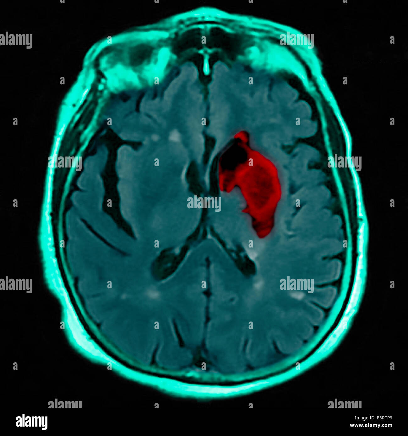 Coloured magnetic resonance imaging (MRI) scan and CT of an axial section through the brain of patient, showing the damage Stock Photohttps://www.alamy.com/image-license-details/?v=1https://www.alamy.com/stock-photo-coloured-magnetic-resonance-imaging-mri-scan-and-ct-of-an-axial-section-72439083.html
RME5RTP3–Coloured magnetic resonance imaging (MRI) scan and CT of an axial section through the brain of patient, showing the damage
 Infrared thermography (IRT), thermal imaging, and thermal video are examples of infrared imaging science. When viewed through a thermal imaging camera, warm objects stand out well against cooler backgrounds; humans and other warm-blooded animals become easily visible against the environment, day or night. Stock Photohttps://www.alamy.com/image-license-details/?v=1https://www.alamy.com/stock-image-infrared-thermography-irt-thermal-imaging-and-thermal-video-are-examples-162596281.html
RMKCEW61–Infrared thermography (IRT), thermal imaging, and thermal video are examples of infrared imaging science. When viewed through a thermal imaging camera, warm objects stand out well against cooler backgrounds; humans and other warm-blooded animals become easily visible against the environment, day or night.
 Many people at Nikon stand at Photokina digital imaging trade show in Cologne Germany Stock Photohttps://www.alamy.com/image-license-details/?v=1https://www.alamy.com/stock-photo-many-people-at-nikon-stand-at-photokina-digital-imaging-trade-show-31657471.html
RMBRE3BY–Many people at Nikon stand at Photokina digital imaging trade show in Cologne Germany
 Brain tumour. Coloured Magnetic Resonance Imaging (MRI) scan of an axial section through the brain showing a metastatic tumour. At bottom left is the tumour (red-yellow) This tumour occurs within one cerebral hemisphere; the other hemisphere is at right. The eyeballs - not visible -are at top. Metastatic cancer is a secondary disease spread from cancer elsewhere in the body. Metastatic brain tumours are malignant. Typically they cause brain compression and nerve damage Stock Photohttps://www.alamy.com/image-license-details/?v=1https://www.alamy.com/brain-tumour-coloured-magnetic-resonance-imaging-mri-scan-of-an-axial-image158728426.html
RFK66KMA–Brain tumour. Coloured Magnetic Resonance Imaging (MRI) scan of an axial section through the brain showing a metastatic tumour. At bottom left is the tumour (red-yellow) This tumour occurs within one cerebral hemisphere; the other hemisphere is at right. The eyeballs - not visible -are at top. Metastatic cancer is a secondary disease spread from cancer elsewhere in the body. Metastatic brain tumours are malignant. Typically they cause brain compression and nerve damage
 The Focus on Imaging show at The National Exhibition Centre in Birmingham. Stock Photohttps://www.alamy.com/image-license-details/?v=1https://www.alamy.com/stock-photo-the-focus-on-imaging-show-at-the-national-exhibition-centre-in-birmingham-112408995.html
RMGETJPY–The Focus on Imaging show at The National Exhibition Centre in Birmingham.
 Forensics expert using RUVIS Reflected Ultra-violet Imaging Systems to examine evidence. Nebraska State patrol Crime Lab. Stock Photohttps://www.alamy.com/image-license-details/?v=1https://www.alamy.com/stock-photo-forensics-expert-using-ruvis-reflected-ultra-violet-imaging-systems-29362178.html
RMBKNFN6–Forensics expert using RUVIS Reflected Ultra-violet Imaging Systems to examine evidence. Nebraska State patrol Crime Lab.
 The Cancer Imaging Centre at The University of Manchester Wolfson Molecular Imaging Centre. Stock Photohttps://www.alamy.com/image-license-details/?v=1https://www.alamy.com/the-cancer-imaging-centre-at-the-university-of-manchester-wolfson-image62107412.html
RMDH16HT–The Cancer Imaging Centre at The University of Manchester Wolfson Molecular Imaging Centre.
 Aptina Imaging Corporation Executive Office in San Jose, California Stock Photohttps://www.alamy.com/image-license-details/?v=1https://www.alamy.com/stock-photo-aptina-imaging-corporation-executive-office-in-san-jose-california-51144315.html
RMCY5R2K–Aptina Imaging Corporation Executive Office in San Jose, California
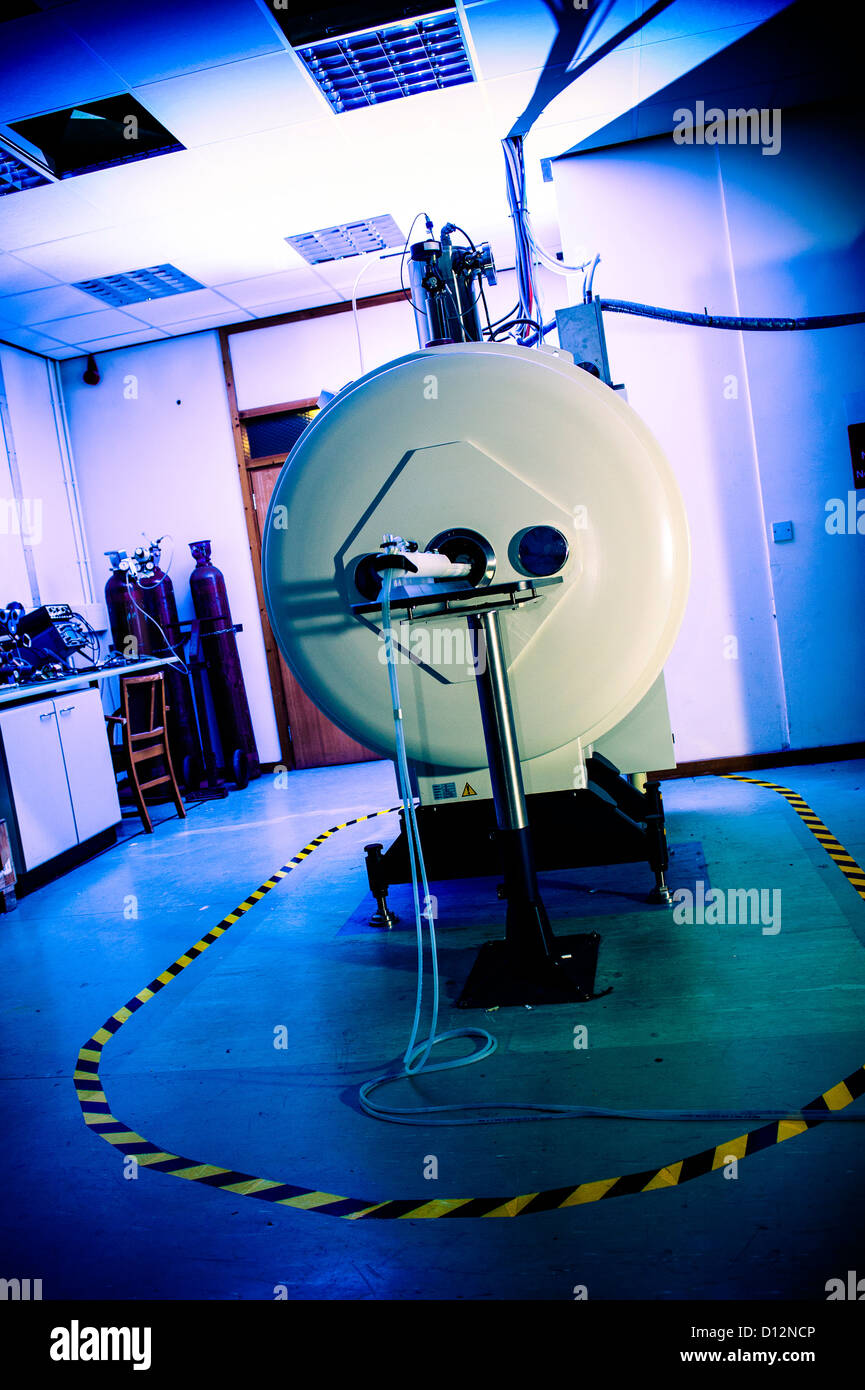 Small bore Magnetic Resonance Imaging (MRI) scanner, in which small subjects are scanned. Stock Photohttps://www.alamy.com/image-license-details/?v=1https://www.alamy.com/stock-photo-small-bore-magnetic-resonance-imaging-mri-scanner-in-which-small-subjects-52306486.html
RMD12NCP–Small bore Magnetic Resonance Imaging (MRI) scanner, in which small subjects are scanned.
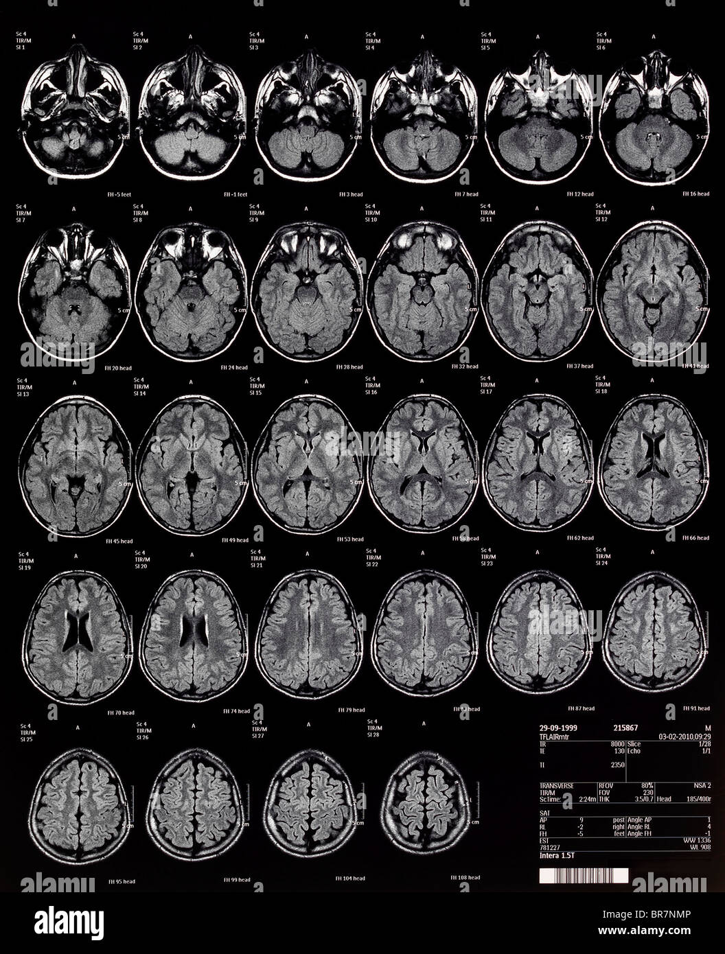 children's Brain MRI magnetic resonance imaging or NMRI nuclear magnetic resonance imaging of Stock Photohttps://www.alamy.com/image-license-details/?v=1https://www.alamy.com/stock-photo-childrens-brain-mri-magnetic-resonance-imaging-or-nmri-nuclear-magnetic-31518166.html
RMBR7NMP–children's Brain MRI magnetic resonance imaging or NMRI nuclear magnetic resonance imaging of
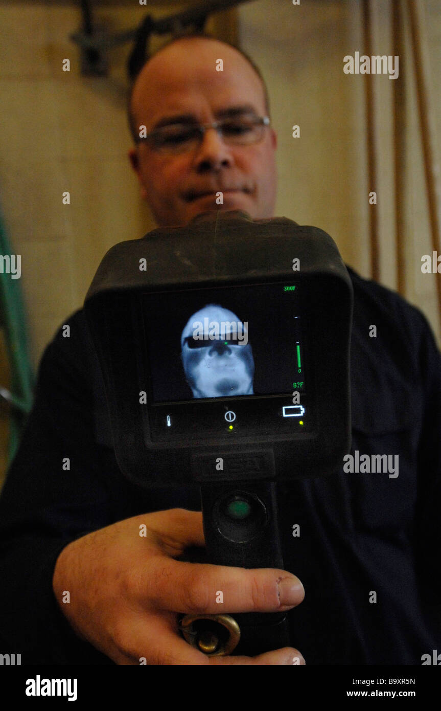 Fireman showing display on MSA thermal imaging Camera. Note glasses block out some of heat signature. Stock Photohttps://www.alamy.com/image-license-details/?v=1https://www.alamy.com/stock-photo-fireman-showing-display-on-msa-thermal-imaging-camera-note-glasses-23331217.html
RMB9XR5N–Fireman showing display on MSA thermal imaging Camera. Note glasses block out some of heat signature.
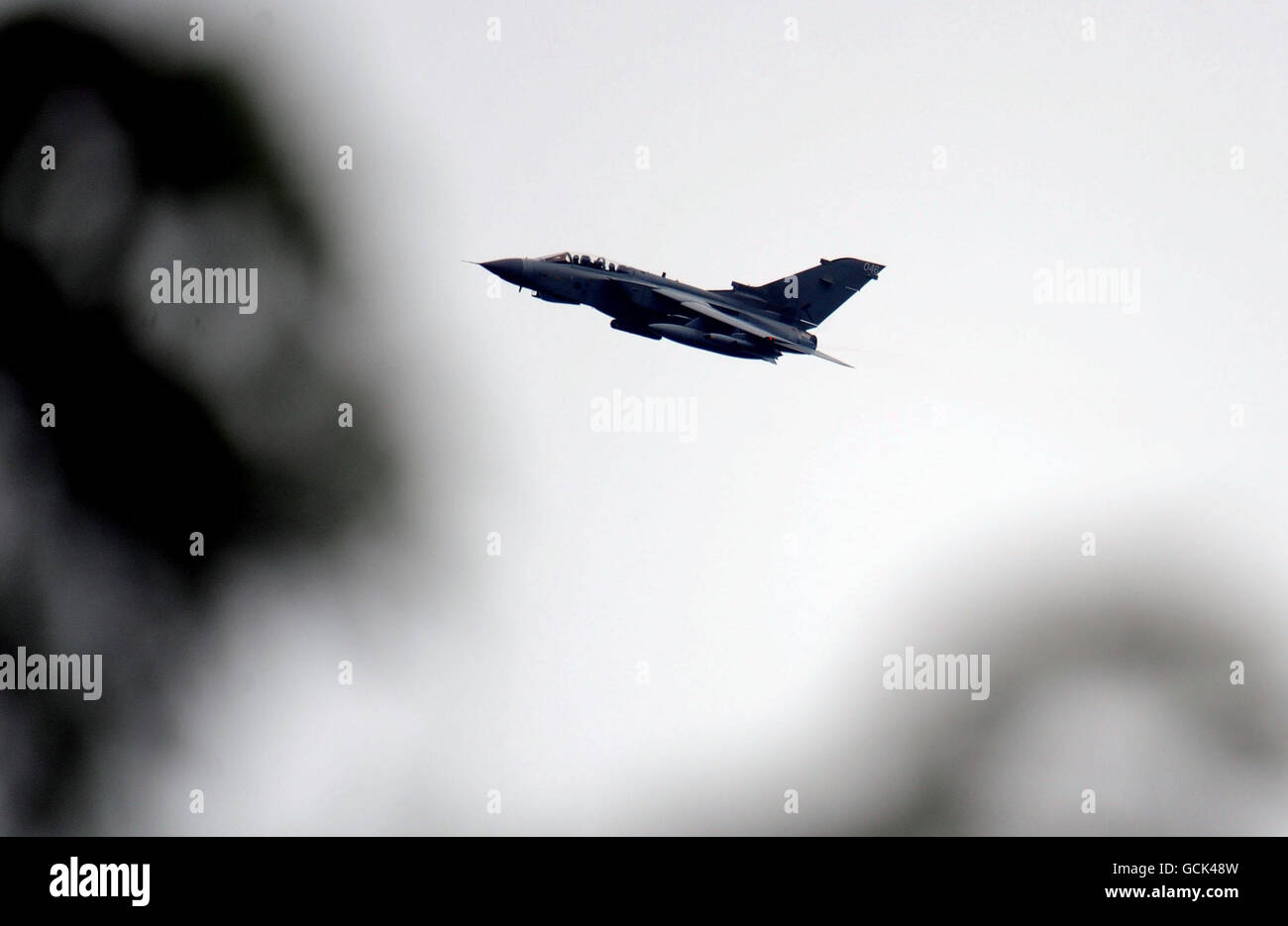 Gateshead shootings. A RAF Tornado with imaging equipment flies over Rothbury as the search for Raoul Moat continues. Stock Photohttps://www.alamy.com/image-license-details/?v=1https://www.alamy.com/stock-photo-gateshead-shootings-a-raf-tornado-with-imaging-equipment-flies-over-111058553.html
RMGCK48W–Gateshead shootings. A RAF Tornado with imaging equipment flies over Rothbury as the search for Raoul Moat continues.
 Hunting a deer in a forest at night using thermal imaging. Scope view with crosshair. Stock Photohttps://www.alamy.com/image-license-details/?v=1https://www.alamy.com/hunting-a-deer-in-a-forest-at-night-using-thermal-imaging-scope-view-with-crosshair-image328002648.html
RF2A1HPM8–Hunting a deer in a forest at night using thermal imaging. Scope view with crosshair.
 MRI scanner Magnetic Resonance Imaging Hospital Medical Imaging department in hospital Stock Photohttps://www.alamy.com/image-license-details/?v=1https://www.alamy.com/stock-photo-mri-scanner-magnetic-resonance-imaging-hospital-medical-imaging-department-82516269.html
RMEP6X9H–MRI scanner Magnetic Resonance Imaging Hospital Medical Imaging department in hospital
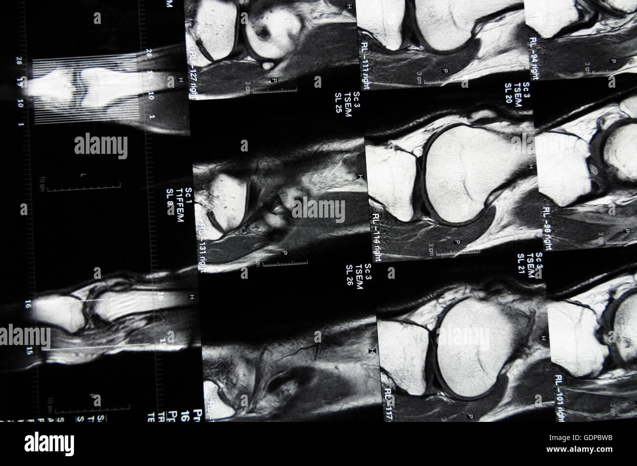 : Magnetic Resonance Imaging ( MRI ) : cross-sectional images of a knee, , , Stock Photohttps://www.alamy.com/image-license-details/?v=1https://www.alamy.com/stock-photo-magnetic-resonance-imaging-mri-cross-sectional-images-of-a-knee-111745015.html
RMGDPBWB–: Magnetic Resonance Imaging ( MRI ) : cross-sectional images of a knee, , ,
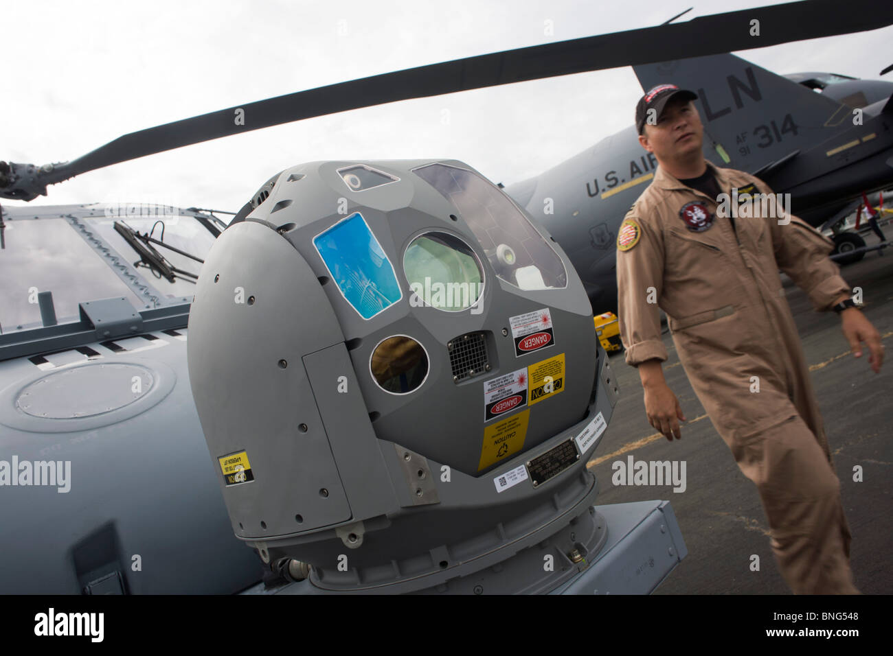 US Navy pilot and infra-red imaging camera on nose of a Sikorsky MH-60R helicopter at the Farnborough Airshow. Stock Photohttps://www.alamy.com/image-license-details/?v=1https://www.alamy.com/stock-photo-us-navy-pilot-and-infra-red-imaging-camera-on-nose-of-a-sikorsky-mh-30473416.html
RMBNG548–US Navy pilot and infra-red imaging camera on nose of a Sikorsky MH-60R helicopter at the Farnborough Airshow.
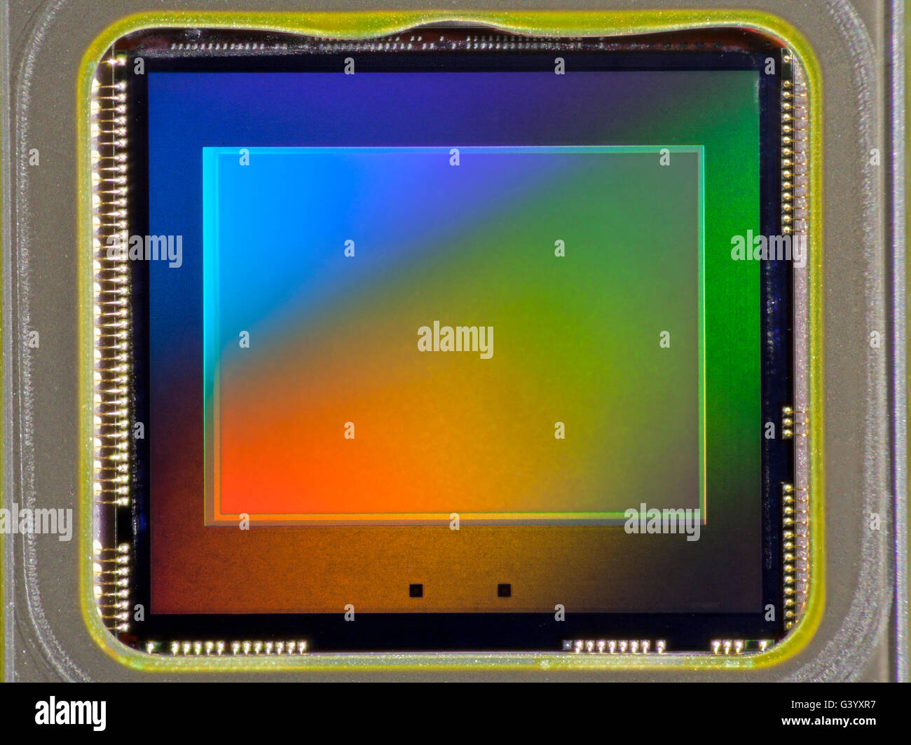 Tiny CCD imaging chip from an amateur consumer camera (approx 3mp) Stock Photohttps://www.alamy.com/image-license-details/?v=1https://www.alamy.com/stock-photo-tiny-ccd-imaging-chip-from-an-amateur-consumer-camera-approx-3mp-105719915.html
RMG3YXR7–Tiny CCD imaging chip from an amateur consumer camera (approx 3mp)
 Nanoparticles Deliver Theranostic Imaging Agent Stock Photohttps://www.alamy.com/image-license-details/?v=1https://www.alamy.com/nanoparticles-deliver-theranostic-imaging-agent-image352771879.html
RM2BDX43K–Nanoparticles Deliver Theranostic Imaging Agent
 Equipment in an Imaging Lab. Stock Photohttps://www.alamy.com/image-license-details/?v=1https://www.alamy.com/stock-photo-equipment-in-an-imaging-lab-56551243.html
RMD803K7–Equipment in an Imaging Lab.
 Coloured magnetic resonance imaging (MRI) scan and CT of an axial section through the brain of patient, showing the damage Stock Photohttps://www.alamy.com/image-license-details/?v=1https://www.alamy.com/stock-photo-coloured-magnetic-resonance-imaging-mri-scan-and-ct-of-an-axial-section-72439080.html
RME5RTP0–Coloured magnetic resonance imaging (MRI) scan and CT of an axial section through the brain of patient, showing the damage
 Infrared thermography (IRT), thermal imaging, and thermal video are examples of infrared imaging science. When viewed through a thermal imaging camera, warm objects stand out well against cooler backgrounds; humans and other warm-blooded animals become easily visible against the environment, day or night. Stock Photohttps://www.alamy.com/image-license-details/?v=1https://www.alamy.com/stock-image-infrared-thermography-irt-thermal-imaging-and-thermal-video-are-examples-162595483.html
RMKCET5F–Infrared thermography (IRT), thermal imaging, and thermal video are examples of infrared imaging science. When viewed through a thermal imaging camera, warm objects stand out well against cooler backgrounds; humans and other warm-blooded animals become easily visible against the environment, day or night.
 Crowds passing Fujifilm stand at Photokina digital imaging trade show in Cologne Germany Stock Photohttps://www.alamy.com/image-license-details/?v=1https://www.alamy.com/stock-photo-crowds-passing-fujifilm-stand-at-photokina-digital-imaging-trade-show-31656882.html
RMBRE2JX–Crowds passing Fujifilm stand at Photokina digital imaging trade show in Cologne Germany
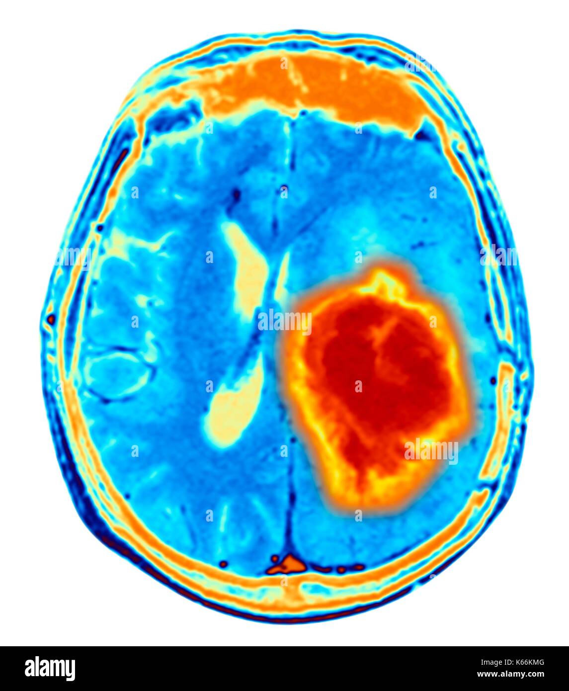 Brain tumour. Coloured Magnetic Resonance Imaging (MRI) scan of an axial section through the brain showing a metastatic tumour. At bottom left is the tumour (red-yellow) This tumour occurs within one cerebral hemisphere; the other hemisphere is at right. The eyeballs - not visible -are at top. Metastatic cancer is a secondary disease spread from cancer elsewhere in the body. Metastatic brain tumours are malignant. Typically they cause brain compression and nerve damage Stock Photohttps://www.alamy.com/image-license-details/?v=1https://www.alamy.com/brain-tumour-coloured-magnetic-resonance-imaging-mri-scan-of-an-axial-image158728432.html
RFK66KMG–Brain tumour. Coloured Magnetic Resonance Imaging (MRI) scan of an axial section through the brain showing a metastatic tumour. At bottom left is the tumour (red-yellow) This tumour occurs within one cerebral hemisphere; the other hemisphere is at right. The eyeballs - not visible -are at top. Metastatic cancer is a secondary disease spread from cancer elsewhere in the body. Metastatic brain tumours are malignant. Typically they cause brain compression and nerve damage
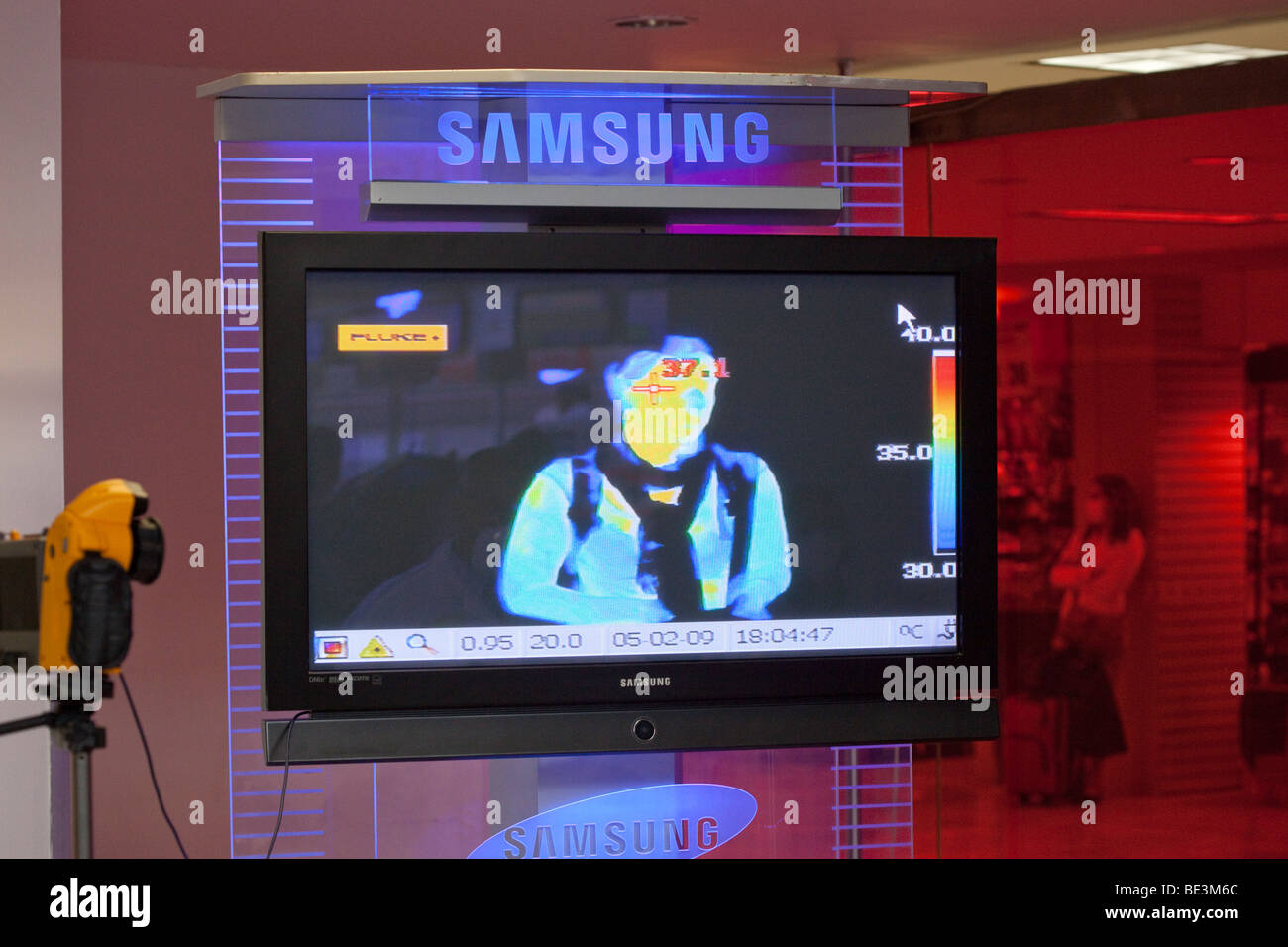 Test for elevated body temperature with a thermal imaging camera at the international airport of Mexico City, Mexico / DF, Mexi Stock Photohttps://www.alamy.com/image-license-details/?v=1https://www.alamy.com/stock-photo-test-for-elevated-body-temperature-with-a-thermal-imaging-camera-at-25897268.html
RMBE3M6C–Test for elevated body temperature with a thermal imaging camera at the international airport of Mexico City, Mexico / DF, Mexi
 Forensics expert using RUVIS Reflected Ultra-violet Imaging Systems to examine evidence. Nebraska State patrol Crime Lab. Stock Photohttps://www.alamy.com/image-license-details/?v=1https://www.alamy.com/stock-photo-forensics-expert-using-ruvis-reflected-ultra-violet-imaging-systems-29366540.html
RMBKNN90–Forensics expert using RUVIS Reflected Ultra-violet Imaging Systems to examine evidence. Nebraska State patrol Crime Lab.
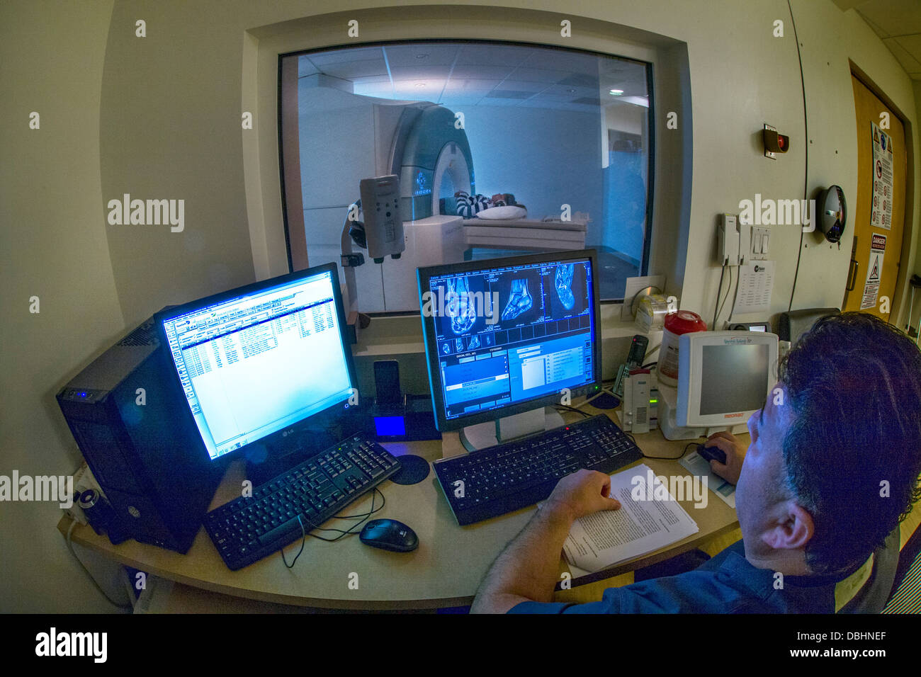 Viewed through safety glass window teenage girll's injured ankle is examined using a Siemens 3T Vario magnetic resonance imaging Stock Photohttps://www.alamy.com/image-license-details/?v=1https://www.alamy.com/stock-photo-viewed-through-safety-glass-window-teenage-girlls-injured-ankle-is-58782375.html
RMDBHNEF–Viewed through safety glass window teenage girll's injured ankle is examined using a Siemens 3T Vario magnetic resonance imaging
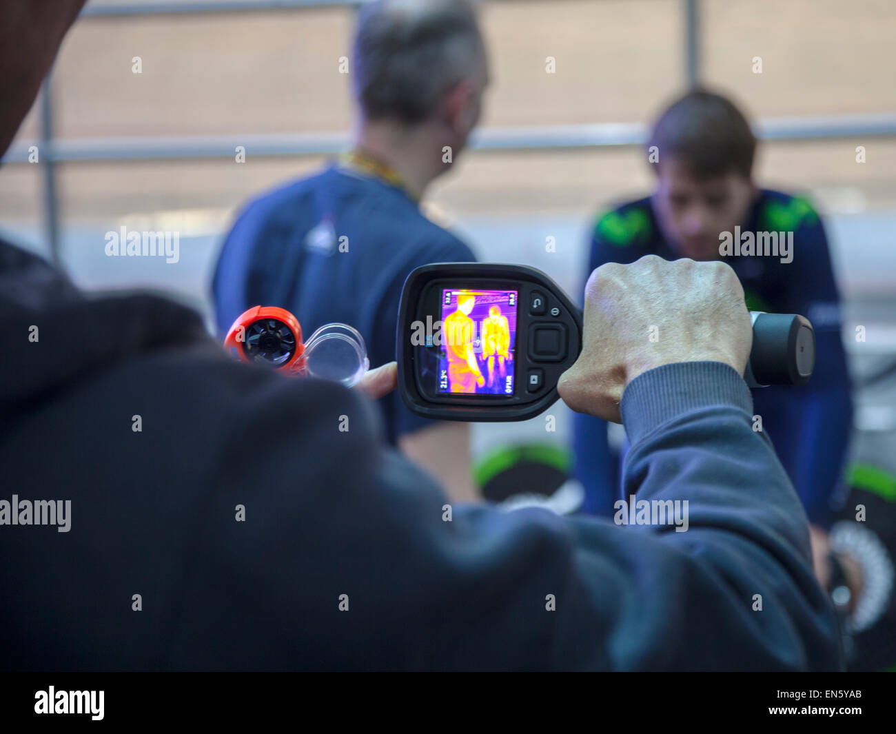 Manchester Velodrome, UK. 28th April, 2015. Alex Dowsett Team Movistar carrying out tests with infra red imaging prior to his attempt on the Hour record on Saturday 2nd May 2015. Credit: Anthony Collins/Alamy Live News Stock Photohttps://www.alamy.com/image-license-details/?v=1https://www.alamy.com/stock-photo-manchester-velodrome-uk-28th-april-2015-alex-dowsett-team-movistar-81880467.html
RMEN5YAB–Manchester Velodrome, UK. 28th April, 2015. Alex Dowsett Team Movistar carrying out tests with infra red imaging prior to his attempt on the Hour record on Saturday 2nd May 2015. Credit: Anthony Collins/Alamy Live News
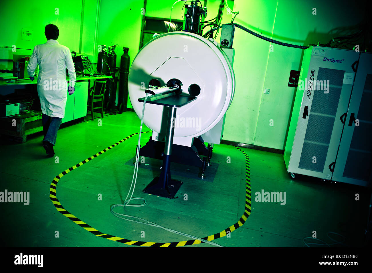 Technician or scientist in room with small bore Magnetic Resonance Imaging (MRI) scanner, in which small subjects are scanned. Stock Photohttps://www.alamy.com/image-license-details/?v=1https://www.alamy.com/stock-photo-technician-or-scientist-in-room-with-small-bore-magnetic-resonance-52306436.html
RMD12NB0–Technician or scientist in room with small bore Magnetic Resonance Imaging (MRI) scanner, in which small subjects are scanned.
 children's Brain MRI magnetic resonance imaging or NMRI nuclear magnetic resonance imaging of Stock Photohttps://www.alamy.com/image-license-details/?v=1https://www.alamy.com/stock-photo-childrens-brain-mri-magnetic-resonance-imaging-or-nmri-nuclear-magnetic-31518162.html
RMBR7NMJ–children's Brain MRI magnetic resonance imaging or NMRI nuclear magnetic resonance imaging of
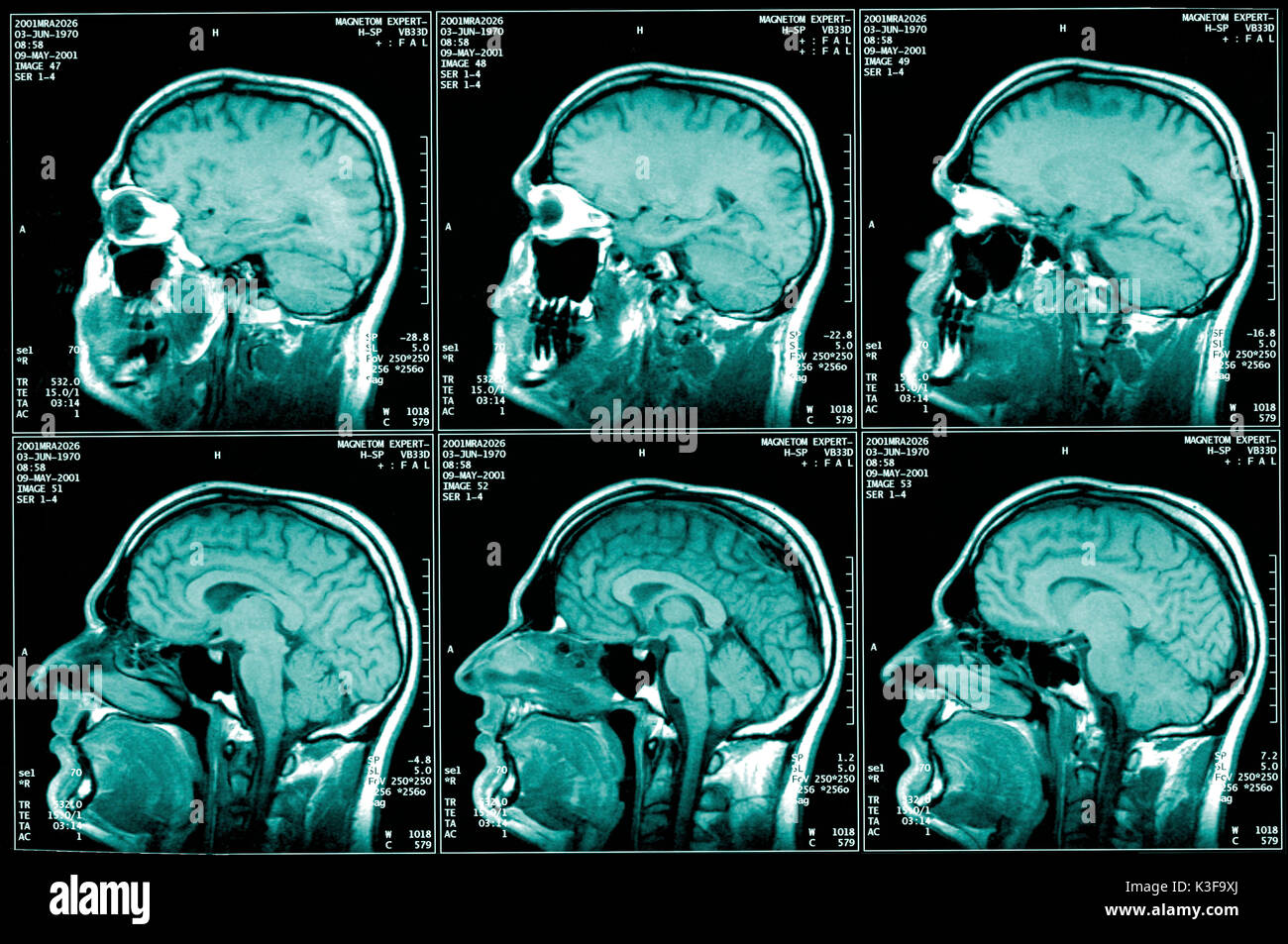 nuclear magnetic resonance imaging, magnetic resonance imaging Stock Photohttps://www.alamy.com/image-license-details/?v=1https://www.alamy.com/nuclear-magnetic-resonance-imaging-magnetic-resonance-imaging-image157074362.html
RFK3F9XJ–nuclear magnetic resonance imaging, magnetic resonance imaging
 General view of the Cardiff University Brain Research Imaging Centre, in Cardiff, Wales. Stock Photohttps://www.alamy.com/image-license-details/?v=1https://www.alamy.com/general-view-of-the-cardiff-university-brain-research-imaging-centre-in-cardiff-wales-image372083996.html
RM2CH9TX4–General view of the Cardiff University Brain Research Imaging Centre, in Cardiff, Wales.
 Miami Beach Florida,PillCam Endoscopy camera,ingestible imaging capsule,FL190331058 Stock Photohttps://www.alamy.com/image-license-details/?v=1https://www.alamy.com/miami-beach-floridapillcam-endoscopy-cameraingestible-imaging-capsulefl190331058-image248491627.html
RMTC7NGY–Miami Beach Florida,PillCam Endoscopy camera,ingestible imaging capsule,FL190331058
 New medical imaging device, doctors examining monitor Stock Photohttps://www.alamy.com/image-license-details/?v=1https://www.alamy.com/stock-photo-new-medical-imaging-device-doctors-examining-monitor-26668402.html
RMBFARPX–New medical imaging device, doctors examining monitor
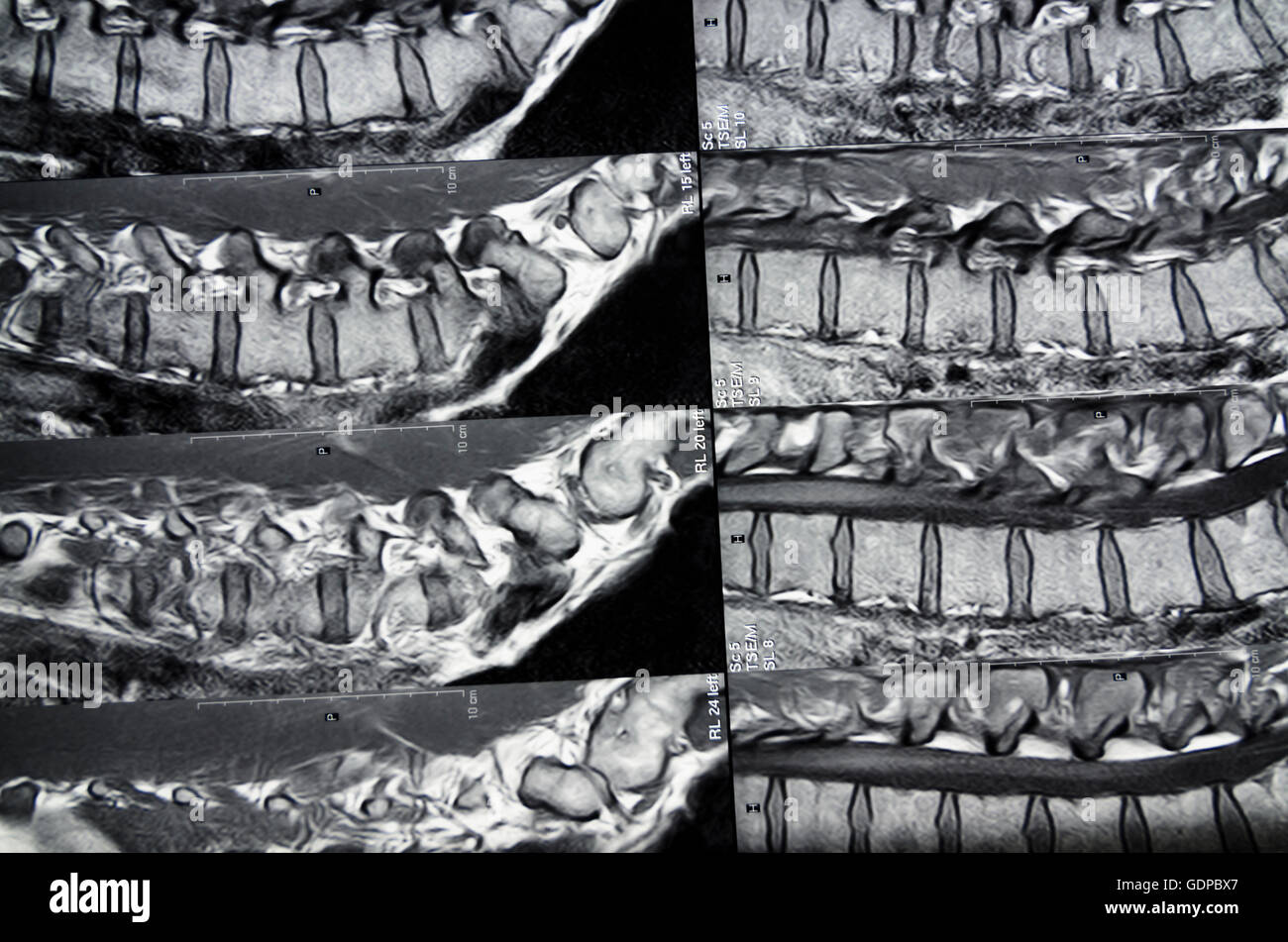 : Magnetic Resonance Imaging ( MRI ) : cross-sectional images of the lumbar spine, , , Stock Photohttps://www.alamy.com/image-license-details/?v=1https://www.alamy.com/stock-photo-magnetic-resonance-imaging-mri-cross-sectional-images-of-the-lumbar-111745039.html
RMGDPBX7–: Magnetic Resonance Imaging ( MRI ) : cross-sectional images of the lumbar spine, , ,
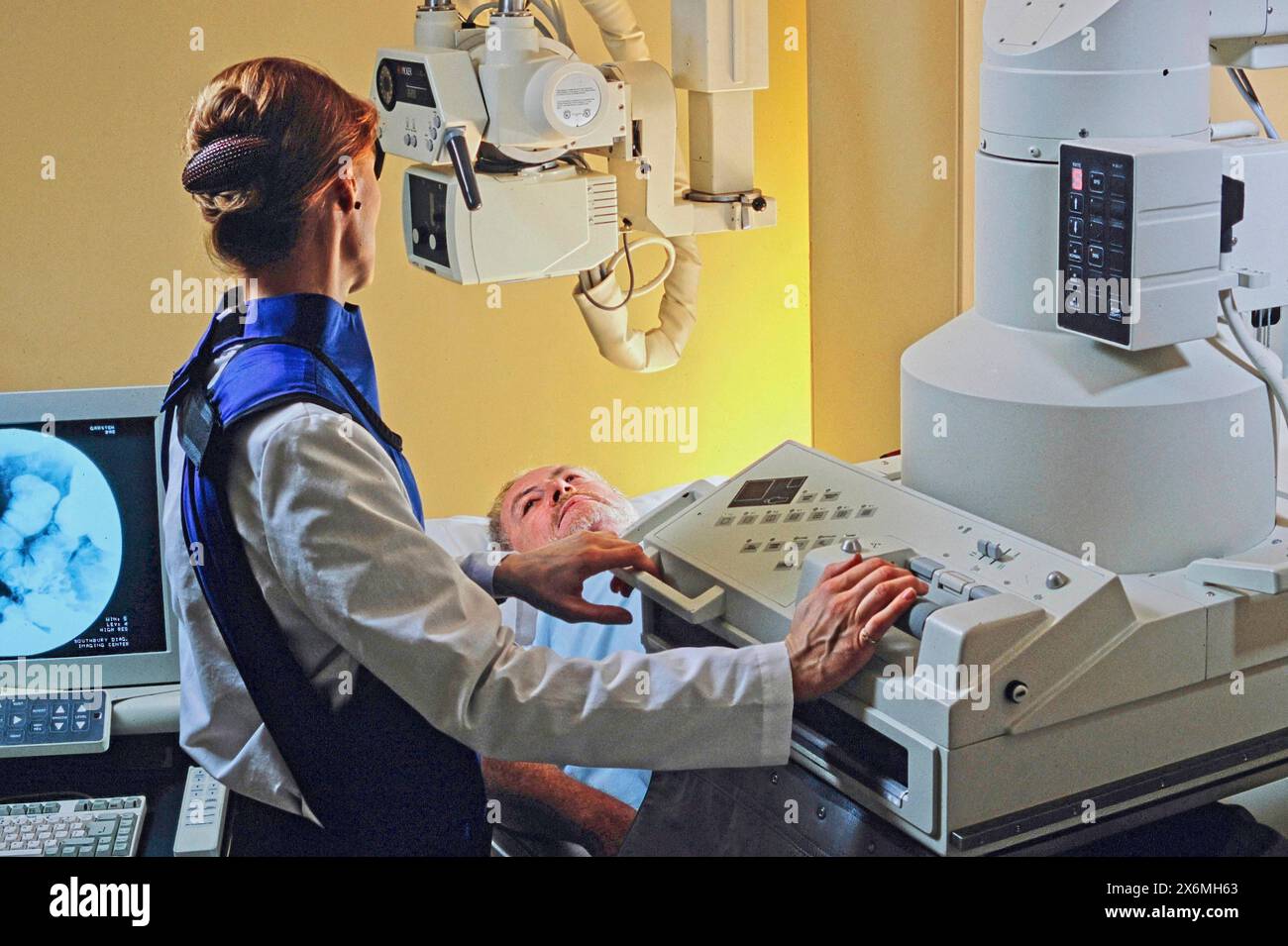 A medical technician conducts an X-ray examination for an elderly patient in a modern medical facility using advanced imaging equipment. The technicia Stock Photohttps://www.alamy.com/image-license-details/?v=1https://www.alamy.com/a-medical-technician-conducts-an-x-ray-examination-for-an-elderly-patient-in-a-modern-medical-facility-using-advanced-imaging-equipment-the-technicia-image606503355.html
RF2X6MH63–A medical technician conducts an X-ray examination for an elderly patient in a modern medical facility using advanced imaging equipment. The technicia
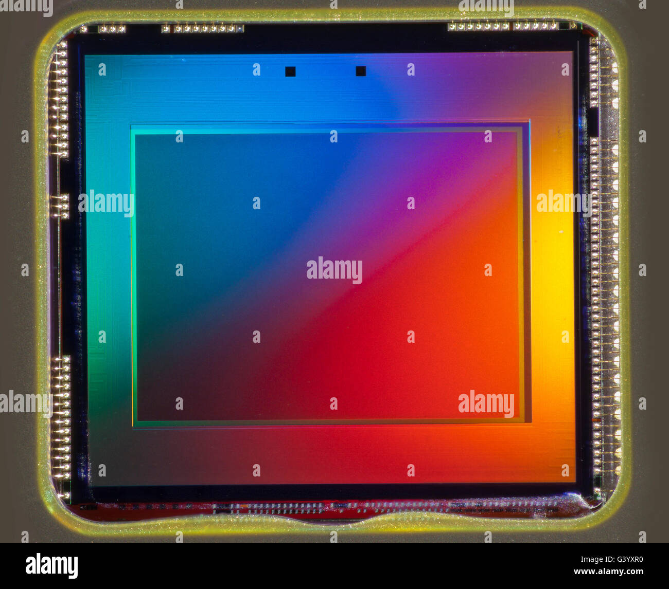 Tiny CCD imaging chip from an amateur consumer camera (approx 3mp) Stock Photohttps://www.alamy.com/image-license-details/?v=1https://www.alamy.com/stock-photo-tiny-ccd-imaging-chip-from-an-amateur-consumer-camera-approx-3mp-105719908.html
RMG3YXR0–Tiny CCD imaging chip from an amateur consumer camera (approx 3mp)
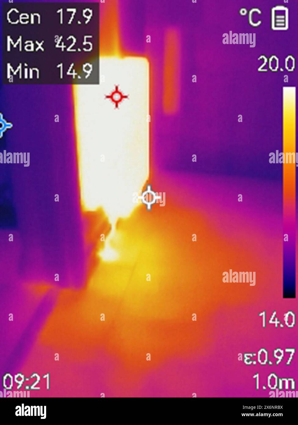 Leak detection using thermal imaging camera. Infrared image of a radiator and a heat plume in the floor from a leak in a hot water pipe. Central heati Stock Photohttps://www.alamy.com/image-license-details/?v=1https://www.alamy.com/leak-detection-using-thermal-imaging-camera-infrared-image-of-a-radiator-and-a-heat-plume-in-the-floor-from-a-leak-in-a-hot-water-pipe-central-heati-image606530174.html
RF2X6NRBX–Leak detection using thermal imaging camera. Infrared image of a radiator and a heat plume in the floor from a leak in a hot water pipe. Central heati
 Reseachers check the monitor image in the Imaging Lab. Stock Photohttps://www.alamy.com/image-license-details/?v=1https://www.alamy.com/stock-photo-reseachers-check-the-monitor-image-in-the-imaging-lab-56551225.html
RMD803JH–Reseachers check the monitor image in the Imaging Lab.
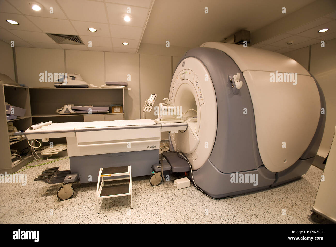 Closed geometry Magnetic Resonance Imaging (MRI) unit, 3 tesla. Stock Photohttps://www.alamy.com/image-license-details/?v=1https://www.alamy.com/stock-photo-closed-geometry-magnetic-resonance-imaging-mri-unit-3-tesla-72424617.html
RME5R69D–Closed geometry Magnetic Resonance Imaging (MRI) unit, 3 tesla.
 The Visible Infrared Imaging Radiometer Suite (VIIRS) on the Suomi NPP satellite, captured this nightime view of the Americas at night Stock Photohttps://www.alamy.com/image-license-details/?v=1https://www.alamy.com/stock-image-the-visible-infrared-imaging-radiometer-suite-viirs-on-the-suomi-npp-165992276.html
RMKJ1GRG–The Visible Infrared Imaging Radiometer Suite (VIIRS) on the Suomi NPP satellite, captured this nightime view of the Americas at night
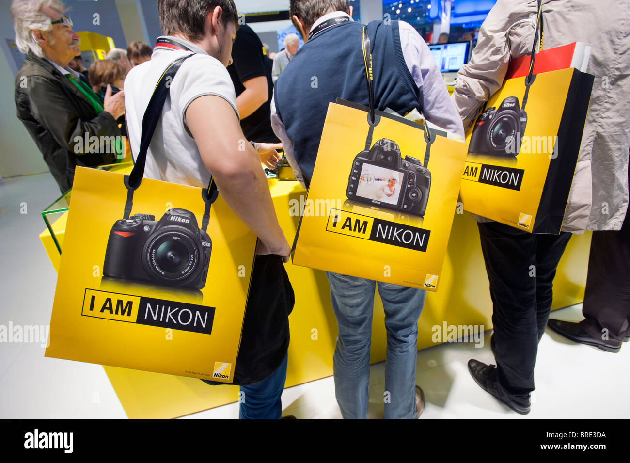 Many people at Nikon stand at Photokina digital imaging trade show in Cologne Germany Stock Photohttps://www.alamy.com/image-license-details/?v=1https://www.alamy.com/stock-photo-many-people-at-nikon-stand-at-photokina-digital-imaging-trade-show-31657510.html
RMBRE3DA–Many people at Nikon stand at Photokina digital imaging trade show in Cologne Germany
 Normal brain. Coloured magnetic resonance imaging (MRI) scan of an axial section through a healthy brain, The images shows the cortex and lateral ventricle (X-shaped in the middle). Stock Photohttps://www.alamy.com/image-license-details/?v=1https://www.alamy.com/stock-photo-normal-brain-coloured-magnetic-resonance-imaging-mri-scan-of-an-axial-134062886.html
RFHP32G6–Normal brain. Coloured magnetic resonance imaging (MRI) scan of an axial section through a healthy brain, The images shows the cortex and lateral ventricle (X-shaped in the middle).
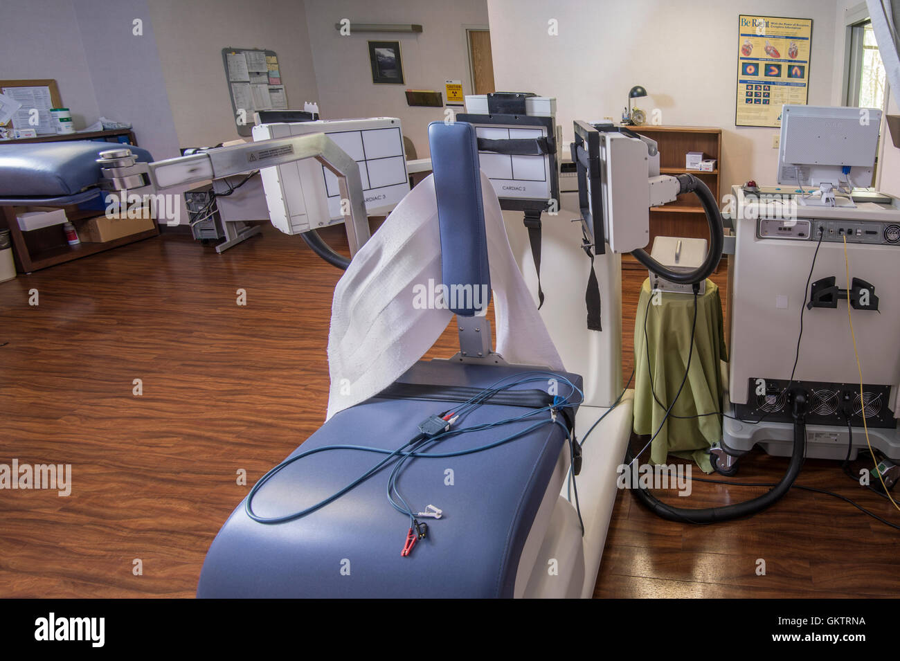 Nuclear Imaging Equipment Stock Photohttps://www.alamy.com/image-license-details/?v=1https://www.alamy.com/stock-photo-nuclear-imaging-equipment-115486150.html
RMGKTRNA–Nuclear Imaging Equipment
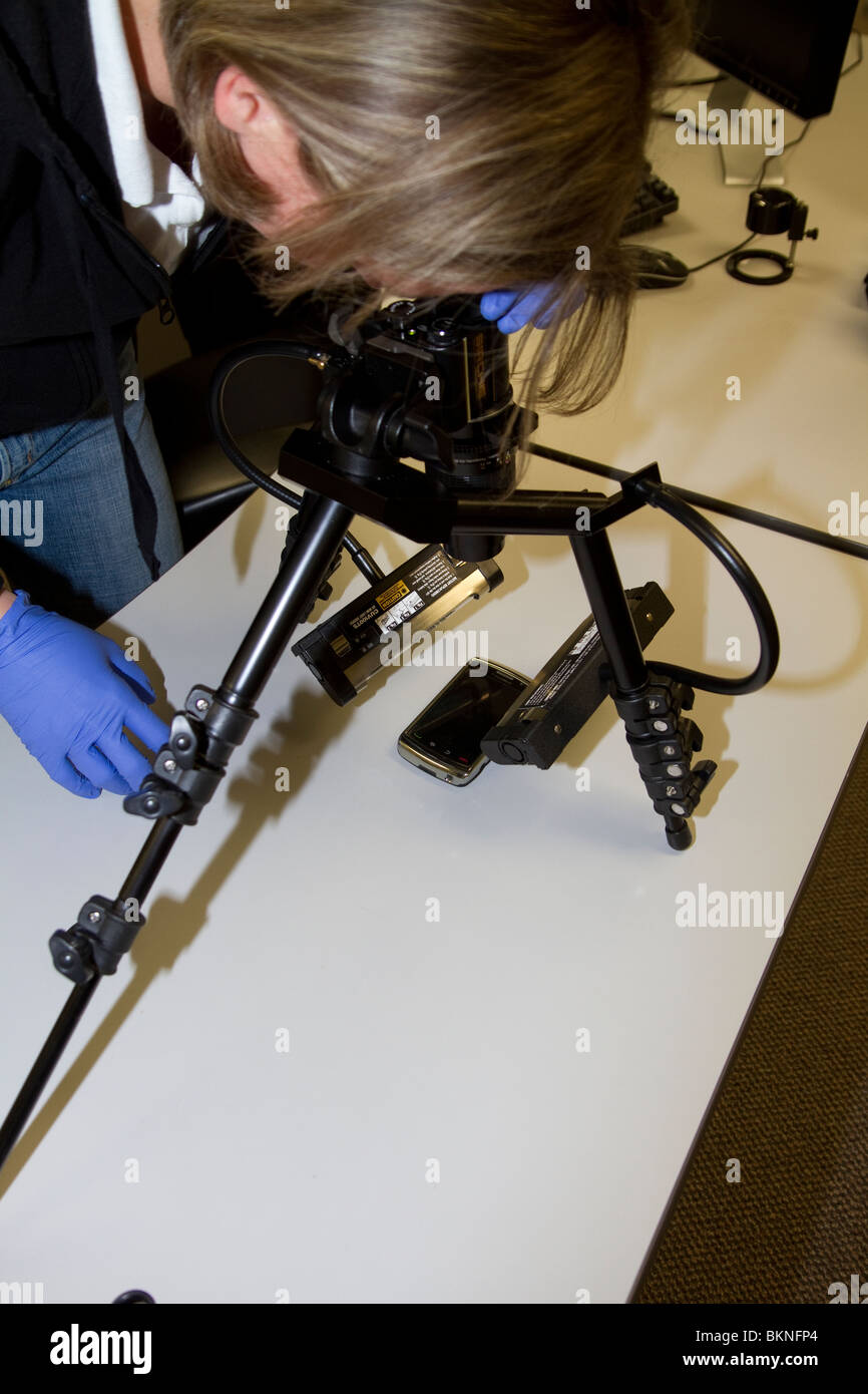 Forensics expert using RUVIS Reflected Ultra-violet Imaging Systems to examine evidence. Nebraska State patrol Crime Lab. Stock Photohttps://www.alamy.com/image-license-details/?v=1https://www.alamy.com/stock-photo-forensics-expert-using-ruvis-reflected-ultra-violet-imaging-systems-29362204.html
RMBKNFP4–Forensics expert using RUVIS Reflected Ultra-violet Imaging Systems to examine evidence. Nebraska State patrol Crime Lab.
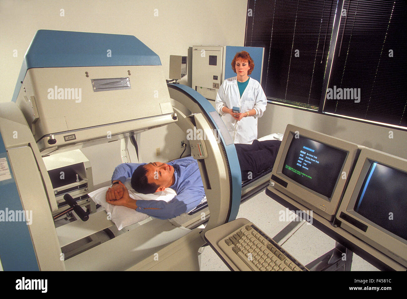 A radiology technician demonstrates an 'open' MRI machine in Laguna Hills, CA. Magnetic resonance imaging is used in radiology to investigate the anatomy and physiology of the body in both health and disease. MRI scanners use magnetic fields and radio wav Stock Photohttps://www.alamy.com/image-license-details/?v=1https://www.alamy.com/stock-photo-a-radiology-technician-demonstrates-an-open-mri-machine-in-laguna-88626536.html
RMF4581C–A radiology technician demonstrates an 'open' MRI machine in Laguna Hills, CA. Magnetic resonance imaging is used in radiology to investigate the anatomy and physiology of the body in both health and disease. MRI scanners use magnetic fields and radio wav
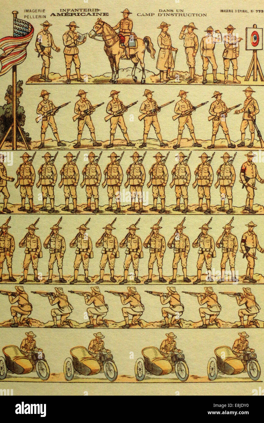 Imaging of Epinal. American infantry. France. The Museum of the Great War. Meaux. Stock Photohttps://www.alamy.com/image-license-details/?v=1https://www.alamy.com/stock-photo-imaging-of-epinal-american-infantry-france-the-museum-of-the-great-74164804.html
RME8JDY0–Imaging of Epinal. American infantry. France. The Museum of the Great War. Meaux.
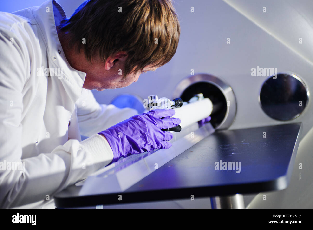 Scientist or technician loads a specimen on a tray into a small bore Magnetic Resonance Imaging (MRI) scanner Stock Photohttps://www.alamy.com/image-license-details/?v=1https://www.alamy.com/stock-photo-scientist-or-technician-loads-a-specimen-on-a-tray-into-a-small-bore-52306555.html
RMD12NF7–Scientist or technician loads a specimen on a tray into a small bore Magnetic Resonance Imaging (MRI) scanner
 children's Brain MRI magnetic resonance imaging or NMRI nuclear magnetic resonance imaging of Stock Photohttps://www.alamy.com/image-license-details/?v=1https://www.alamy.com/stock-photo-childrens-brain-mri-magnetic-resonance-imaging-or-nmri-nuclear-magnetic-31518156.html
RMBR7NMC–children's Brain MRI magnetic resonance imaging or NMRI nuclear magnetic resonance imaging of
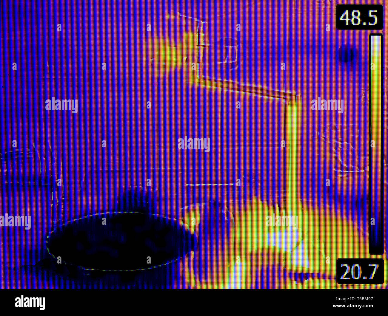 Tube Thermal Imaging Stock Photohttps://www.alamy.com/image-license-details/?v=1https://www.alamy.com/tube-thermal-imaging-image244890499.html
RMT6BM97–Tube Thermal Imaging
 The Duke of Edinburgh at the Cardiff University Brain Research Imaging Centre during a visit to Cardiff. Stock Photohttps://www.alamy.com/image-license-details/?v=1https://www.alamy.com/stock-photo-the-duke-of-edinburgh-at-the-cardiff-university-brain-research-imaging-105189822.html
RMG33PKA–The Duke of Edinburgh at the Cardiff University Brain Research Imaging Centre during a visit to Cardiff.
 Miami Beach Florida,41st Street,Arthur Godfrey Boulevard,MRI machine,magnetic resonance imaging,radiology,medical,scanner,FL110228009 Stock Photohttps://www.alamy.com/image-license-details/?v=1https://www.alamy.com/miami-beach-florida41st-streetarthur-godfrey-boulevardmri-machinemagnetic-resonance-imagingradiologymedicalscannerfl110228009-image350839072.html
RM2BAP2PT–Miami Beach Florida,41st Street,Arthur Godfrey Boulevard,MRI machine,magnetic resonance imaging,radiology,medical,scanner,FL110228009
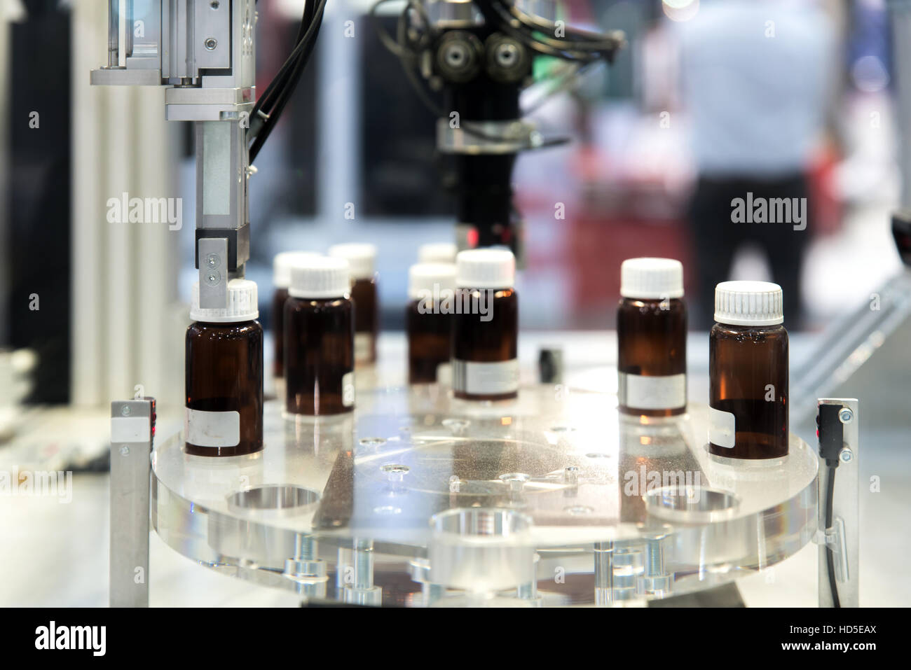 Automatic robot arm with imaging sensor in assembly line working in factory. Smart factory industry 4.0 concept. Stock Photohttps://www.alamy.com/image-license-details/?v=1https://www.alamy.com/stock-photo-automatic-robot-arm-with-imaging-sensor-in-assembly-line-working-in-128584146.html
RFHD5EAX–Automatic robot arm with imaging sensor in assembly line working in factory. Smart factory industry 4.0 concept.
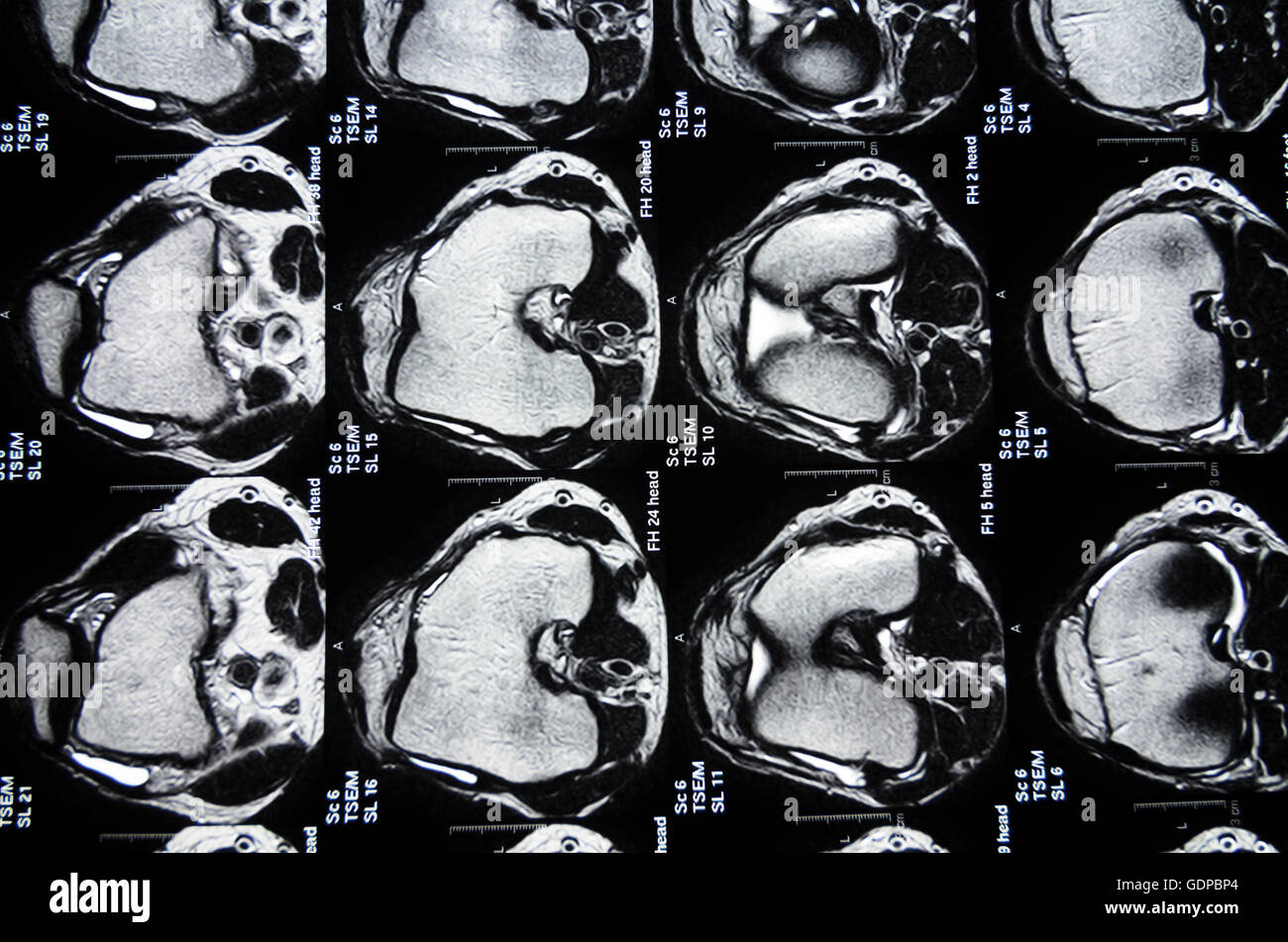 : Magnetic Resonance Imaging ( MRI ) : cross-sectional images of a knee, , , Stock Photohttps://www.alamy.com/image-license-details/?v=1https://www.alamy.com/stock-photo-magnetic-resonance-imaging-mri-cross-sectional-images-of-a-knee-111744924.html
RMGDPBP4–: Magnetic Resonance Imaging ( MRI ) : cross-sectional images of a knee, , ,
 A boy playing with a video imaging exhibit at a science museum Stock Photohttps://www.alamy.com/image-license-details/?v=1https://www.alamy.com/stock-photo-a-boy-playing-with-a-video-imaging-exhibit-at-a-science-museum-59725708.html
RMDD4MN0–A boy playing with a video imaging exhibit at a science museum
 A laser imaging system set up in a science research laboratory. Stock Photohttps://www.alamy.com/image-license-details/?v=1https://www.alamy.com/stock-photo-a-laser-imaging-system-set-up-in-a-science-research-laboratory-72522481.html
RME5YK4H–A laser imaging system set up in a science research laboratory.
