Quick filters:
Blood vessels pelvis Stock Photos and Images
 Human hip blood vessels computer artwork. Stock Photohttps://www.alamy.com/image-license-details/?v=1https://www.alamy.com/human-hip-blood-vessels-computer-artwork-image69880824.html
RFE1K9KM–Human hip blood vessels computer artwork.
 3/4 upper body front view of human skeletal and vascular systems, white background. Stock Photohttps://www.alamy.com/image-license-details/?v=1https://www.alamy.com/34-upper-body-front-view-of-human-skeletal-and-vascular-systems-white-background-image350066097.html
RF2B9ETTH–3/4 upper body front view of human skeletal and vascular systems, white background.
 Sagittal section of the left kidney with the renal arteries and veins. Stock Photohttps://www.alamy.com/image-license-details/?v=1https://www.alamy.com/sagittal-section-of-the-left-kidney-with-the-renal-arteries-and-veins-image476923739.html
RF2JKWN2K–Sagittal section of the left kidney with the renal arteries and veins.
 Human body model with organs on black background Stock Photohttps://www.alamy.com/image-license-details/?v=1https://www.alamy.com/human-body-model-with-organs-on-black-background-image274328432.html
RMWX8MM0–Human body model with organs on black background
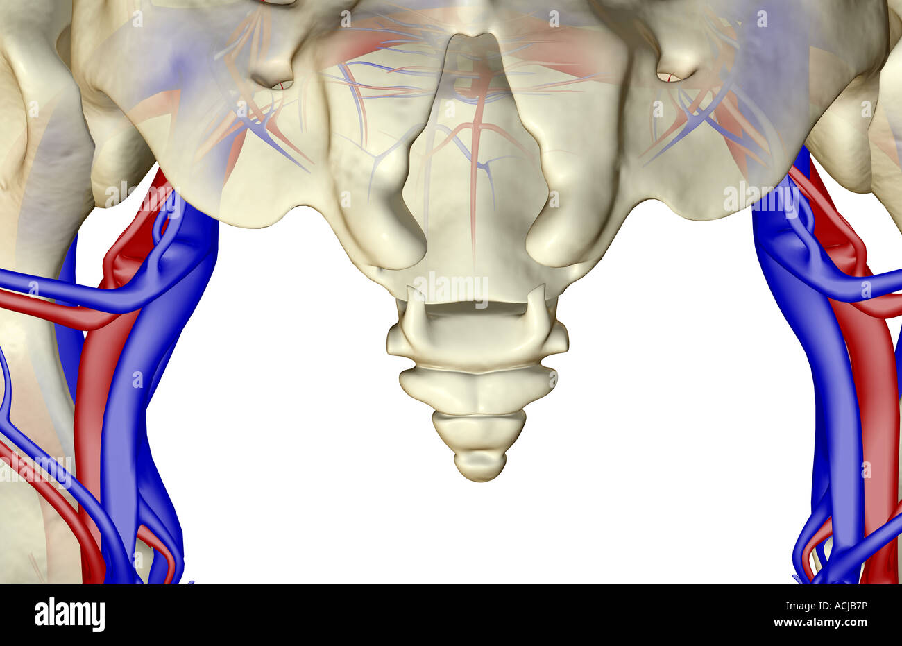 The blood vessels of the pelvis Stock Photohttps://www.alamy.com/image-license-details/?v=1https://www.alamy.com/stock-photo-the-blood-vessels-of-the-pelvis-13168713.html
RFACJB7P–The blood vessels of the pelvis
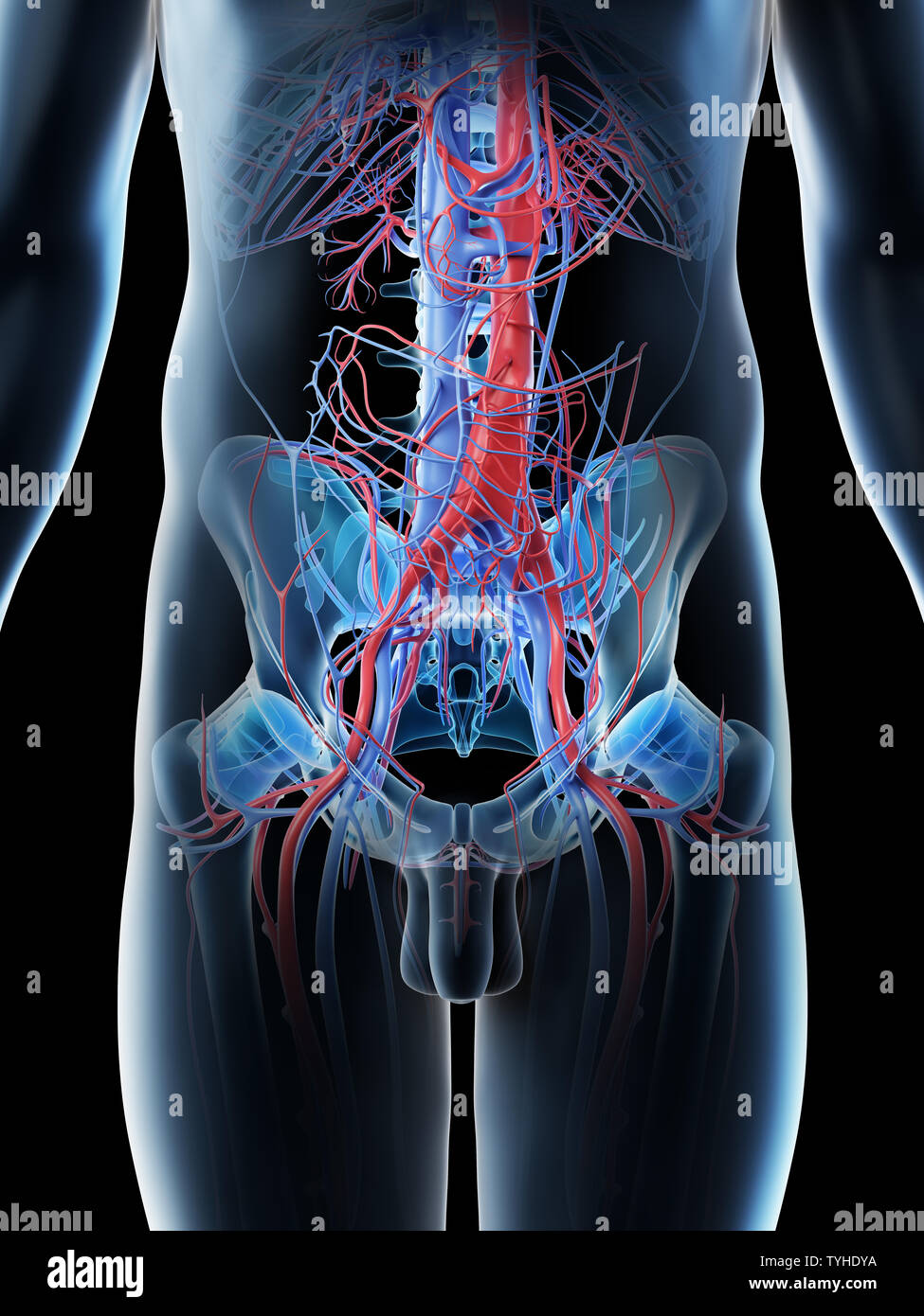 3d rendered illustration of a mans abdominal blood vessels Stock Photohttps://www.alamy.com/image-license-details/?v=1https://www.alamy.com/3d-rendered-illustration-of-a-mans-abdominal-blood-vessels-image257925006.html
RFTYHDYA–3d rendered illustration of a mans abdominal blood vessels
 Cutaneous Nerves and Veins of Lower Limb Anterior and Posterior View Stock Photohttps://www.alamy.com/image-license-details/?v=1https://www.alamy.com/cutaneous-nerves-and-veins-of-lower-limb-anterior-and-posterior-view-image490198303.html
RF2KDECX7–Cutaneous Nerves and Veins of Lower Limb Anterior and Posterior View
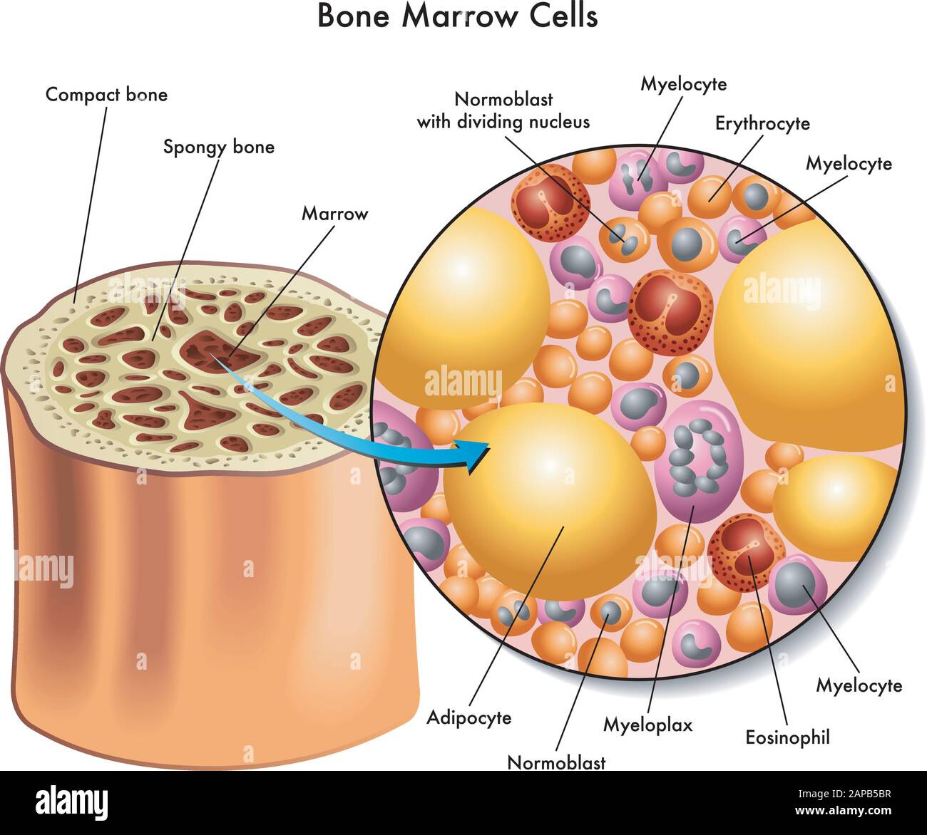 Medical illustration of the composition of bone marrow cells. Stock Vectorhttps://www.alamy.com/image-license-details/?v=1https://www.alamy.com/medical-illustration-of-the-composition-of-bone-marrow-cells-image340765147.html
RF2APB5BR–Medical illustration of the composition of bone marrow cells.
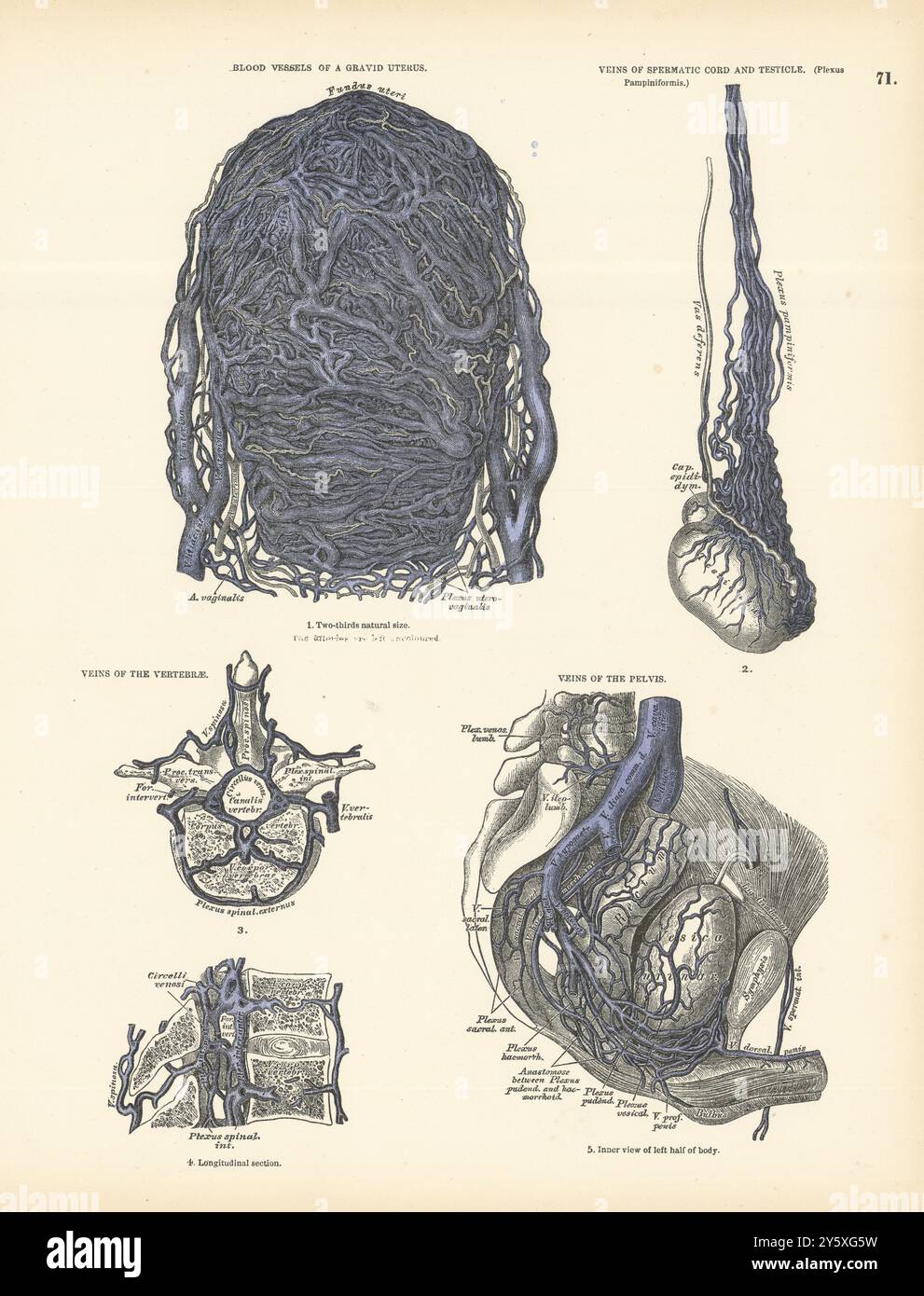 Anatomy. Gravid Uterus Blood Vessels, Testicle, Vertebrae, Pelvis Veins 1880 Stock Photohttps://www.alamy.com/image-license-details/?v=1https://www.alamy.com/anatomy-gravid-uterus-blood-vessels-testicle-vertebrae-pelvis-veins-1880-image623229989.html
RF2Y5XG5W–Anatomy. Gravid Uterus Blood Vessels, Testicle, Vertebrae, Pelvis Veins 1880
 Front view of the bones and major blood vessels of the pelvic region. Stock Photohttps://www.alamy.com/image-license-details/?v=1https://www.alamy.com/stock-photo-front-view-of-the-bones-and-major-blood-vessels-of-the-pelvic-region-52076816.html
RMD0M8E8–Front view of the bones and major blood vessels of the pelvic region.
 Anatomy of male urinary system Stock Photohttps://www.alamy.com/image-license-details/?v=1https://www.alamy.com/anatomy-of-male-urinary-system-image619441097.html
RM2XYNYC9–Anatomy of male urinary system
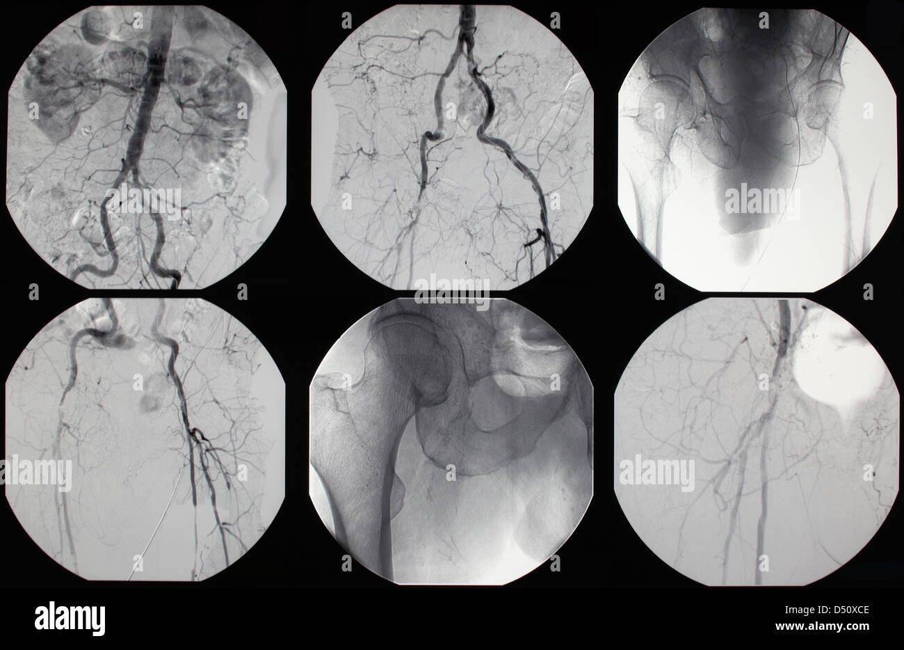 Berlin, Germany, a digital subtraction Stock Photohttps://www.alamy.com/image-license-details/?v=1https://www.alamy.com/stock-photo-berlin-germany-a-digital-subtraction-54725118.html
RMD50XCE–Berlin, Germany, a digital subtraction
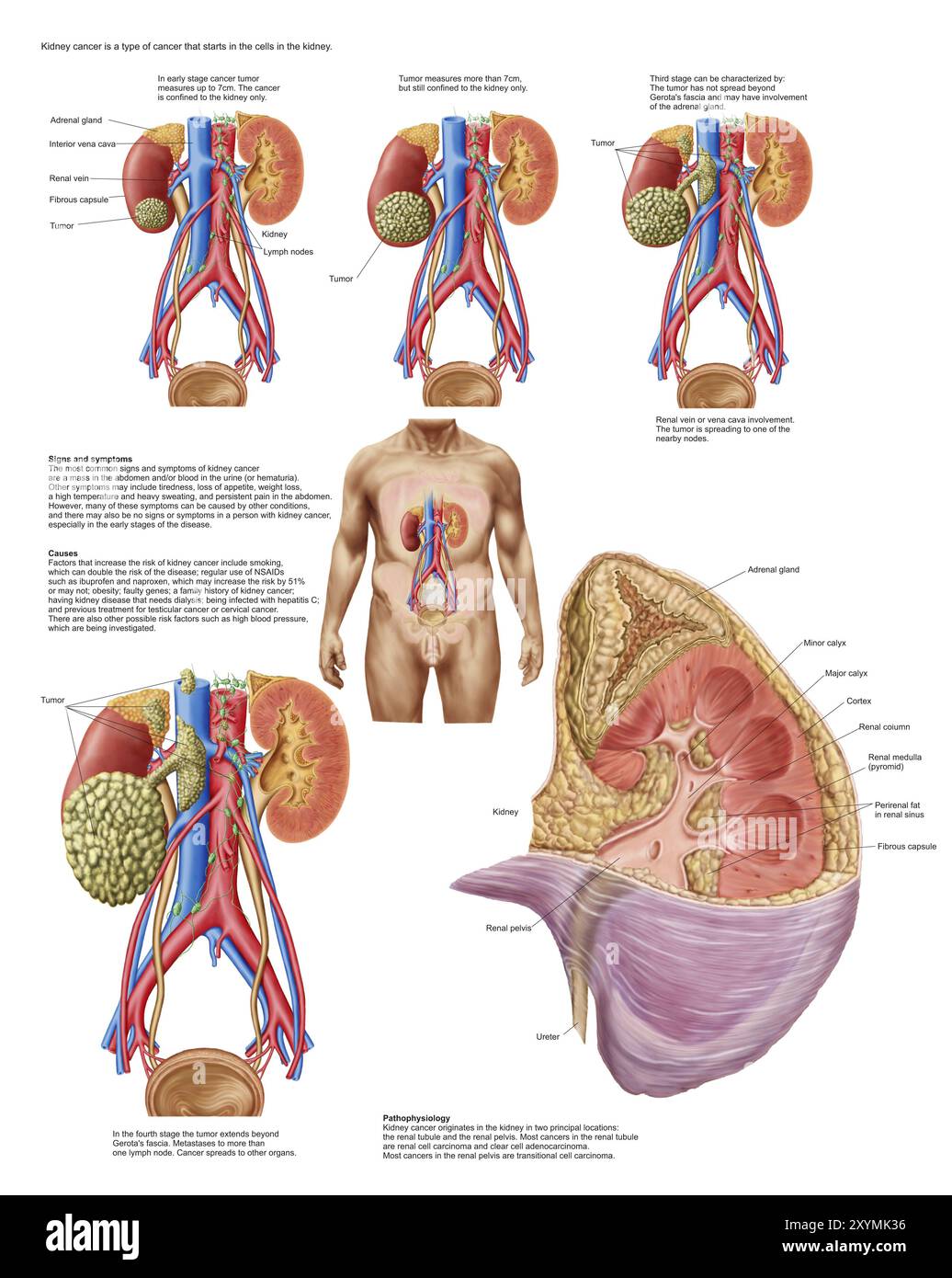 Medical chart showing the signs and symptoms of kidney cancer Stock Photohttps://www.alamy.com/image-license-details/?v=1https://www.alamy.com/medical-chart-showing-the-signs-and-symptoms-of-kidney-cancer-image619412618.html
RM2XYMK36–Medical chart showing the signs and symptoms of kidney cancer
 Male & Female Human Skeletons Stock Photohttps://www.alamy.com/image-license-details/?v=1https://www.alamy.com/stock-photo-male-female-human-skeletons-173239612.html
RMM1RMW0–Male & Female Human Skeletons
 Anatomy of female urinary system Stock Photohttps://www.alamy.com/image-license-details/?v=1https://www.alamy.com/anatomy-of-female-urinary-system-image619256439.html
RM2XYDFWB–Anatomy of female urinary system
 Iliac arteries. The main veins and arteries of the lower body, blood vessels that provide blood to the legs, pelvis and reproductive organs. Flat vector illustration Stock Vectorhttps://www.alamy.com/image-license-details/?v=1https://www.alamy.com/iliac-arteries-the-main-veins-and-arteries-of-the-lower-body-blood-vessels-that-provide-blood-to-the-legs-pelvis-and-reproductive-organs-flat-vector-illustration-image609352421.html
RF2XBAB6D–Iliac arteries. The main veins and arteries of the lower body, blood vessels that provide blood to the legs, pelvis and reproductive organs. Flat vector illustration
 Anatomy of male urinary system Stock Photohttps://www.alamy.com/image-license-details/?v=1https://www.alamy.com/anatomy-of-male-urinary-system-image619220034.html
RM2XYBWD6–Anatomy of male urinary system
 The external iliac veins are blood vessels in your pelvis 3d illustration Stock Photohttps://www.alamy.com/image-license-details/?v=1https://www.alamy.com/the-external-iliac-veins-are-blood-vessels-in-your-pelvis-3d-illustration-image596591059.html
RF2WJH1YF–The external iliac veins are blood vessels in your pelvis 3d illustration
 Human midsection with internal organs Stock Photohttps://www.alamy.com/image-license-details/?v=1https://www.alamy.com/human-midsection-with-internal-organs-image619556963.html
RM2XYY76B–Human midsection with internal organs
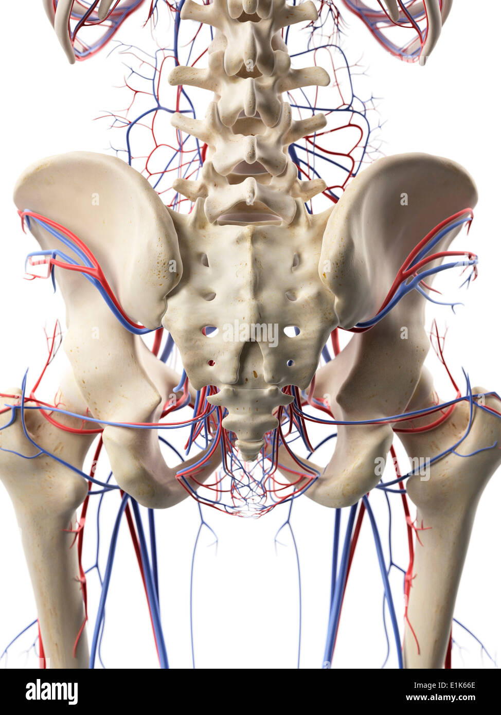 Human blood vessels in the hips computer artwork. Stock Photohttps://www.alamy.com/image-license-details/?v=1https://www.alamy.com/human-blood-vessels-in-the-hips-computer-artwork-image69878102.html
RFE1K66E–Human blood vessels in the hips computer artwork.
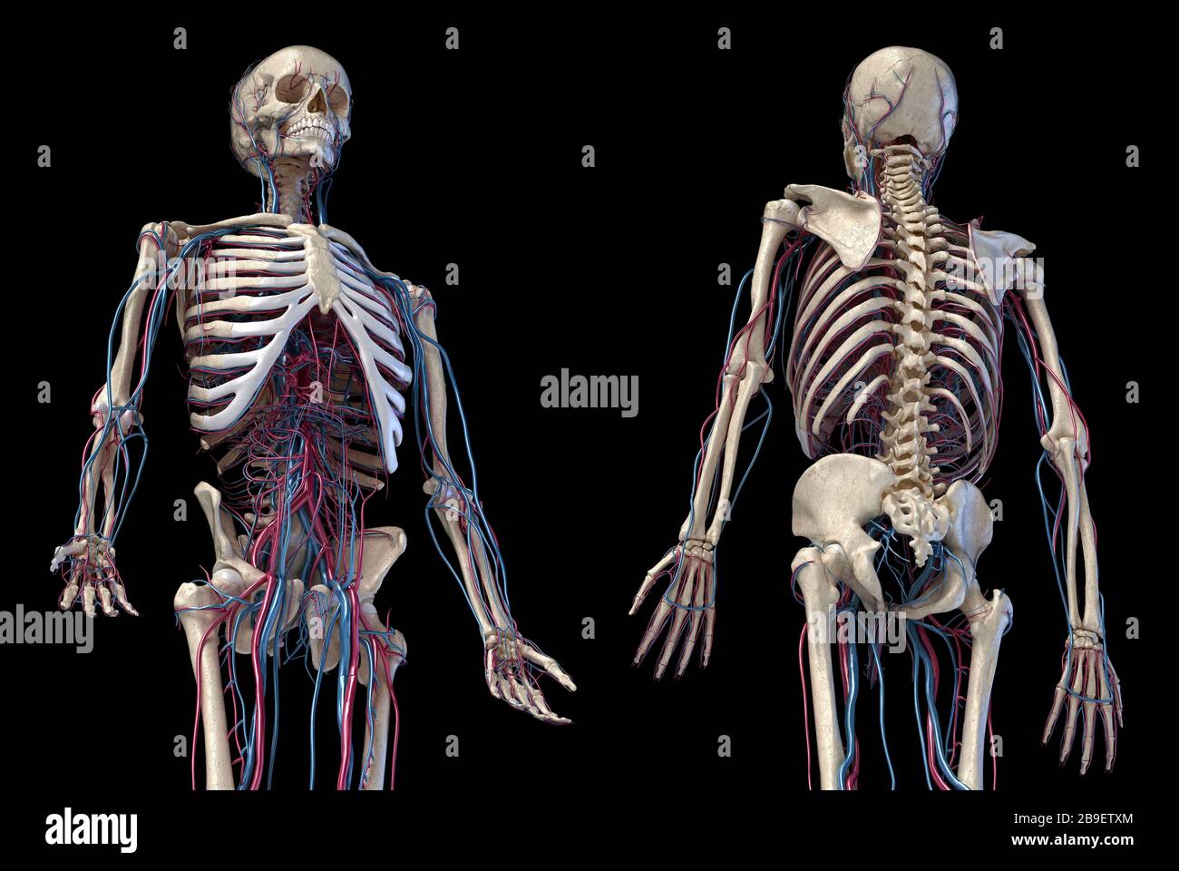 3/4 upper body view of human skeletal and vascular systems, black background. Stock Photohttps://www.alamy.com/image-license-details/?v=1https://www.alamy.com/34-upper-body-view-of-human-skeletal-and-vascular-systems-black-background-image350066156.html
RF2B9ETXM–3/4 upper body view of human skeletal and vascular systems, black background.
 Sagittal section of the left kidney with the renal arteries and veins. Stock Photohttps://www.alamy.com/image-license-details/?v=1https://www.alamy.com/sagittal-section-of-the-left-kidney-with-the-renal-arteries-and-veins-image476923695.html
RF2JKWN13–Sagittal section of the left kidney with the renal arteries and veins.
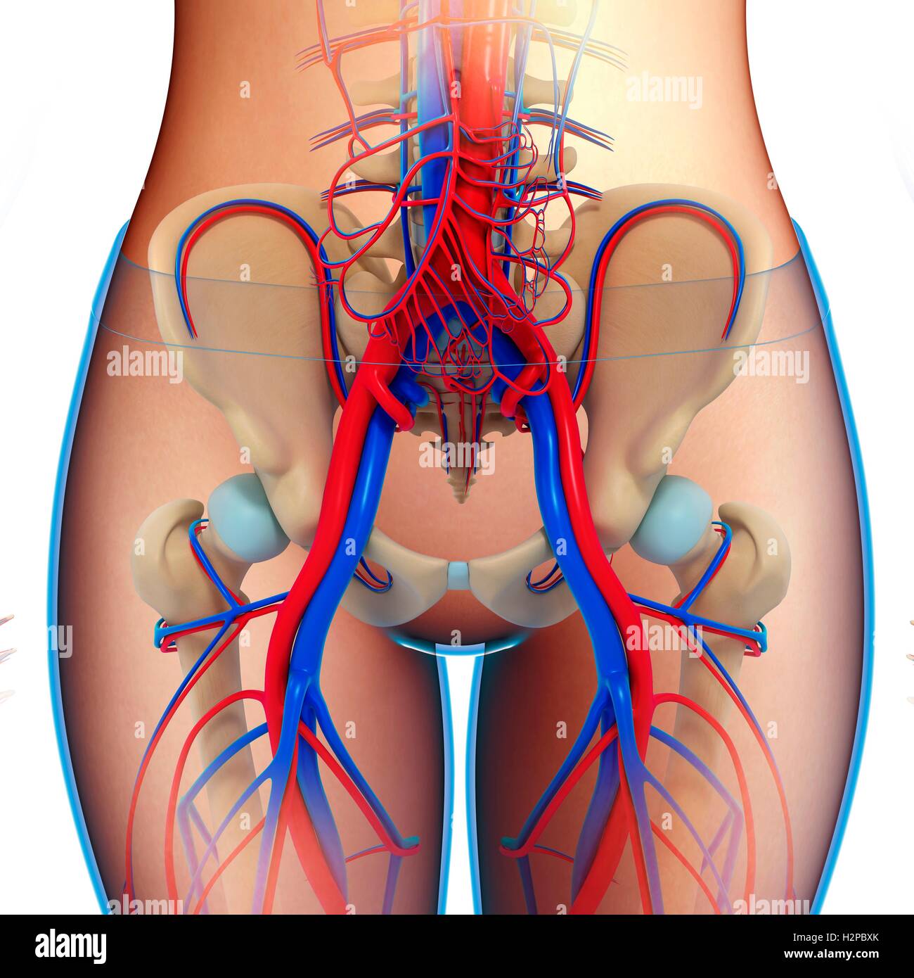 Illustration of female pelvic blood vessels. Stock Photohttps://www.alamy.com/image-license-details/?v=1https://www.alamy.com/stock-photo-illustration-of-female-pelvic-blood-vessels-122194203.html
RFH2PBXK–Illustration of female pelvic blood vessels.
 The blood vessels of the pelvis Stock Photohttps://www.alamy.com/image-license-details/?v=1https://www.alamy.com/stock-photo-the-blood-vessels-of-the-pelvis-13170040.html
RFACJF6H–The blood vessels of the pelvis
 Blood vessels of the hip, illustration. Stock Photohttps://www.alamy.com/image-license-details/?v=1https://www.alamy.com/blood-vessels-of-the-hip-illustration-image559786910.html
RF2REMDWJ–Blood vessels of the hip, illustration.
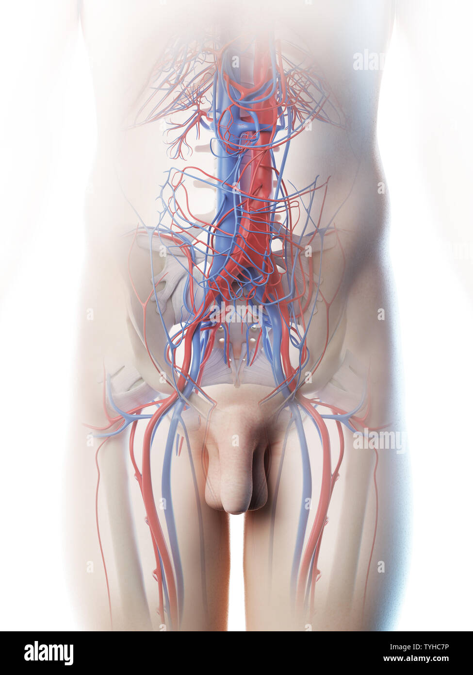 3d rendered illustration of a mans abdominal blood vessels Stock Photohttps://www.alamy.com/image-license-details/?v=1https://www.alamy.com/3d-rendered-illustration-of-a-mans-abdominal-blood-vessels-image257923674.html
RFTYHC7P–3d rendered illustration of a mans abdominal blood vessels
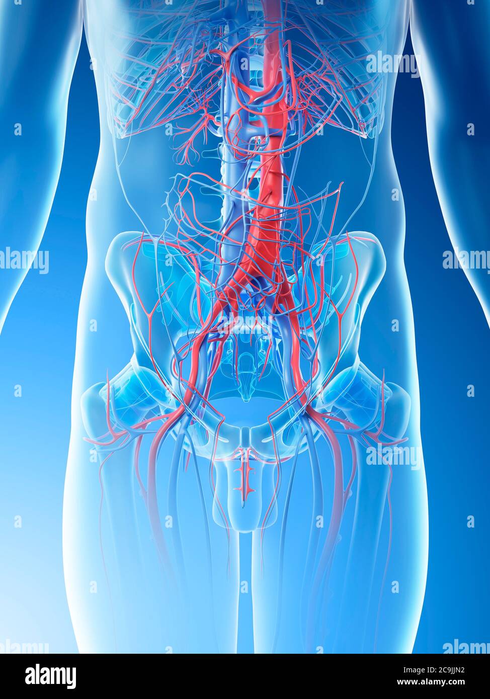 Abdominal blood vessels, computer illustration. Stock Photohttps://www.alamy.com/image-license-details/?v=1https://www.alamy.com/abdominal-blood-vessels-computer-illustration-image367359470.html
RF2C9JJN2–Abdominal blood vessels, computer illustration.
 Medical Illustration of Articularis Genus Muscle Stock Photohttps://www.alamy.com/image-license-details/?v=1https://www.alamy.com/medical-illustration-of-articularis-genus-muscle-image490198510.html
RF2KDED5J–Medical Illustration of Articularis Genus Muscle
 Standing posed female with the bones of the skeleton, blood vessels and nerves visible. Stock Photohttps://www.alamy.com/image-license-details/?v=1https://www.alamy.com/stock-photo-standing-posed-female-with-the-bones-of-the-skeleton-blood-vessels-52077250.html
RMD0M91P–Standing posed female with the bones of the skeleton, blood vessels and nerves visible.
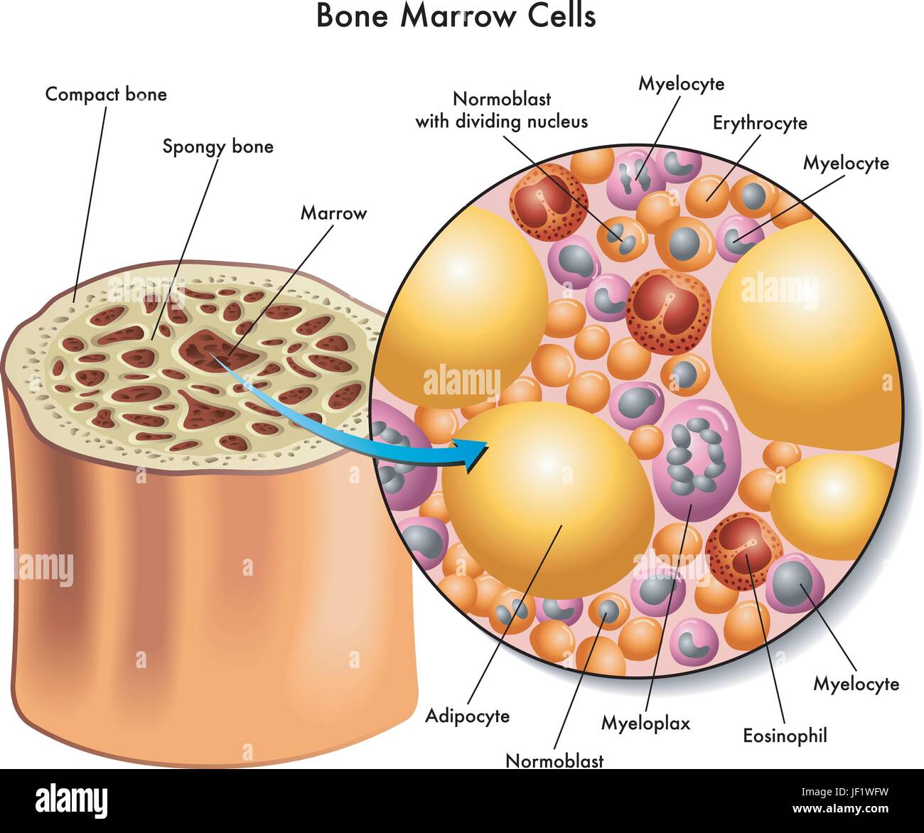 skull, bone, blood, ribs, cells, immune system, leukemia, adipose tissue, Stock Vectorhttps://www.alamy.com/image-license-details/?v=1https://www.alamy.com/stock-photo-skull-bone-blood-ribs-cells-immune-system-leukemia-adipose-tissue-146944781.html
RFJF1WFW–skull, bone, blood, ribs, cells, immune system, leukemia, adipose tissue,
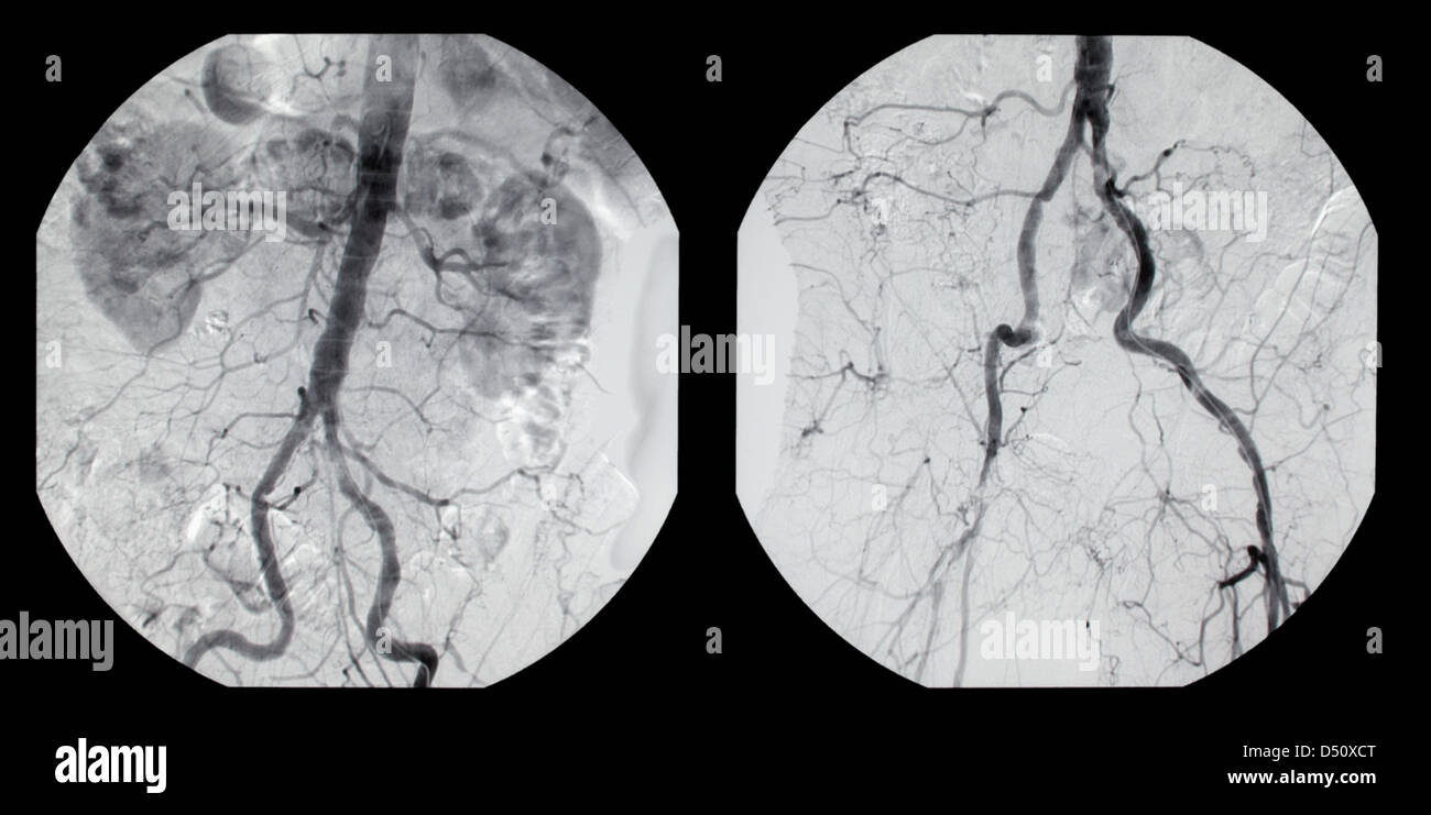 Berlin, Germany, a digital subtraction Stock Photohttps://www.alamy.com/image-license-details/?v=1https://www.alamy.com/stock-photo-berlin-germany-a-digital-subtraction-54725128.html
RMD50XCT–Berlin, Germany, a digital subtraction
 Musculature, bones and blood vessels of the pelvis and thigh, c1805. Stock Photohttps://www.alamy.com/image-license-details/?v=1https://www.alamy.com/musculature-bones-and-blood-vessels-of-the-pelvis-and-thigh-c1805-image635653475.html
RM2YX4EDR–Musculature, bones and blood vessels of the pelvis and thigh, c1805.
 Male Human Skeleton Stock Photohttps://www.alamy.com/image-license-details/?v=1https://www.alamy.com/stock-photo-male-human-skeleton-173225912.html
RMM1R3BM–Male Human Skeleton
 . Internal medicine; a work for the practicing physician on diagnosis and treatment, with a complete Desk index. upernumerary kidneys, atrophyof one kidney. They may be anomalous in form: general departuresfrom type, as lobulation; hypertrophy of one or both organs, and fusion—horse-shoe kidney, sigmoid kidney,disk-shaped kidney. Finally, theremay be variations in the blood-vessels, pelvis, and ureters. Of these abnormal conditionsthe hypertrophied kidney can bediagnosticated only when the affectedorgan is movable and is recognizedupon palpation through the abdom-inal wall; the horse-shoe kidn Stock Photohttps://www.alamy.com/image-license-details/?v=1https://www.alamy.com/internal-medicine-a-work-for-the-practicing-physician-on-diagnosis-and-treatment-with-a-complete-desk-index-upernumerary-kidneys-atrophyof-one-kidney-they-may-be-anomalous-in-form-general-departuresfrom-type-as-lobulation-hypertrophy-of-one-or-both-organs-and-fusionhorse-shoe-kidney-sigmoid-kidneydisk-shaped-kidney-finally-theremay-be-variations-in-the-blood-vessels-pelvis-and-ureters-of-these-abnormal-conditionsthe-hypertrophied-kidney-can-bediagnosticated-only-when-the-affectedorgan-is-movable-and-is-recognizedupon-palpation-through-the-abdom-inal-wall-the-horse-shoe-kidn-image336711445.html
RM2AFPETN–. Internal medicine; a work for the practicing physician on diagnosis and treatment, with a complete Desk index. upernumerary kidneys, atrophyof one kidney. They may be anomalous in form: general departuresfrom type, as lobulation; hypertrophy of one or both organs, and fusion—horse-shoe kidney, sigmoid kidney,disk-shaped kidney. Finally, theremay be variations in the blood-vessels, pelvis, and ureters. Of these abnormal conditionsthe hypertrophied kidney can bediagnosticated only when the affectedorgan is movable and is recognizedupon palpation through the abdom-inal wall; the horse-shoe kidn
 CTA ABDOMINAL AORTA 3D rendering fusion with X-ray Abdomen image. Stock Photohttps://www.alamy.com/image-license-details/?v=1https://www.alamy.com/cta-abdominal-aorta-3d-rendering-fusion-with-x-ray-abdomen-image-image518157427.html
RF2N3032B–CTA ABDOMINAL AORTA 3D rendering fusion with X-ray Abdomen image.
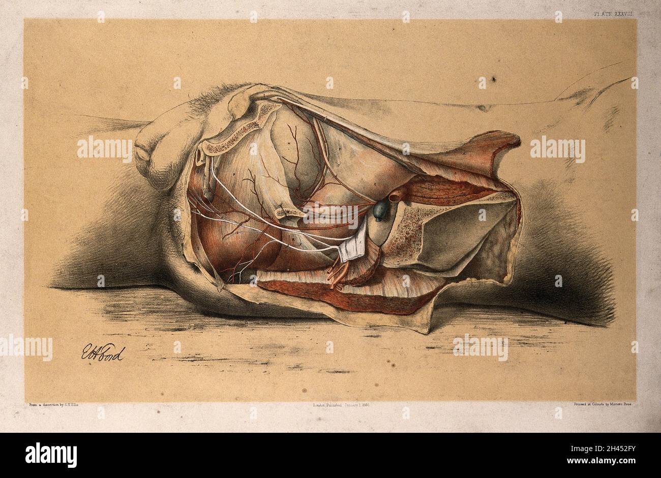 Dissection of the pelvis and abdomen of a man, showing the arteries, blood vessels and muscles: side view. Lithograph by G.H. Ford, 1866. Stock Photohttps://www.alamy.com/image-license-details/?v=1https://www.alamy.com/dissection-of-the-pelvis-and-abdomen-of-a-man-showing-the-arteries-blood-vessels-and-muscles-side-view-lithograph-by-gh-ford-1866-image450039967.html
RM2H452FY–Dissection of the pelvis and abdomen of a man, showing the arteries, blood vessels and muscles: side view. Lithograph by G.H. Ford, 1866.
 The external iliac veins are blood vessels in your pelvis 3d illustration Stock Photohttps://www.alamy.com/image-license-details/?v=1https://www.alamy.com/the-external-iliac-veins-are-blood-vessels-in-your-pelvis-3d-illustration-image596590573.html
RF2WJH1A5–The external iliac veins are blood vessels in your pelvis 3d illustration
 Archive image from page 356 of The cyclopædia of anatomy and. The cyclopædia of anatomy and physiology cyclopdiaofana01todd Year: 1836 AVES. 341 contribute to the secretory vessels of the liver, but proceed to the superior part of that viscus, to terminate in the vena cava, as does also the umbilical vein. ' The vein which returns the blood of the inferior extremities is divided in the pelvis into two branches, which correspond with the femoral and ischiadic arteries; the one passes through the ischiadic foramen, and the other through the hole upo/i the anterior margin of the pelvis; but the Stock Photohttps://www.alamy.com/image-license-details/?v=1https://www.alamy.com/archive-image-from-page-356-of-the-cyclopdia-of-anatomy-and-the-cyclopdia-of-anatomy-and-physiology-cyclopdiaofana01todd-year-1836-aves-341-contribute-to-the-secretory-vessels-of-the-liver-but-proceed-to-the-superior-part-of-that-viscus-to-terminate-in-the-vena-cava-as-does-also-the-umbilical-vein-the-vein-which-returns-the-blood-of-the-inferior-extremities-is-divided-in-the-pelvis-into-two-branches-which-correspond-with-the-femoral-and-ischiadic-arteries-the-one-passes-through-the-ischiadic-foramen-and-the-other-through-the-hole-upoi-the-anterior-margin-of-the-pelvis-but-the-image259563783.html
RMW28473–Archive image from page 356 of The cyclopædia of anatomy and. The cyclopædia of anatomy and physiology cyclopdiaofana01todd Year: 1836 AVES. 341 contribute to the secretory vessels of the liver, but proceed to the superior part of that viscus, to terminate in the vena cava, as does also the umbilical vein. ' The vein which returns the blood of the inferior extremities is divided in the pelvis into two branches, which correspond with the femoral and ischiadic arteries; the one passes through the ischiadic foramen, and the other through the hole upo/i the anterior margin of the pelvis; but the
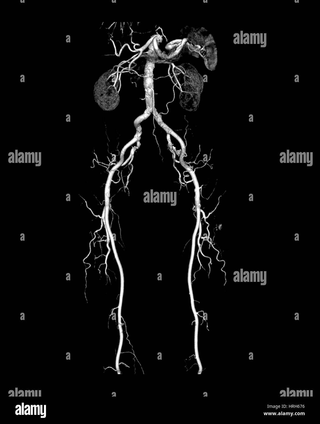 CT Angiogram of Abdomen and Legs Stock Photohttps://www.alamy.com/image-license-details/?v=1https://www.alamy.com/stock-photo-ct-angiogram-of-abdomen-and-legs-134987754.html
RMHRH676–CT Angiogram of Abdomen and Legs
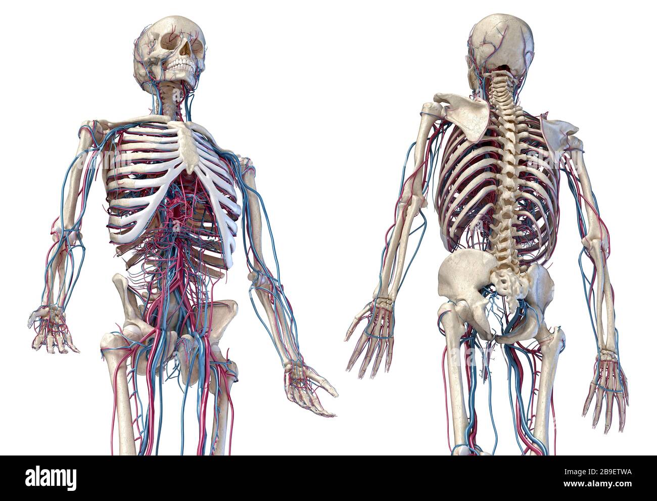 3/4 upper body view of human skeletal and vascular systems, black background. Stock Photohttps://www.alamy.com/image-license-details/?v=1https://www.alamy.com/34-upper-body-view-of-human-skeletal-and-vascular-systems-black-background-image350066118.html
RF2B9ETWA–3/4 upper body view of human skeletal and vascular systems, black background.
 Urinary system from kidney to glomerulus with structures of kidney and ureter. Stock Photohttps://www.alamy.com/image-license-details/?v=1https://www.alamy.com/urinary-system-from-kidney-to-glomerulus-with-structures-of-kidney-and-ureter-image476924450.html
RF2JKWP02–Urinary system from kidney to glomerulus with structures of kidney and ureter.
RF2PNXFFM–CT scan line icons collection. Radiology, Imaging, Diagnosis, Cancer, Tumor, Brain, Spine vector and linear illustration. Heart,Abdomen,Chest outline
 The blood vessels of the pelvis Stock Photohttps://www.alamy.com/image-license-details/?v=1https://www.alamy.com/stock-photo-the-blood-vessels-of-the-pelvis-13195519.html
RFACN71M–The blood vessels of the pelvis
 . The cyclopædia of anatomy and physiology. Anatomy; Physiology; Zoology. AVES. 341 contribute to the secretory vessels of the liver, but proceed to the superior part of that viscus, to terminate in the vena cava, as does also the umbilical vein. " The vein which returns the blood of the inferior extremities is divided in the pelvis into two branches, which correspond with the femoral and ischiadic arteries; the one passes through the ischiadic foramen, and the other through the hole upo/i the anterior margin of the pelvis; but the proportion they bear to each other in magnitude is the ve Stock Photohttps://www.alamy.com/image-license-details/?v=1https://www.alamy.com/the-cyclopdia-of-anatomy-and-physiology-anatomy-physiology-zoology-aves-341-contribute-to-the-secretory-vessels-of-the-liver-but-proceed-to-the-superior-part-of-that-viscus-to-terminate-in-the-vena-cava-as-does-also-the-umbilical-vein-quot-the-vein-which-returns-the-blood-of-the-inferior-extremities-is-divided-in-the-pelvis-into-two-branches-which-correspond-with-the-femoral-and-ischiadic-arteries-the-one-passes-through-the-ischiadic-foramen-and-the-other-through-the-hole-upoi-the-anterior-margin-of-the-pelvis-but-the-proportion-they-bear-to-each-other-in-magnitude-is-the-ve-image216211481.html
RMPFN7XH–. The cyclopædia of anatomy and physiology. Anatomy; Physiology; Zoology. AVES. 341 contribute to the secretory vessels of the liver, but proceed to the superior part of that viscus, to terminate in the vena cava, as does also the umbilical vein. " The vein which returns the blood of the inferior extremities is divided in the pelvis into two branches, which correspond with the femoral and ischiadic arteries; the one passes through the ischiadic foramen, and the other through the hole upo/i the anterior margin of the pelvis; but the proportion they bear to each other in magnitude is the ve
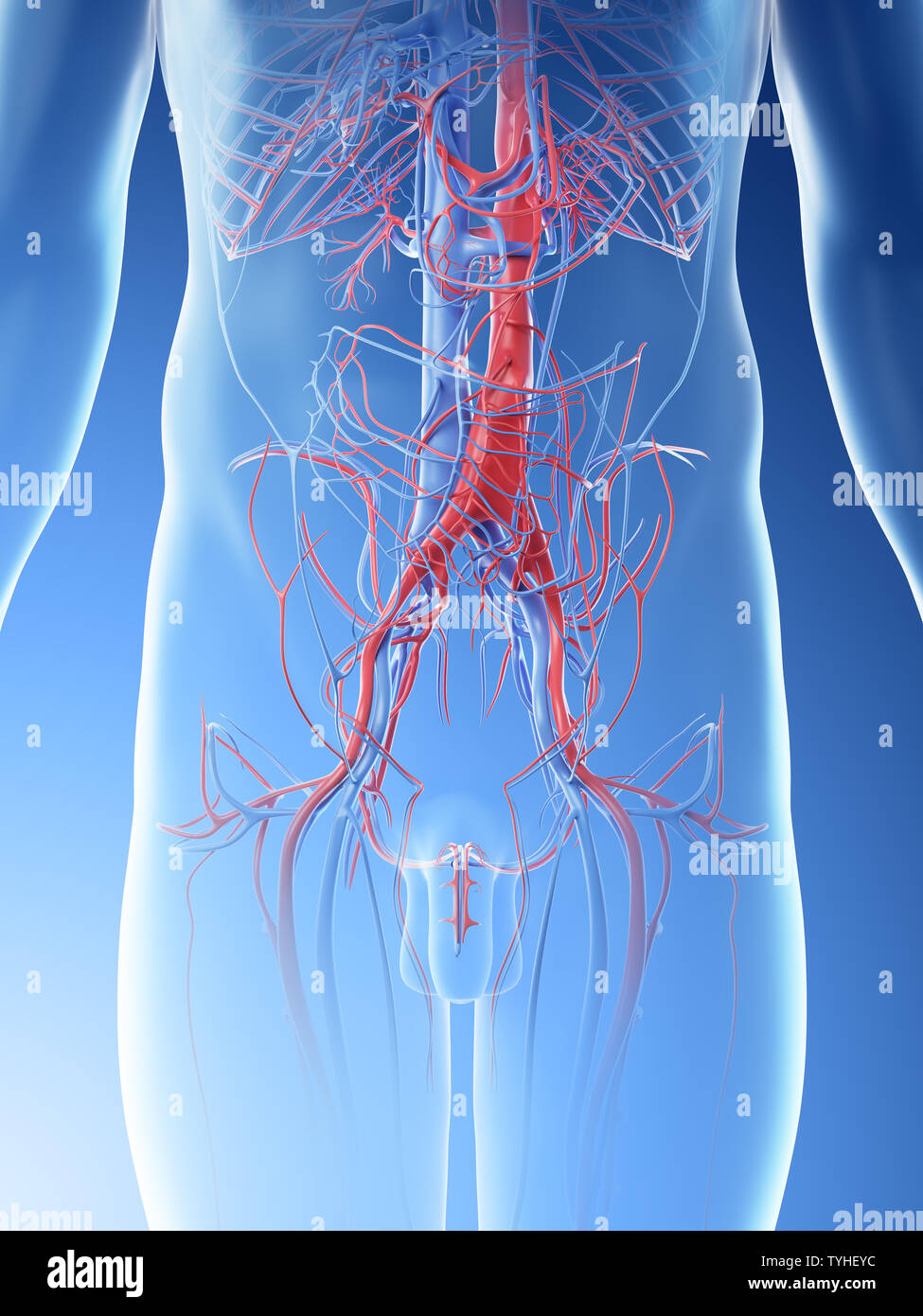 3d rendered illustration of a mans abdominal blood vessels Stock Photohttps://www.alamy.com/image-license-details/?v=1https://www.alamy.com/3d-rendered-illustration-of-a-mans-abdominal-blood-vessels-image257925792.html
RFTYHEYC–3d rendered illustration of a mans abdominal blood vessels
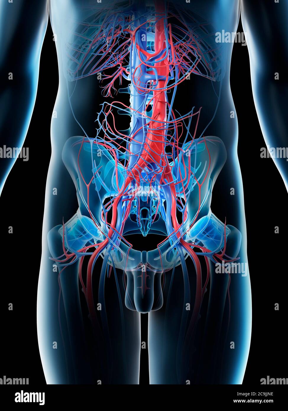 Abdominal blood vessels, computer illustration. Stock Photohttps://www.alamy.com/image-license-details/?v=1https://www.alamy.com/abdominal-blood-vessels-computer-illustration-image367359482.html
RF2C9JJNE–Abdominal blood vessels, computer illustration.
 Text sign showing Abdominal Aortic Aneurysmquestion. Conceptual photo getting to know the enlargement of aorta Keyboard key Intention to create comput Stock Photohttps://www.alamy.com/image-license-details/?v=1https://www.alamy.com/text-sign-showing-abdominal-aortic-aneurysmquestion-conceptual-photo-getting-to-know-the-enlargement-of-aorta-keyboard-key-intention-to-create-comput-image229924724.html
RFRA1Y98–Text sign showing Abdominal Aortic Aneurysmquestion. Conceptual photo getting to know the enlargement of aorta Keyboard key Intention to create comput
 Female Human Skeleton Stock Photohttps://www.alamy.com/image-license-details/?v=1https://www.alamy.com/stock-photo-female-human-skeleton-173207808.html
RMM1P894–Female Human Skeleton
 The practice of obstetrics, designed for the use of students and practitioners of medicine . Fig. 151.—Shape of the Abdomen in a Multigravida with a Moderate GenerallyContracted Pelvis at the Thirty-eighth Week and in the Standing Posture. on account of the close connection between them and the surrounding connectivetissue. They are obliterated after labor by the contraction of the uterinemuscle, which surrounds them. These blood-vessels penetrate the minutestdivisions of the chorion frondosum, and consist of the end ramifications of theumbilical arteries and veins. The arteries and veins purs Stock Photohttps://www.alamy.com/image-license-details/?v=1https://www.alamy.com/the-practice-of-obstetrics-designed-for-the-use-of-students-and-practitioners-of-medicine-fig-151shape-of-the-abdomen-in-a-multigravida-with-a-moderate-generallycontracted-pelvis-at-the-thirty-eighth-week-and-in-the-standing-posture-on-account-of-the-close-connection-between-them-and-the-surrounding-connectivetissue-they-are-obliterated-after-labor-by-the-contraction-of-the-uterinemuscle-which-surrounds-them-these-blood-vessels-penetrate-the-minutestdivisions-of-the-chorion-frondosum-and-consist-of-the-end-ramifications-of-theumbilical-arteries-and-veins-the-arteries-and-veins-purs-image343342731.html
RM2AXGH4B–The practice of obstetrics, designed for the use of students and practitioners of medicine . Fig. 151.—Shape of the Abdomen in a Multigravida with a Moderate GenerallyContracted Pelvis at the Thirty-eighth Week and in the Standing Posture. on account of the close connection between them and the surrounding connectivetissue. They are obliterated after labor by the contraction of the uterinemuscle, which surrounds them. These blood-vessels penetrate the minutestdivisions of the chorion frondosum, and consist of the end ramifications of theumbilical arteries and veins. The arteries and veins purs
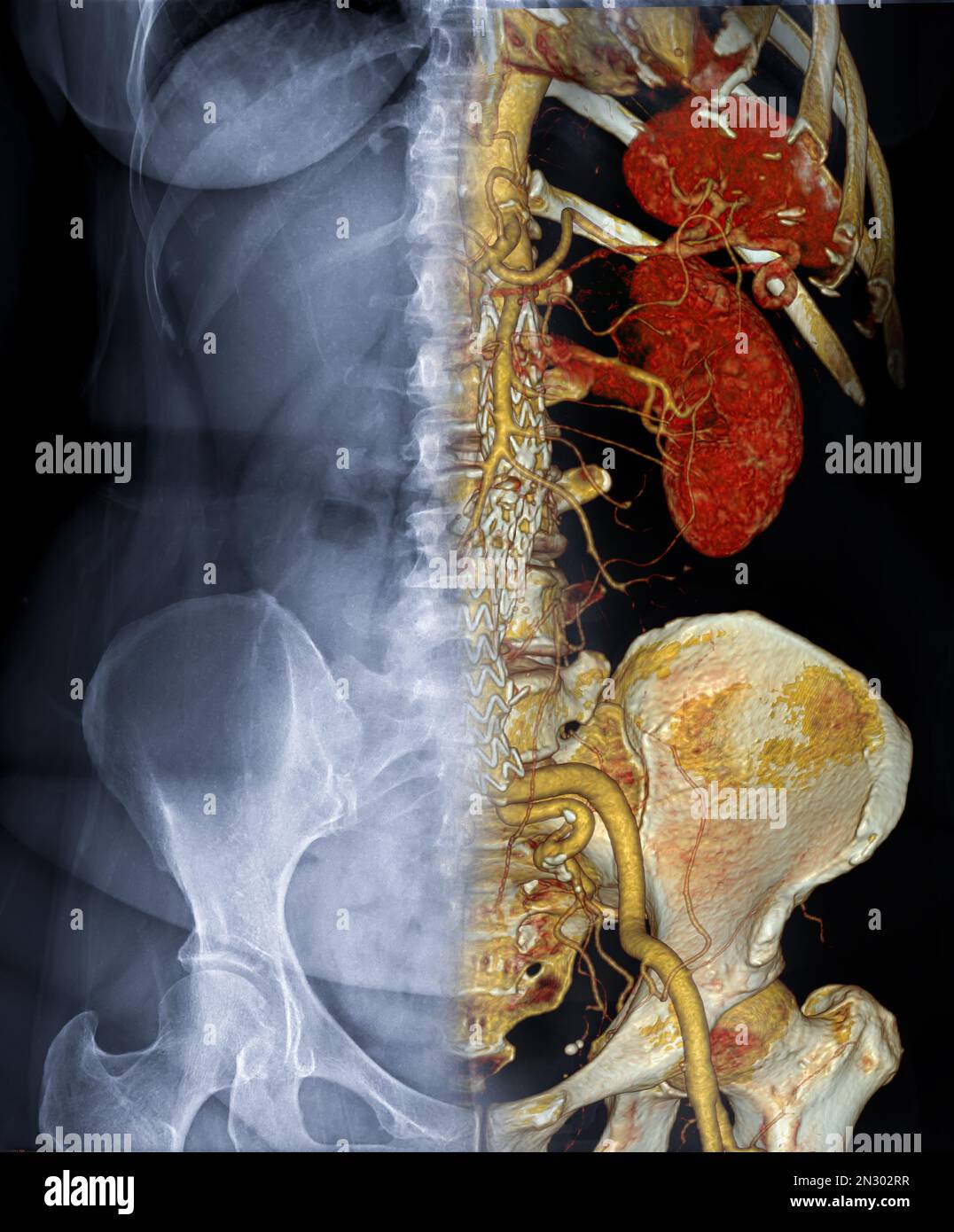 CTA ABDOMINAL AORTA 3D rendering fusion with X-ray Abdomen image. Stock Photohttps://www.alamy.com/image-license-details/?v=1https://www.alamy.com/cta-abdominal-aorta-3d-rendering-fusion-with-x-ray-abdomen-image-image518157243.html
RF2N302RR–CTA ABDOMINAL AORTA 3D rendering fusion with X-ray Abdomen image.
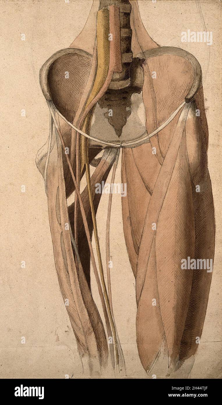 Muscles, bones and blood-vessels (?) of the pelvis. Pen and ink, with pink, yellow and brown watercolour washes, by C. Landseer, ca. 1815. Stock Photohttps://www.alamy.com/image-license-details/?v=1https://www.alamy.com/muscles-bones-and-blood-vessels-of-the-pelvis-pen-and-ink-with-pink-yellow-and-brown-watercolour-washes-by-c-landseer-ca-1815-image450035335.html
RM2H44TJF–Muscles, bones and blood-vessels (?) of the pelvis. Pen and ink, with pink, yellow and brown watercolour washes, by C. Landseer, ca. 1815.
 The external iliac veins are blood vessels in your pelvis 3d illustration Stock Photohttps://www.alamy.com/image-license-details/?v=1https://www.alamy.com/the-external-iliac-veins-are-blood-vessels-in-your-pelvis-3d-illustration-image596590760.html
RF2WJH1GT–The external iliac veins are blood vessels in your pelvis 3d illustration
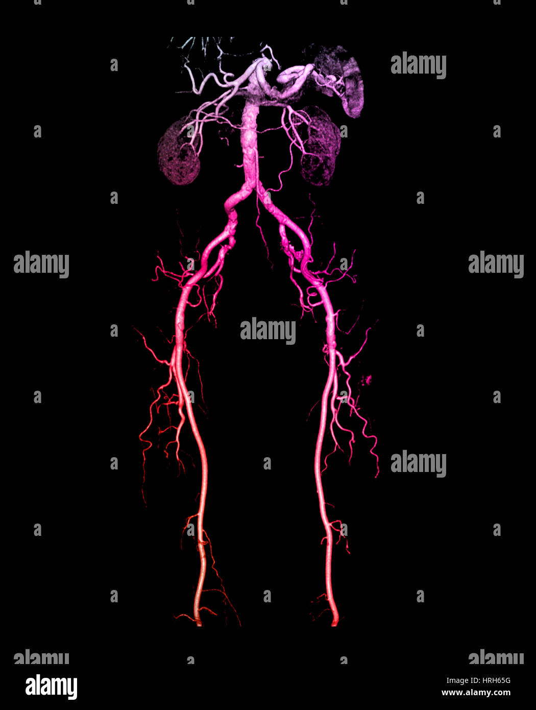 CT Angiogram of Abdomen and Legs Stock Photohttps://www.alamy.com/image-license-details/?v=1https://www.alamy.com/stock-photo-ct-angiogram-of-abdomen-and-legs-134987708.html
RMHRH65G–CT Angiogram of Abdomen and Legs
 3/4 upper body rear view of human skeletal and vascular systems, black background. Stock Photohttps://www.alamy.com/image-license-details/?v=1https://www.alamy.com/34-upper-body-rear-view-of-human-skeletal-and-vascular-systems-black-background-image350065947.html
RF2B9ETK7–3/4 upper body rear view of human skeletal and vascular systems, black background.
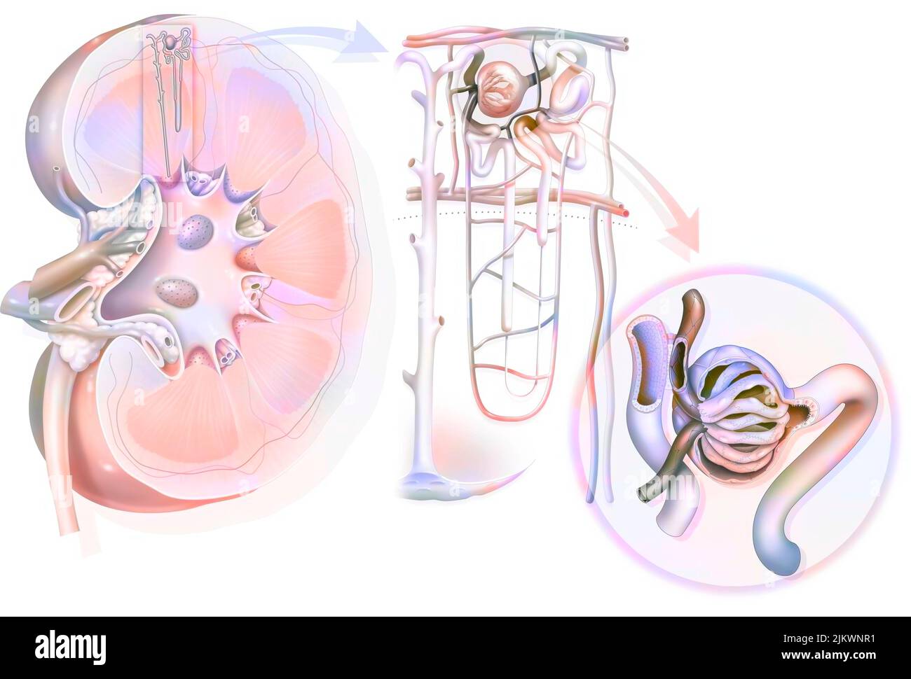 Urinary system from kidney to glomerulus with structures of kidney and ureter. Stock Photohttps://www.alamy.com/image-license-details/?v=1https://www.alamy.com/urinary-system-from-kidney-to-glomerulus-with-structures-of-kidney-and-ureter-image476924309.html
RF2JKWNR1–Urinary system from kidney to glomerulus with structures of kidney and ureter.
RF2PNYYYK–Questions and asnwers line icons collection. Radiology, Imaging, Diagnosis, Cancer, Tumor, Brain, Spine vector and linear illustration. Heart,Abdomen
 The blood vessels of the pelvis Stock Photohttps://www.alamy.com/image-license-details/?v=1https://www.alamy.com/stock-photo-the-blood-vessels-of-the-pelvis-13174710.html
RFACK13K–The blood vessels of the pelvis
 3d rendered illustration of a mans abdominal blood vessels Stock Photohttps://www.alamy.com/image-license-details/?v=1https://www.alamy.com/3d-rendered-illustration-of-a-mans-abdominal-blood-vessels-image257925416.html
RFTYHEE0–3d rendered illustration of a mans abdominal blood vessels
 Abdominal blood vessels, computer illustration. Stock Photohttps://www.alamy.com/image-license-details/?v=1https://www.alamy.com/abdominal-blood-vessels-computer-illustration-image367359389.html
RF2C9JJJ5–Abdominal blood vessels, computer illustration.
 Handwriting text Abdominal Aortic Aneurysmquestion. Concept meaning getting to know the enlargement of aorta Keyboard key Intention to create computer Stock Photohttps://www.alamy.com/image-license-details/?v=1https://www.alamy.com/handwriting-text-abdominal-aortic-aneurysmquestion-concept-meaning-getting-to-know-the-enlargement-of-aorta-keyboard-key-intention-to-create-computer-image229969391.html
RFRA408F–Handwriting text Abdominal Aortic Aneurysmquestion. Concept meaning getting to know the enlargement of aorta Keyboard key Intention to create computer
 Manual of gynecology . d blood-vessels, be removed on one side of the pelvis,say the right, the two muscles known as the coccygeus and levator aniwill be exposed. .These spring from the middle of the inner side of thetrue pelvis, and, blending partly directly and partly indirectly with oneanother, form what may be termed the diaphragmatic muscles of thepelvic floor. If looked at through the pelvic brim, they are seen toform on both sides a concave arrangement analogous to the thoracic dia-phragm (Fig. 11). The Coccygeus sjDrings from the spine of the ischium and is insertedinto the side of the Stock Photohttps://www.alamy.com/image-license-details/?v=1https://www.alamy.com/manual-of-gynecology-d-blood-vessels-be-removed-on-one-side-of-the-pelvissay-the-right-the-two-muscles-known-as-the-coccygeus-and-levator-aniwill-be-exposed-these-spring-from-the-middle-of-the-inner-side-of-thetrue-pelvis-and-blending-partly-directly-and-partly-indirectly-with-oneanother-form-what-may-be-termed-the-diaphragmatic-muscles-of-thepelvic-floor-if-looked-at-through-the-pelvic-brim-they-are-seen-toform-on-both-sides-a-concave-arrangement-analogous-to-the-thoracic-dia-phragm-fig-11-the-coccygeus-sjdrings-from-the-spine-of-the-ischium-and-is-insertedinto-the-side-of-the-image340177529.html
RM2ANCBWD–Manual of gynecology . d blood-vessels, be removed on one side of the pelvis,say the right, the two muscles known as the coccygeus and levator aniwill be exposed. .These spring from the middle of the inner side of thetrue pelvis, and, blending partly directly and partly indirectly with oneanother, form what may be termed the diaphragmatic muscles of thepelvic floor. If looked at through the pelvic brim, they are seen toform on both sides a concave arrangement analogous to the thoracic dia-phragm (Fig. 11). The Coccygeus sjDrings from the spine of the ischium and is insertedinto the side of the
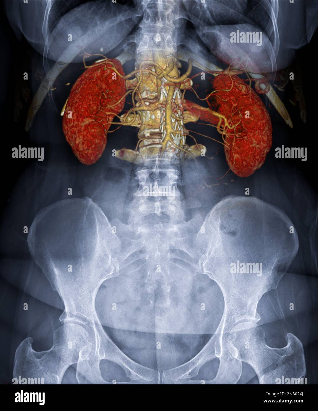 CTA ABDOMINAL AORTA 3D rendering fusion with X-ray Abdomen image. Stock Photohttps://www.alamy.com/image-license-details/?v=1https://www.alamy.com/cta-abdominal-aorta-3d-rendering-fusion-with-x-ray-abdomen-image-image518157322.html
RF2N302XJ–CTA ABDOMINAL AORTA 3D rendering fusion with X-ray Abdomen image.
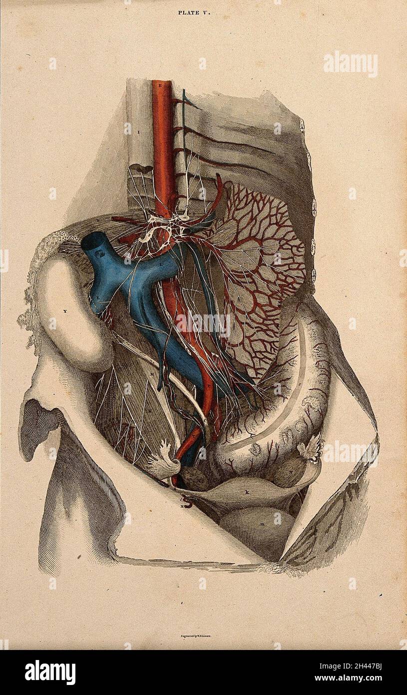 Female pelvis: dissection, with blood-vessels and nerves indicated in red and blue. Coloured line engraving by W.H. Lizars, 1822/1826. Stock Photohttps://www.alamy.com/image-license-details/?v=1https://www.alamy.com/female-pelvis-dissection-with-blood-vessels-and-nerves-indicated-in-red-and-blue-coloured-line-engraving-by-wh-lizars-18221826-image450021814.html
RM2H447BJ–Female pelvis: dissection, with blood-vessels and nerves indicated in red and blue. Coloured line engraving by W.H. Lizars, 1822/1826.
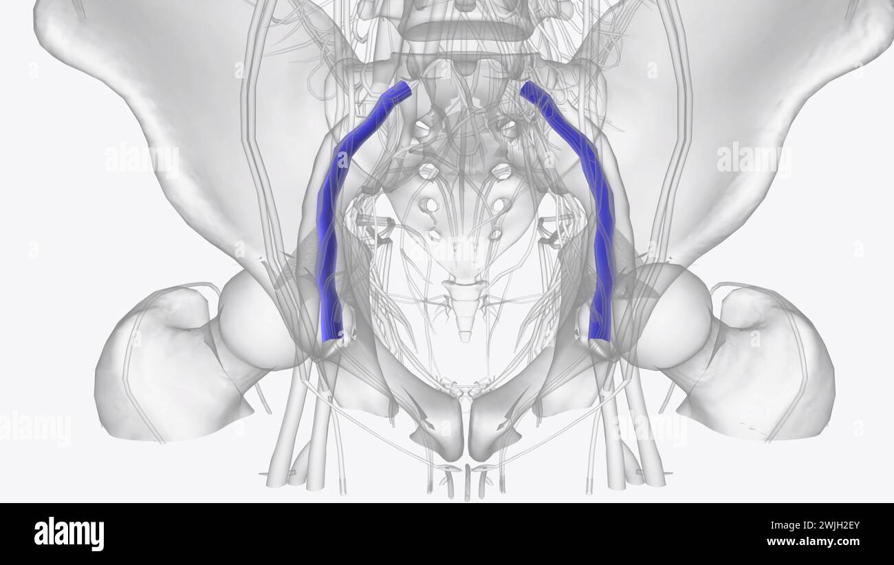 The external iliac veins are blood vessels in your pelvis 3d illustration Stock Photohttps://www.alamy.com/image-license-details/?v=1https://www.alamy.com/the-external-iliac-veins-are-blood-vessels-in-your-pelvis-3d-illustration-image596591491.html
RF2WJH2EY–The external iliac veins are blood vessels in your pelvis 3d illustration
 CT Angiogram of Abdomen and Legs Stock Photohttps://www.alamy.com/image-license-details/?v=1https://www.alamy.com/stock-photo-ct-angiogram-of-abdomen-and-legs-134987707.html
RMHRH65F–CT Angiogram of Abdomen and Legs
 3/4 upper body front view of human skeletal and vascular systems, black background. Stock Photohttps://www.alamy.com/image-license-details/?v=1https://www.alamy.com/34-upper-body-front-view-of-human-skeletal-and-vascular-systems-black-background-image350065913.html
RF2B9ETJ1–3/4 upper body front view of human skeletal and vascular systems, black background.
 Urinary system from kidney to glomerulus with structures of kidney and ureter. Stock Photohttps://www.alamy.com/image-license-details/?v=1https://www.alamy.com/urinary-system-from-kidney-to-glomerulus-with-structures-of-kidney-and-ureter-image476924471.html
RF2JKWP0R–Urinary system from kidney to glomerulus with structures of kidney and ureter.
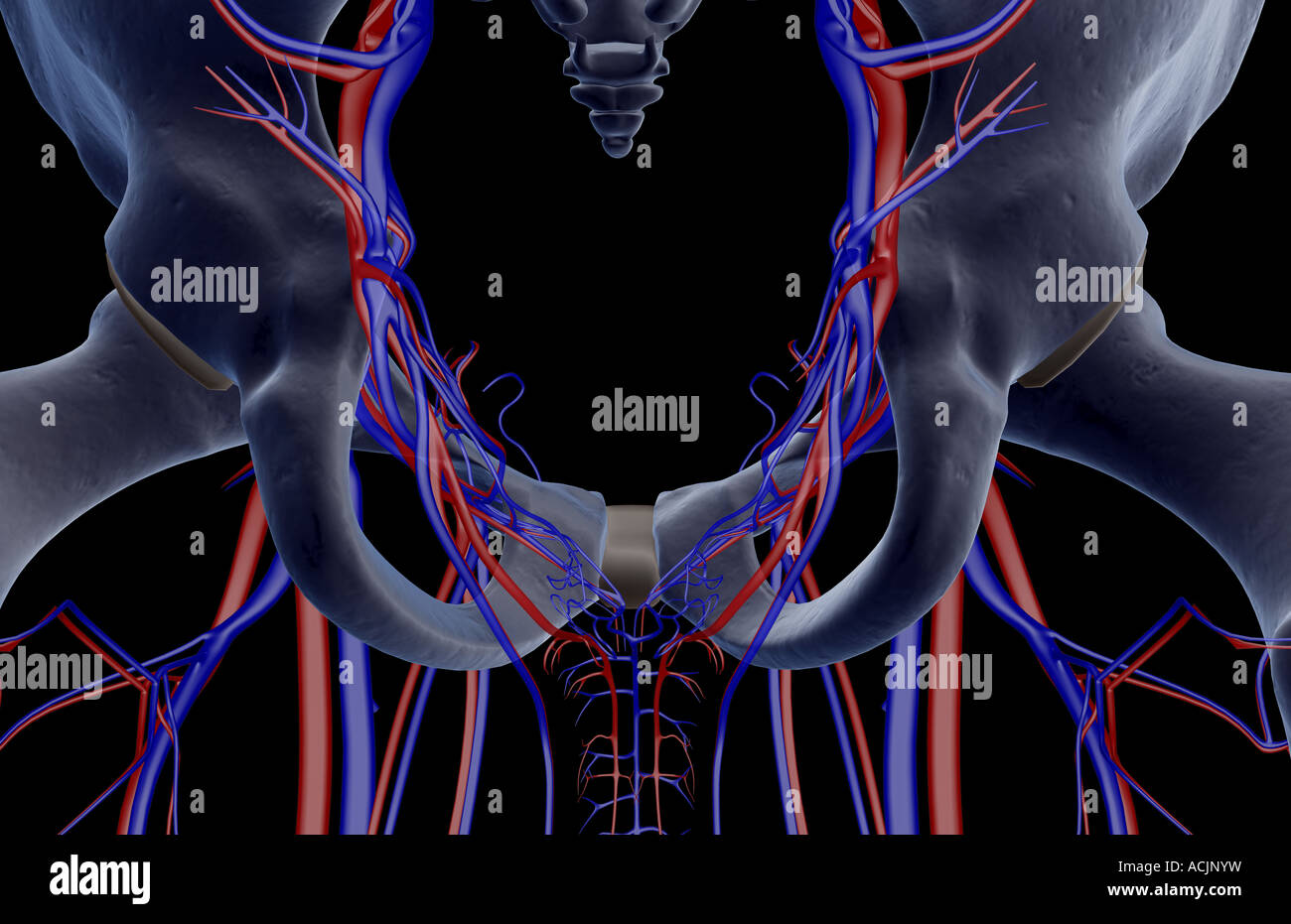 The blood vessels of the pelvis Stock Photohttps://www.alamy.com/image-license-details/?v=1https://www.alamy.com/stock-photo-the-blood-vessels-of-the-pelvis-13172316.html
RFACJNYW–The blood vessels of the pelvis
 3d rendered illustration of a mans abdominal blood vessels Stock Photohttps://www.alamy.com/image-license-details/?v=1https://www.alamy.com/3d-rendered-illustration-of-a-mans-abdominal-blood-vessels-image257923662.html
RFTYHC7A–3d rendered illustration of a mans abdominal blood vessels
 Abdominal blood vessels, computer illustration. Stock Photohttps://www.alamy.com/image-license-details/?v=1https://www.alamy.com/abdominal-blood-vessels-computer-illustration-image367359383.html
RF2C9JJHY–Abdominal blood vessels, computer illustration.
 Text sign showing Abdominal Aortic Aneurysmquestion. Conceptual photo getting to know the enlargement of aorta Keyboard key Intention to create comput Stock Photohttps://www.alamy.com/image-license-details/?v=1https://www.alamy.com/text-sign-showing-abdominal-aortic-aneurysmquestion-conceptual-photo-getting-to-know-the-enlargement-of-aorta-keyboard-key-intention-to-create-comput-image229923464.html
RFRA1WM8–Text sign showing Abdominal Aortic Aneurysmquestion. Conceptual photo getting to know the enlargement of aorta Keyboard key Intention to create comput
 Textbook of normal histology: including an account of the development of the tissues and of the organs . and pass tothe pelvis, where they aid in forming the large renal veins. Thevessels collecting the blood from the peripheral zone of the cortex Transverse section of papillary region of medulla of hu-man kidney, more highly magnified : C, large collectingtubules ; x and y, descending and ascending limbs ofHenles loops ; v, blood-vessels. 200 NORMAL HISTOLOGY. converge to certain points, where they form the venae stellatae ; these veins afterwards pass into the labyrinth and follow the inter- Stock Photohttps://www.alamy.com/image-license-details/?v=1https://www.alamy.com/textbook-of-normal-histology-including-an-account-of-the-development-of-the-tissues-and-of-the-organs-and-pass-tothe-pelvis-where-they-aid-in-forming-the-large-renal-veins-thevessels-collecting-the-blood-from-the-peripheral-zone-of-the-cortex-transverse-section-of-papillary-region-of-medulla-of-hu-man-kidney-more-highly-magnified-c-large-collectingtubules-x-and-y-descending-and-ascending-limbs-ofhenles-loops-v-blood-vessels-200-normal-histology-converge-to-certain-points-where-they-form-the-venae-stellatae-these-veins-afterwards-pass-into-the-labyrinth-and-follow-the-inter-image338951640.html
RM2AKCG7M–Textbook of normal histology: including an account of the development of the tissues and of the organs . and pass tothe pelvis, where they aid in forming the large renal veins. Thevessels collecting the blood from the peripheral zone of the cortex Transverse section of papillary region of medulla of hu-man kidney, more highly magnified : C, large collectingtubules ; x and y, descending and ascending limbs ofHenles loops ; v, blood-vessels. 200 NORMAL HISTOLOGY. converge to certain points, where they form the venae stellatae ; these veins afterwards pass into the labyrinth and follow the inter-
 Dissection of the pelvis of a man, showing the arteries, blood vessels, muscles and internal organs: side view. Lithograph by G.H. Ford, 1866. Stock Photohttps://www.alamy.com/image-license-details/?v=1https://www.alamy.com/dissection-of-the-pelvis-of-a-man-showing-the-arteries-blood-vessels-muscles-and-internal-organs-side-view-lithograph-by-gh-ford-1866-image450040260.html
RM2H452XC–Dissection of the pelvis of a man, showing the arteries, blood vessels, muscles and internal organs: side view. Lithograph by G.H. Ford, 1866.
 The external iliac veins are blood vessels in your pelvis 3d illustration Stock Photohttps://www.alamy.com/image-license-details/?v=1https://www.alamy.com/the-external-iliac-veins-are-blood-vessels-in-your-pelvis-3d-illustration-image596591911.html
RF2WJH31Y–The external iliac veins are blood vessels in your pelvis 3d illustration
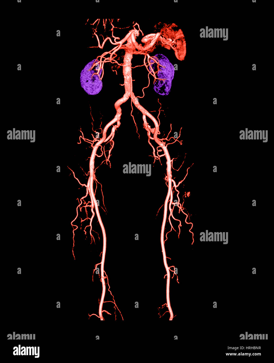 CT Angiogram of Abdomen and Legs Stock Photohttps://www.alamy.com/image-license-details/?v=1https://www.alamy.com/stock-photo-ct-angiogram-of-abdomen-and-legs-134992083.html
RMHRHBNR–CT Angiogram of Abdomen and Legs
 Front view of hip, limbs and hands of skeletal system with veins and arteries, black background. Stock Photohttps://www.alamy.com/image-license-details/?v=1https://www.alamy.com/front-view-of-hip-limbs-and-hands-of-skeletal-system-with-veins-and-arteries-black-background-image350068768.html
RF2B9F080–Front view of hip, limbs and hands of skeletal system with veins and arteries, black background.
 Urinary system from kidney to glomerulus with structures of kidney and ureter. Stock Photohttps://www.alamy.com/image-license-details/?v=1https://www.alamy.com/urinary-system-from-kidney-to-glomerulus-with-structures-of-kidney-and-ureter-image476924340.html
RF2JKWNT4–Urinary system from kidney to glomerulus with structures of kidney and ureter.
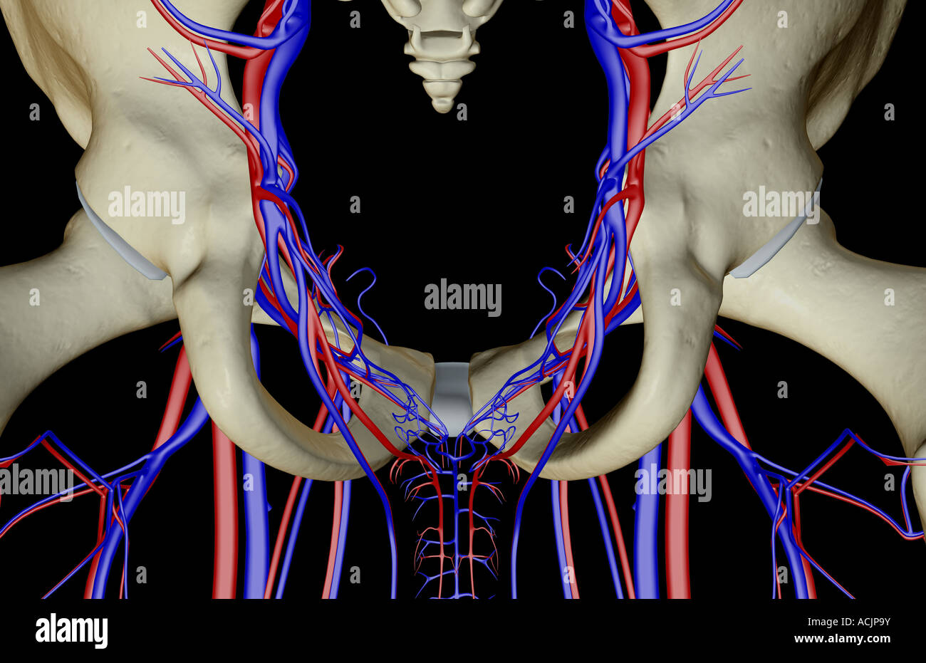 The blood vessels of the pelvis Stock Photohttps://www.alamy.com/image-license-details/?v=1https://www.alamy.com/stock-photo-the-blood-vessels-of-the-pelvis-13172438.html
RFACJP9Y–The blood vessels of the pelvis
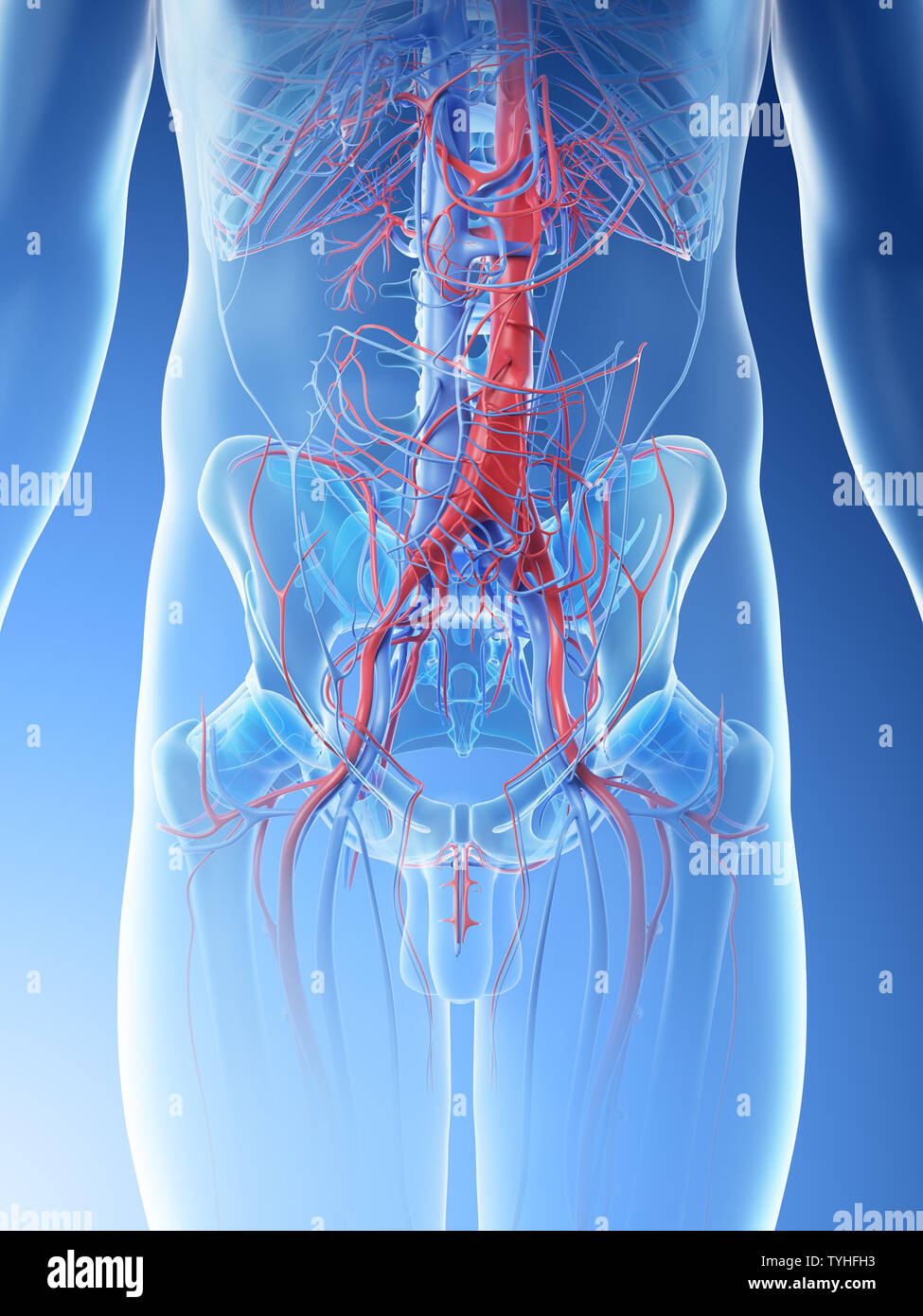 3d rendered illustration of a mans abdominal blood vessels Stock Photohttps://www.alamy.com/image-license-details/?v=1https://www.alamy.com/3d-rendered-illustration-of-a-mans-abdominal-blood-vessels-image257926287.html
RFTYHFH3–3d rendered illustration of a mans abdominal blood vessels
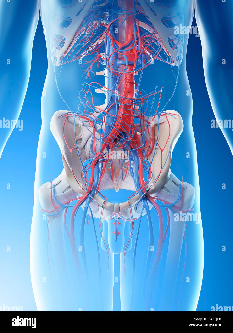 Abdominal blood vessels, computer illustration. Stock Photohttps://www.alamy.com/image-license-details/?v=1https://www.alamy.com/abdominal-blood-vessels-computer-illustration-image367359496.html
RF2C9JJP0–Abdominal blood vessels, computer illustration.
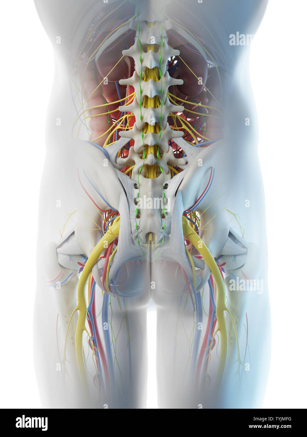 3d rendered illustration of a mans anatomy of the pelvis Stock Photohttps://www.alamy.com/image-license-details/?v=1https://www.alamy.com/3d-rendered-illustration-of-a-mans-anatomy-of-the-pelvis-image257952116.html
RFTYJMFG–3d rendered illustration of a mans anatomy of the pelvis
 Word writing text Abdominal Aortic Aneurysmquestion. Business concept for getting to know the enlargement of aorta Keyboard key Intention to create co Stock Photohttps://www.alamy.com/image-license-details/?v=1https://www.alamy.com/word-writing-text-abdominal-aortic-aneurysmquestion-business-concept-for-getting-to-know-the-enlargement-of-aorta-keyboard-key-intention-to-create-co-image229969102.html
RFRA3YX6–Word writing text Abdominal Aortic Aneurysmquestion. Business concept for getting to know the enlargement of aorta Keyboard key Intention to create co
 Lectures on ectopic pregnancy and pelvic haematocele . extra-peritoneal tissue, with its blood-vessels, is therefore not onlycapable of forming anastomoses in abdominal aneurism, as Turnerand Chiene have shown, but may attempt to carry on the functionsof the maternal portion of the placenta. We have here what may be termed slow displacement of tlieplacenta. At first it lay in the Fallopian tube, but the growingovum has slowly pushed it up (a process attended with bloodextravasation) from pelvis to abdominal cavity, until at last itsupper edge is about ten inches from its original site. Part of Stock Photohttps://www.alamy.com/image-license-details/?v=1https://www.alamy.com/lectures-on-ectopic-pregnancy-and-pelvic-haematocele-extra-peritoneal-tissue-with-its-blood-vessels-is-therefore-not-onlycapable-of-forming-anastomoses-in-abdominal-aneurism-as-turnerand-chiene-have-shown-but-may-attempt-to-carry-on-the-functionsof-the-maternal-portion-of-the-placenta-we-have-here-what-may-be-termed-slow-displacement-of-tlieplacenta-at-first-it-lay-in-the-fallopian-tube-but-the-growingovum-has-slowly-pushed-it-up-a-process-attended-with-bloodextravasation-from-pelvis-to-abdominal-cavity-until-at-last-itsupper-edge-is-about-ten-inches-from-its-original-site-part-of-image343192338.html
RM2AX9N96–Lectures on ectopic pregnancy and pelvic haematocele . extra-peritoneal tissue, with its blood-vessels, is therefore not onlycapable of forming anastomoses in abdominal aneurism, as Turnerand Chiene have shown, but may attempt to carry on the functionsof the maternal portion of the placenta. We have here what may be termed slow displacement of tlieplacenta. At first it lay in the Fallopian tube, but the growingovum has slowly pushed it up (a process attended with bloodextravasation) from pelvis to abdominal cavity, until at last itsupper edge is about ten inches from its original site. Part of
 Dissection of the pelvis and abdomen of a man, showing the arteries, blood vessels, muscles and internal organs: side view. Lithograph by G.H. Ford, 1866. Stock Photohttps://www.alamy.com/image-license-details/?v=1https://www.alamy.com/dissection-of-the-pelvis-and-abdomen-of-a-man-showing-the-arteries-blood-vessels-muscles-and-internal-organs-side-view-lithograph-by-gh-ford-1866-image450114654.html
RM2H48DRA–Dissection of the pelvis and abdomen of a man, showing the arteries, blood vessels, muscles and internal organs: side view. Lithograph by G.H. Ford, 1866.
 The external iliac veins are blood vessels in your pelvis 3d illustration Stock Photohttps://www.alamy.com/image-license-details/?v=1https://www.alamy.com/the-external-iliac-veins-are-blood-vessels-in-your-pelvis-3d-illustration-image596591920.html
RF2WJH328–The external iliac veins are blood vessels in your pelvis 3d illustration
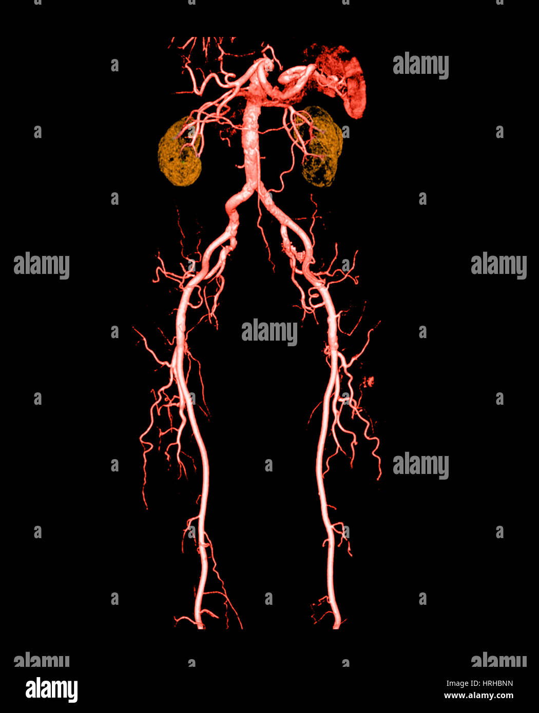 CT Angiogram of Abdomen and Legs Stock Photohttps://www.alamy.com/image-license-details/?v=1https://www.alamy.com/stock-photo-ct-angiogram-of-abdomen-and-legs-134992081.html
RMHRHBNN–CT Angiogram of Abdomen and Legs
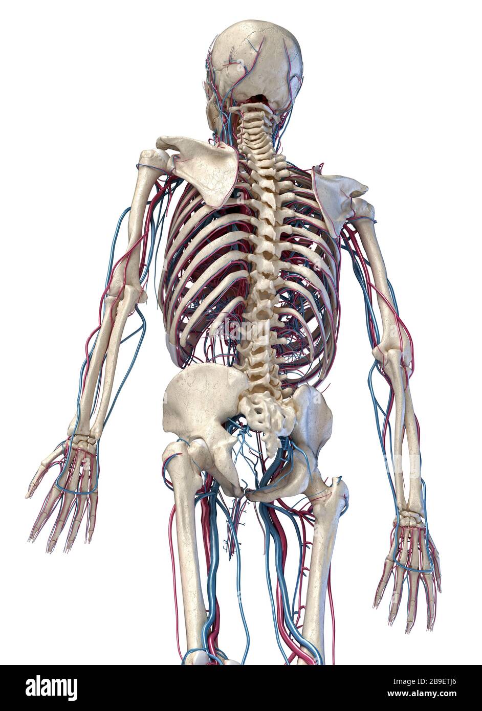 3/4 upper body rear view of human skeletal and vascular systems, white background. Stock Photohttps://www.alamy.com/image-license-details/?v=1https://www.alamy.com/34-upper-body-rear-view-of-human-skeletal-and-vascular-systems-white-background-image350065918.html
RF2B9ETJ6–3/4 upper body rear view of human skeletal and vascular systems, white background.
 Urinary system from kidney to glomerulus with structures of kidney and ureter. Stock Photohttps://www.alamy.com/image-license-details/?v=1https://www.alamy.com/urinary-system-from-kidney-to-glomerulus-with-structures-of-kidney-and-ureter-image476924294.html
RF2JKWNPE–Urinary system from kidney to glomerulus with structures of kidney and ureter.
 Liver-biliary bladder-infant Stock Photohttps://www.alamy.com/image-license-details/?v=1https://www.alamy.com/liver-biliary-bladder-infant-image466107157.html
RM2J290C5–Liver-biliary bladder-infant
 Blood supply of the pelvis Stock Photohttps://www.alamy.com/image-license-details/?v=1https://www.alamy.com/stock-photo-blood-supply-of-the-pelvis-13236042.html
RFACWFJK–Blood supply of the pelvis
 PELVIS, ANGIOGRAPHY Stock Photohttps://www.alamy.com/image-license-details/?v=1https://www.alamy.com/stock-photo-pelvis-angiography-49255775.html
RMCT3P6R–PELVIS, ANGIOGRAPHY
 Abdominal blood vessels, computer illustration. Stock Photohttps://www.alamy.com/image-license-details/?v=1https://www.alamy.com/abdominal-blood-vessels-computer-illustration-image367359494.html
RF2C9JJNX–Abdominal blood vessels, computer illustration.
 3d rendered illustration of a mans anatomy of the pelvis Stock Photohttps://www.alamy.com/image-license-details/?v=1https://www.alamy.com/3d-rendered-illustration-of-a-mans-anatomy-of-the-pelvis-image257953562.html
RFTYJPB6–3d rendered illustration of a mans anatomy of the pelvis
 Handwriting text Abdominal Aortic Aneurysmquestion. Concept meaning getting to know the enlargement of aorta Keyboard key Intention to create computer Stock Photohttps://www.alamy.com/image-license-details/?v=1https://www.alamy.com/handwriting-text-abdominal-aortic-aneurysmquestion-concept-meaning-getting-to-know-the-enlargement-of-aorta-keyboard-key-intention-to-create-computer-image229925993.html
RFRA20XH–Handwriting text Abdominal Aortic Aneurysmquestion. Concept meaning getting to know the enlargement of aorta Keyboard key Intention to create computer
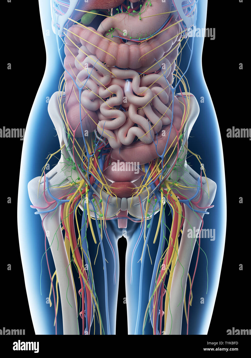 3d rendered illustration of a females abdominal anatomy Stock Photohttps://www.alamy.com/image-license-details/?v=1https://www.alamy.com/3d-rendered-illustration-of-a-females-abdominal-anatomy-image257967009.html
RFTYKBFD–3d rendered illustration of a females abdominal anatomy
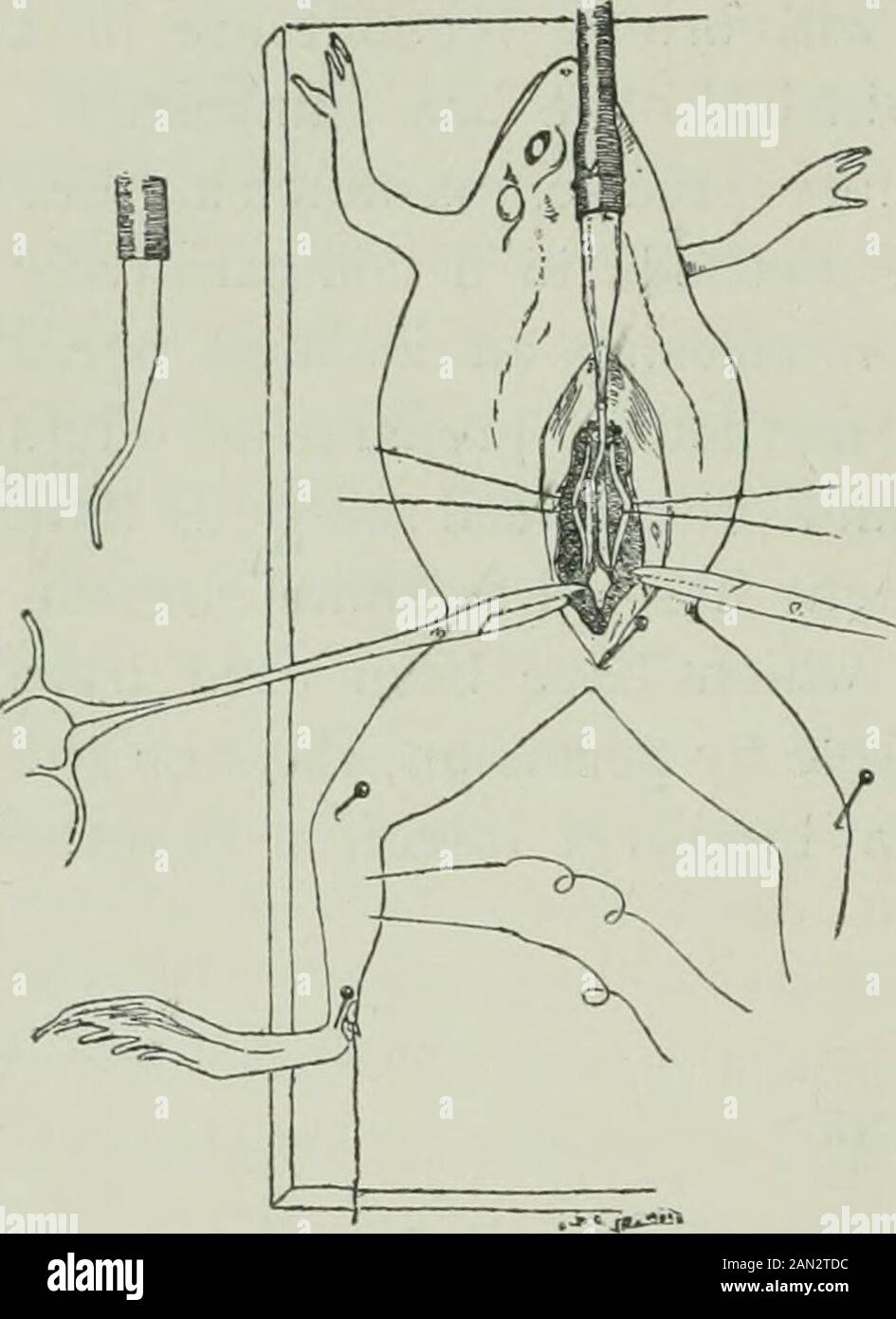 American journal of physiology . rigation. The abdominal and iliac veinsare then opened to allow the per-fused fluid to pass out, and so tolargely prevent any subsequentoedema. The animal is fastened to a boardby sharp pins passing through thejoints and pelvis in such a waythat the muscle contractions arenot interfered with nor the blood-vessels compressed, although thepreparation is made immovable. The irrigation should be started assoon as possible lest coagulation occur in the cannula or in the smallcapillaries and thus prevent a successful circulation. The circulationhas usually been contr Stock Photohttps://www.alamy.com/image-license-details/?v=1https://www.alamy.com/american-journal-of-physiology-rigation-the-abdominal-and-iliac-veinsare-then-opened-to-allow-the-per-fused-fluid-to-pass-out-and-so-tolargely-prevent-any-subsequentoedema-the-animal-is-fastened-to-a-boardby-sharp-pins-passing-through-thejoints-and-pelvis-in-such-a-waythat-the-muscle-contractions-arenot-interfered-with-nor-the-blood-vessels-compressed-although-thepreparation-is-made-immovable-the-irrigation-should-be-started-assoon-as-possible-lest-coagulation-occur-in-the-cannula-or-in-the-smallcapillaries-and-thus-prevent-a-successful-circulation-the-circulationhas-usually-been-contr-image339967864.html
RM2AN2TDC–American journal of physiology . rigation. The abdominal and iliac veinsare then opened to allow the per-fused fluid to pass out, and so tolargely prevent any subsequentoedema. The animal is fastened to a boardby sharp pins passing through thejoints and pelvis in such a waythat the muscle contractions arenot interfered with nor the blood-vessels compressed, although thepreparation is made immovable. The irrigation should be started assoon as possible lest coagulation occur in the cannula or in the smallcapillaries and thus prevent a successful circulation. The circulationhas usually been contr
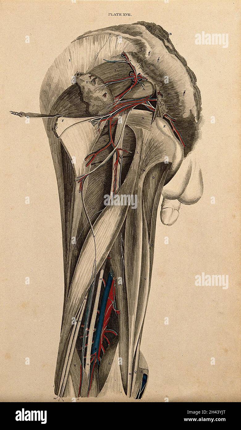 Male pelvis and thigh: dissection, with blood vessels and nerves indicated in red and blue. Coloured line engraving by W.H. Lizars, 1822/1826. Stock Photohttps://www.alamy.com/image-license-details/?v=1https://www.alamy.com/male-pelvis-and-thigh-dissection-with-blood-vessels-and-nerves-indicated-in-red-and-blue-coloured-line-engraving-by-wh-lizars-18221826-image450015744.html
RM2H43YJT–Male pelvis and thigh: dissection, with blood vessels and nerves indicated in red and blue. Coloured line engraving by W.H. Lizars, 1822/1826.
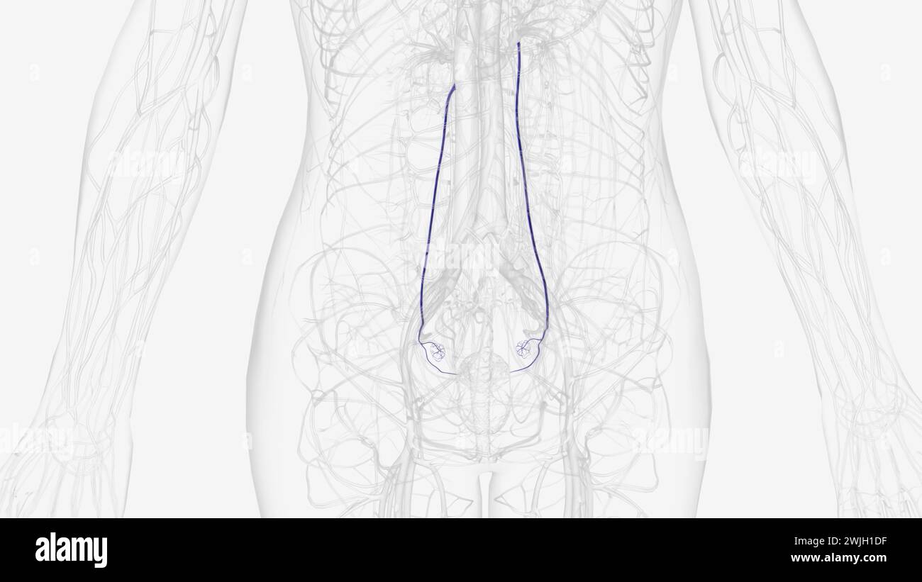 Each ovarian vein drains the deoxygenated venous blood from the region of the corresponding ovary in the pelvis up into the abdominal area Stock Photohttps://www.alamy.com/image-license-details/?v=1https://www.alamy.com/each-ovarian-vein-drains-the-deoxygenated-venous-blood-from-the-region-of-the-corresponding-ovary-in-the-pelvis-up-into-the-abdominal-area-image596590667.html
RF2WJH1DF–Each ovarian vein drains the deoxygenated venous blood from the region of the corresponding ovary in the pelvis up into the abdominal area
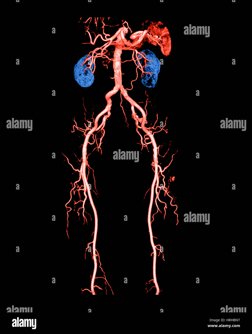 CT Angiogram of Abdomen and Legs Stock Photohttps://www.alamy.com/image-license-details/?v=1https://www.alamy.com/stock-photo-ct-angiogram-of-abdomen-and-legs-134992084.html
RMHRHBNT–CT Angiogram of Abdomen and Legs
 Front view of hip, limbs and hands of skeletal system with veins and arteries, white background. Stock Photohttps://www.alamy.com/image-license-details/?v=1https://www.alamy.com/front-view-of-hip-limbs-and-hands-of-skeletal-system-with-veins-and-arteries-white-background-image350068777.html
RF2B9F089–Front view of hip, limbs and hands of skeletal system with veins and arteries, white background.