JP4912738B2 - Laser scanning microscope - Google Patents
Laser scanning microscope Download PDFInfo
- Publication number
- JP4912738B2 JP4912738B2 JP2006132675A JP2006132675A JP4912738B2 JP 4912738 B2 JP4912738 B2 JP 4912738B2 JP 2006132675 A JP2006132675 A JP 2006132675A JP 2006132675 A JP2006132675 A JP 2006132675A JP 4912738 B2 JP4912738 B2 JP 4912738B2
- Authority
- JP
- Japan
- Prior art keywords
- laser
- laser beam
- observation
- laser light
- specimen
- Prior art date
- Legal status (The legal status is an assumption and is not a legal conclusion. Google has not performed a legal analysis and makes no representation as to the accuracy of the status listed.)
- Expired - Fee Related
Links
Images
Landscapes
- Microscoopes, Condenser (AREA)
Description
本発明は、レーザ走査型顕微鏡に関し、特に、観察用レーザ光と操作用レーザ光とを合波して同一の対物レンズにより標本に照射するレーザ走査型顕微鏡に関するものである。 The present invention relates to a laser scanning microscope, and more particularly to a laser scanning microscope that combines observation laser light and operation laser light and irradiates a specimen with the same objective lens.
従来、観察用レーザ光源と操作用レーザ光源とを備え、観察用レーザ光および操作用レーザ光を別個の走査手段により標本上で2次元的に走査するレーザ走査型顕微鏡が知られている(例えば、特許文献1参照。)。
このレーザ走査型顕微鏡においては、観察用レーザ光および操作用レーザ光が合波された後、同一の対物レンズによって標本上に集光されるようになっている。
In this laser scanning microscope, the observation laser beam and the operation laser beam are combined and then focused on the specimen by the same objective lens.
しかしながら、標本において発生した蛍光は観察用レーザ光の光路を走査手段を経由して戻った後に観察用レーザ光から分岐されるため、観察用レーザ光に操作用レーザ光を合波する合波手段を通過する際に蛍光が失われる。特に、操作用レーザ光の数が増加していくと、蛍光のロスが大きくなり、明るい蛍光画像を得ることができないという不都合が考えられる。 However, since the fluorescence generated in the specimen is branched from the observation laser light after returning the optical path of the observation laser light via the scanning means, the multiplexing means for combining the operation laser light with the observation laser light Fluorescence is lost when passing through. In particular, when the number of operating laser beams increases, the loss of fluorescence increases, and there is a disadvantage that a bright fluorescent image cannot be obtained.
本発明は上述した事情に鑑みてなされたものであって、観察用レーザ光に合波される操作用レーザ光の数が増加しても、蛍光のロスを低減して明るい蛍光画像を取得することができるレーザ走査型顕微鏡を提供することを目的としている。 The present invention has been made in view of the above-described circumstances, and even if the number of operation laser beams combined with the observation laser beam is increased, a fluorescence loss is reduced and a bright fluorescence image is acquired. It is an object of the present invention to provide a laser scanning microscope that can be used.
上記目的を達成するために、本発明は以下の手段を提供する。
本発明は、観察用レーザ光を発する観察用レーザ光源と、標本を操作するための複数の操作用レーザ光を発する操作用レーザ光源と、該操作用レーザ光源から発せられた複数の操作用レーザ光を合波する操作用レーザ光合波手段と、観察用レーザ光を2次元的に走査する走査手段と、複数の前記操作用レーザ光に対応して設けられ、複数の前記操作用レーザ光の照射位置をそれぞれ2次元的に調節する複数のレーザ光位置調節手段と、走査手段により走査された観察用レーザ光と、レーザ光位置調節手段により位置調節された操作用レーザ光とを合波する合波手段と、該合波手段により合波された観察用レーザ光および操作用レーザ光を集光して標本に照射する一方、標本において発生した蛍光を集光する対物レンズと、該対物レンズにより集光された蛍光を検出する光検出器とを備え、前記操作用レーザ光合波手段が、前記合波手段による観察用レーザ光との合波前に、前記複数の操作用レーザ光を合波する位置に配置されているレーザ走査型顕微鏡を提供する。
In order to achieve the above object, the present invention provides the following means.
The present invention relates to an observation laser light source that emits an observation laser light, an operation laser light source that emits a plurality of operation laser lights for operating a sample, and a plurality of operation lasers emitted from the operation laser light source. An operation laser beam combining means for combining the light, a scanning means for two-dimensionally scanning the observation laser light, and a plurality of the operation laser lights. to a plurality of laser beams position adjusting means for adjusting the irradiation position, respectively two-dimensionally, and the observation laser beam scanned by the scanning means and the position adjusted manipulation laser beam by the laser beam positioning means multiplexes An objective lens for condensing the fluorescence generated in the specimen while condensing the observation laser light and the operation laser light multiplexed by the multiplexing means and irradiating the specimen; and the objective lens By A light detector for detecting the emitted fluorescence, and the operation laser light combining means combines the plurality of operation laser lights before combining with the observation laser light by the combining means. A laser scanning microscope is provided that is positioned.
本発明によれば、観察用レーザ光源から発せられた観察用レーザ光が、走査手段により2次元的に走査され、対物レンズによって標本上に集光される。一方、操作用レーザ光源から発せられた複数の操作用レーザ光は、レーザ光位置調節手段の作動により2次元的な配置をそれぞれ調節された後、合波手段の作動により観察用レーザ光と合波され、対物レンズにより標本上に集光される。 According to the present invention, the observation laser light emitted from the observation laser light source is two-dimensionally scanned by the scanning unit and condensed on the sample by the objective lens. On the other hand, the plurality of operation laser beams emitted from the operation laser light source are adjusted to the two-dimensional arrangement by the operation of the laser beam position adjusting unit, and then combined with the observation laser beam by the operation of the combining unit. Waves are collected on the specimen by the objective lens.
観察用レーザ光は、標本に集光されることにより、標本内の蛍光物質を励起して蛍光を発生させる。発生した蛍光は、対物レンズ、合波手段、走査手段を介して戻り、光検出器により検出される。走査手段による標本上の操作位置情報および光検出器により検出された蛍光の光量情報に基づいて、蛍光画像が構築される。 The observation laser light is condensed on the specimen, thereby exciting the fluorescent substance in the specimen and generating fluorescence. The generated fluorescence returns through the objective lens, the multiplexing unit, and the scanning unit, and is detected by the photodetector. A fluorescence image is constructed based on the operation position information on the specimen by the scanning means and the light quantity information of the fluorescence detected by the photodetector.
一方、操作用レーザ光は、対物レンズにより標本に集光されることにより、標本に対し、光刺激、レーザトラップ等の操作を行う。本発明によれば、複数の操作用レーザ光の照射位置がそれぞれ調節されて標本に入射されるので、標本に対し、複数箇所の光刺激やレーザトラップを行うことができる。 On the other hand, the operation laser light is condensed on the specimen by the objective lens, and the specimen is operated for optical stimulation, laser trap, and the like. According to the present invention, since the irradiation positions of the plurality of operation laser beams are adjusted and incident on the specimen, a plurality of light stimulations and laser traps can be performed on the specimen.
この場合において、操作用レーザ光源から発せられた複数の操作用レーザ光は、操作用レーザ光合波手段により合波された後に、合波手段により観察用レーザ光と合波される。したがって、蛍光が辿ることとなる観察用レーザ光の光路上に、複数の操作用レーザ光を合波するための合波手段が配置されず、操作用レーザ光の数が増加しても、蛍光のロスを低く抑えて明るい蛍光画像を取得することが可能となる。 In this case, the plurality of operation laser lights emitted from the operation laser light source are combined by the operation laser light combining means and then combined with the observation laser light by the combining means. Therefore, no multiplexing means for multiplexing a plurality of operation laser lights is arranged on the optical path of the observation laser light that the fluorescence follows, and even if the number of operation laser lights increases, It is possible to obtain a bright fluorescent image with a low loss of the image.
上記発明においては、前記複数の操作用レーザ光が、相互に直交する偏光方向を備え、前記操作用レーザ光合波手段が、偏光ビームスプリッタにより構成されていることが好ましい。
このように構成することで、複数の操作用レーザ光を効率よく合波することができる。
In the above invention, it is preferable that the plurality of operation laser beams have polarization directions orthogonal to each other, and the operation laser beam multiplexing means is constituted by a polarization beam splitter.
By comprising in this way, the several laser beam for operation can be combined efficiently.
また、上記発明においては、前記操作用レーザ光源からの操作用レーザ光を複数に分岐する分岐手段と、分岐された複数の操作用レーザ光の偏光方向を相互に直交させるλ/2板とを備えることが好ましい。
このようにすることで、単一の操作用レーザ光源から複数の操作用レーザ光を得ることができ、また、複数の操作用レーザ光を効率よく合波できる。したがって、装置を簡易かつ安価に構成することができる。
また、上記発明においては、前記複数の操作用レーザ光のそれぞれの光路に、標本における集光位置を光軸方向に変化させる操作用レーザ光集光位置調整手段が備えられていることとしてもよい。
In the above invention, the branching means for branching the operation laser light from the operation laser light source into a plurality of parts, and the λ / 2 plate for making the polarization directions of the plurality of branched operation laser lights orthogonal to each other It is preferable to provide.
By doing in this way, a plurality of operation laser beams can be obtained from a single operation laser light source, and a plurality of operation laser beams can be efficiently combined. Therefore, the apparatus can be configured simply and inexpensively.
Further, in the above invention, an operation laser beam condensing position adjusting means for changing a condensing position in the specimen in the optical axis direction may be provided in each optical path of the plurality of operation laser beams. .
本発明によれば、観察用レーザ光に合波される操作用レーザ光の数が増加しても、蛍光のロスを低減して明るい蛍光画像を取得することができるという効果を奏する。 According to the present invention, even if the number of operation laser beams combined with the observation laser beam is increased, it is possible to obtain a bright fluorescence image by reducing the loss of fluorescence.
以下、本発明の一実施形態に係るレーザ走査型顕微鏡1について、図1〜図7を参照して説明する。
本実施形態に係るレーザ走査型顕微鏡1は、レーザ走査型共焦点顕微鏡である。図1中、各種レンズおよびピンホール等の光学部品は、説明の簡略化のために省略している。
Hereinafter, a
The
本実施形態に係るレーザ走査型顕微鏡1は、図1に示されるように、観察用レーザ光L1を発生する観察用レーザ光発生部2と、操作用レーザ光L2,L3を発生する操作用レーザ光発生部3と、観察用レーザ光L1および操作用レーザ光L2,L3を合波する第1の合波手段4と、合波された観察用レーザ光L1および操作用レーザ光L2,L3を集光して標本Pに照射する一方、観察用レーザ光L1を標本Pに照射することにより、標本P内の蛍光物質が励起されて発生した蛍光Fを集光する対物レンズ5と、該対物レンズ5により集光された蛍光Fを検出する光検出器6とを備えている。
As shown in FIG. 1, the
観察用レーザ光発生部2は、観察用レーザ光L1を出射する観察用レーザ光源7と、該観察用レーザ光源7から発せられた観察用レーザ光L1を光軸に交差する方向に2次元的に走査する第1のスキャナ(走査手段)8とを備えている。符号9はミラーである。
また、観察用レーザ光発生部2の観察用レーザ光源7と第1のスキャナ8との間には、標本Pにおいて発生し、対物レンズ5により集光され、合波手段4、ミラー9および第1のスキャナ8を経由して戻る蛍光Fを観察用レーザ光L1から分岐して光検出器6に向かわせるダイクロイックミラー10が備えられている。
Observation
Further, between the observation laser light source 7 of the observation laser
操作用レーザ光発生部3は、操作用レーザ光L2,L3を出射する操作用レーザ光源11と、該操作用レーザ光源11から発せられた操作用レーザ光L2,L3を分岐するハーフミラー(分岐手段)12と、分岐された2つの操作用レーザ光L2,L3をそれぞれ光軸に交差する方向に2次元的に位置調節する第2のスキャナ(レーザ光位置調節手段)13,14と、第2のスキャナ13,14によりそれぞれ位置調節された2つの操作用レーザ光L2,L3を合波する第2の合波手段(操作用レーザ光合波手段)15とを備えている。符号16はミラーである。
Manipulation laser beam generator 3, branches manipulation
また、操作用レーザ光発生部3には、ハーフミラー12により分岐された一方の操作用レーザ光L3の偏光方向を90°回転させるλ/2板17と、標本P上における操作用レーザ光L2,L3の合焦位置を調節する合焦位置調節手段(操作用レーザ光集光位置調整手段)18,19とが備えられている。該合焦位置調節手段18,19は、波面変換素子あるいは少なくとも一部が光軸方向に移動可能に支持された複数のレンズ群(図示略)により構成されている。
The operation laser light generator 3 includes a λ / 2
また、前記第2の合波手段15は、偏光ビームスプリッタにより構成されている。また、ハーフミラー12により分岐された2つの操作用レーザ光L2,L3の光路には、それぞれ、後述する制御装置20により開閉制御されるシャッタ21,22が設けられている。
シャッタ21,22に代えて、AOTFやAOMのような音響光学素子を用いてもよい。
操作用レーザ光L2,L3は、標本Pに結合されたアクチンまたはミオシンの位置に合焦されることにより、標本Pを保持する光ピンセットとして利用されるようになっている。
The second multiplexing means 15 is constituted by a polarization beam splitter. In addition,
Instead of the
The operating laser beams L 2 and L 3 are used as optical tweezers for holding the specimen P by being focused on the position of actin or myosin bound to the specimen P.
また、前記対物レンズ5は、該対物レンズ5を光軸方向に沿って移動させる合焦機構23により支持されている。
合焦機構23および合焦位置調節手段18,19は、制御装置20に接続されている。制御装置20は、合焦機構23による対物レンズ5の移動量と合焦位置調節手段18,19による2つの操作用レーザ光L2,L3の合焦位置の移動量との関係を記憶している。
The
The
制御装置20は、観察用レーザ光L1および操作用レーザ光L2,L3をいずれも照射している観察モードにおいては、合焦機構23と合焦位置調節手段18,19を連動して作動させ、対物レンズ5の移動方向とは逆方向に同じ距離だけ2つの操作用レーザ光L2,L3の合焦位置を移動させるようになっている。
In the observation mode in which both the observation laser beam L 1 and the operation laser beams L 2 and L 3 are irradiated, the
また、制御装置20は、観察用レーザ光L1のみを照射し、操作用レーザ光L2,L3を停止している準備モードにおいては、観察用レーザ光L1の合焦位置と、停止されている操作用レーザ光L2,L3の合焦位置(操作用レーザ光が出射されたならば達成される合焦位置)とを一致させた状態で、合焦機構23と合焦位置調節手段18,19との連動を停止するようになっている。
これにより、準備モードにおいては、合焦機構23を作動させて対物レンズ5を光軸方向に移動させると、観察用レーザ光L1のみならず停止されている操作用レーザ光L2,L3の合焦位置が対物レンズ5とともに光軸方向に移動させられるようになっている。
Further, in the preparation mode in which only the observation laser beam L 1 is irradiated and the operation laser beams L 2 and L 3 are stopped, the
Thus, in the preparation mode, when the
また、制御装置20には、前記光検出器6、第1、第2のスキャナ8,13、モニタ24およびマウス25が接続されている。
制御装置20は、第1のスキャナ8による観察用レーザ光L1の標本P上における走査位置情報と、光検出器6により検出された蛍光Fの光強度情報とに基づいて、2次元的な蛍光画像を構築し、モニタ24に表示するようになっている。
The
このように構成された本実施形態に係るレーザ走査型顕微鏡1の作用について、図2〜図8を参照しながら以下に説明する。
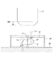
本実施形態に係るレーザ走査型顕微鏡1を用いて、標本P、例えば、シャーレ26内に貯留された培地27内に浮遊する細胞を観察する場合には、図2に示されるように、標本Pの端点A,Bにアクチンまたはミオシンを結合しておき(図8のステップS1)、制御装置20を準備モードに切り替える(図8のステップS2)。
The operation of the
When observing a specimen P, for example, a cell floating in the
これにより、図2に示されるように、観察用レーザ光発生部2からの観察用レーザ光L1のみが標本Pに照射され、2つの操作用レーザ光L2,L3は停止される。操作用レーザ光L2,L3の停止は、操作用レーザ光源11のオフまたはシャッタ21,22の閉鎖あるいは音響光学素子のオフにより行う。ここでは、シャッタ21,22の閉鎖により行うこととして説明する。
As a result, as shown in FIG. 2, only the observation laser light L 1 from the observation
操作者は、合焦機構23を作動させて、例えば、標本Pの最上位近傍に観察用レーザ光L1の焦点面FPが配置されるように対物レンズ5を移動し、第1のスキャナ8を作動させる。これにより、観察用レーザ光L1の焦点面FPにおける標本Pの蛍光画像が取得される(図8のステップS3)。図2に示す例では、標本Pの端点Aが最上位位置であるため、端点Aを含む最上位位置の断面の蛍光画像が取得される。
The operator operates the focusing
操作者は、取得された蛍光画像をモニタ24上で確認しながら、このモニタ24条の蛍光画像に対して端点Aをマウス25等の入力手段により指示する(図8のステップS4)。これにより、一方の操作用レーザ光L2による光ピンセットの把持位置が指定されるので、制御装置20は、第2のスキャナ13を作動させて、一方の操作用レーザ光L2の照射位置を指定された端点Aに一致するように2次元的に位置調整した後(図8のステップS5)、当該一方の操作用レーザ光L2の光路上のシャッタ21を開放し、操作用レーザ光L2を標本Pに照射する。
While confirming the acquired fluorescent image on the
準備モードにおいては、停止されていた操作用レーザ光L2の合焦位置が観察用レーザ光L1の焦点面FPと一致した状態に維持されているので、シャッタ21の開放により照射された操作用レーザ光L2は、図3に示されるように、端点Aに合焦され、標本Pを端点Aにおいて保持することができる。
同時に、制御装置20は、シャッタ21を開放した当該一方の操作用レーザ光L2については準備モードを解除し、観察モードに切り替える(図8のステップS6)。
In the preparation mode, since the in-focus position of the stopped operation laser light L 2 is maintained in a state coincident with the focal plane FP of the observation laser light L 1 , the operation irradiated by opening the
At the same time,
次いで、操作者は、合焦機構23を作動させて、図4に示されるように、観察用レーザ光L1の焦点面FPを下降させていく。このとき、シャッタ22が閉鎖されている操作用レーザ光L3については準備モードに維持されているため、その合焦位置は、観察用レーザ光L1の焦点面FPに一致した状態で合焦機構23の動作に従って下降させられる。
The operator then actuates the focusing
一方、準備モードが解除されて観察モードに切り替えられた操作用レーザ光L2については、制御装置20の作動により、合焦機構23と合焦位置調節手段18とが連動させられる。これにより、操作用レーザ光L2は、合焦機構23により観察用レーザ光L1の焦点面FPとともに移動させられる一方で、その移動量と同一の移動量で逆方向に操作用レーザ光L2の合焦位置が移動するように合焦位置調節手段18が作動させられる。その結果、操作用レーザ光L2は、図4に示されるように、標本Pの端点Aを保持した合焦位置を移動させることなく静止した状態に維持される。
On the other hand, with respect to the operation laser light L 2 that has been released from the preparation mode and switched to the observation mode, the focusing
そして、操作者は、合焦機構23を作動させて、例えば、図5に示されるように、標本Pの最下位近傍に観察用レーザ光L1の焦点面FPが配置されるように対物レンズ5を移動し、第1のスキャナ8を作動させる。図5に示す例では、標本Pの端点Bが最下位位置であるため、端点Bを含む最下位位置の断面の蛍光画像が取得される(図8のステップS7)。
Then, the operator operates the focusing
操作者は、取得された蛍光画像をモニタ24上で確認しながら、端点Bをマウス25等の入力手段により指示する(図8のステップS8)。これにより、他方の操作用レーザ光L3による光ピンセットの把持位置が指定されるので、制御装置20は、第2のスキャナ14を作動させて、他方の操作用レーザ光L3の照射位置を指定された端点Bに一致するように2次元的に位置調整した後(図8のステップS9)、当該他方の操作用レーザ光L3の光路上のシャッタ22を開放し、図6に示されるように、操作用レーザ光L3を標本Pに照射する。
The operator designates the end point B with the input means such as the
シャッタ22の開放により照射された当該他方の操作用レーザ光L3が端点Bに合焦され、標本Pを端点Bにおいて保持することができる。
同時に、制御装置20は、シャッタ22を開放した当該他方の操作用レーザ光L3についても準備モードを解除し、観察モードに切り替える(図8のステップS10)。これにより、図7に示されるように、合焦機構23を作動させて対物レンズ5を光軸方向に移動させても、他方の操作用レーザ光L3も、標本Pの端点Bを保持した合焦位置を移動させることなく静止した状態に維持される。
The other operation laser light L 3 irradiated by opening the
At the same time, the
その後の観察モードにおいては、合焦機構23を作動させることにより対物レンズ5を移動させ、観察用レーザ光L1の焦点面FPを移動させても、端点A,Bを保持している2つの操作用レーザ光L2,L3の合焦位置が静止状態に維持される。したがって、操作用レーザ光L2,L3を用いた光ピンセットにより標本Pを端点A,Bの2カ所で静止した状態に維持しつつ、観察用レーザ光L1の焦点面FPを順次移動させて、蛍光画像の取得位置を対物レンズ5の光軸方向に変位させることができる。そして、対物レンズ5の光軸方向にずれた複数の蛍光画像を取得することにより、これらの蛍光画像を用いて、標本Pの3次元的な蛍光分布を取得することが可能となる(図8のステップS11)。
In the subsequent observation mode, even if the
この場合において、本実施形態に係るレーザ走査型顕微鏡1によれば、2つに分岐された操作用レーザ光の合焦位置L2,L3が、それぞれ、合焦位置調節手段18,19により対物レンズ5の光軸方向に調節され、第2のスキャナ13,14により対物レンズ5の光軸に交差する2次元方向に調節される。そして、合焦位置を調節された2つの操作用レーザ光L2,L3が、第2の合波手段15によって合波された後に、第1の合波手段4により観察用レーザ光L1に合波されるので、対物レンズ5により集光された蛍光Fが第2の合波手段15を通過せずに済む。その結果、2つの操作用レーザ光L2,L3を照射する場合においても、操作用レーザ光L2,L3と観察用レーザ光L1との合波位置における蛍光Fのロスが増大することがなく、明るい蛍光画像を取得することができるという利点がある。
In this case, according to the
また、λ/2板17と偏光ビームスプリッタからなる第2の合波手段15とにより、2つの操作用レーザ光L2,L3を合波することとしているので、操作用レーザ光源11が1つで済み、装置の小型化を図ることができる。また、操作用レーザ光L2,L3のロスを低減することにより無駄をなくし、消費電力を低減することができる。
Further, since the two operation laser beams L 2 and L 3 are combined by the λ / 2
また、本実施形態に係るレーザ走査型顕微鏡1によれば、観察モードにおいて、合焦機構23と合焦位置調節手段18,19とを連動させることにより、単一の対物レンズ5を介して標本Pに照射されている観察用レーザ光L1の焦点面FPのみを対物レンズ5の移動に従って移動させ、操作用レーザ光L2,L3を静止させておくことができる。その結果、標本Pを静止させたままの状態で、観察用レーザ光L1の焦点面FPを対物レンズ5の光軸方向に移動させることができ、標本Pの状態を変化させることなく、また、標本Pに外力を付与することなく、3次元的な蛍光分布を観察することができる。
Further, according to the
なお、本実施形態においては、単一の操作用レーザ光源11から発せられた操作用レーザ光L2,L3をハーフミラー12によって分岐することとしたが、これに代えて、各操作用レーザ光L2,L3毎に、操作用レーザ光源11を配置してもよい。この場合には、操作用レーザ光源11の設置角度を90°異ならせることにより、偏光方向が相互に直交する2つの操作用レーザ光L2,L3を利用することができる。
In the present embodiment, the operation laser beams L 2 and L 3 emitted from the single operation
また、2つの操作用レーザ光L2,L3を用いる場合について説明したが、これに代えて、3以上の操作用レーザ光を用いることにしてもよい。この場合には、2つの操作用レーザ光を上記と同様にして合波した後に合波された操作用レーザ光をさらに他の操作用レーザ光と合波させることとすればよい。 Moreover, although the case where the two operation laser beams L 2 and L 3 are used has been described, three or more operation laser beams may be used instead. In this case, the operation laser light combined after combining the two operation laser lights in the same manner as described above may be combined with another operation laser light.
また、操作用レーザ光L2,L3を光ピンセットとして使用する場合について説明したが、光刺激用に使用することとしてもよい。このようにすることで、2カ所以上の定まった位置に光刺激を付与しながら標本Pの3次元的な蛍光分布を取得することができる。
また、合焦機構23として、対物レンズ5を光軸方向に移動させるものを例示したが、これに代えて、対物レンズ5は固定したままで、標本Pを支持するステージ28を光軸方向に移動させることにしてもよい。
Moreover, although the case where the operation laser beams L 2 and L 3 are used as optical tweezers has been described, they may be used for light stimulation. By doing in this way, the three-dimensional fluorescence distribution of the sample P can be acquired while giving a light stimulus to two or more fixed positions.
Further, as the focusing
また、本実施形態においては、2つの操作用レーザ光L2,L3を光ピンセットとして使用し、標本Pの異なる端点A,Bを静止した状態に保持したままで、合焦機構23の作動により対物レンズ5を光軸方向に移動させて、観察用レーザ光L1の焦点面FPを光軸方向に移動させたが、これに代えて、光ピンセットにより端点A,Bを把持した後は、合焦位置調節手段18,19の作動により標本Pを対物レンズ5の光軸方向に移動させることとしてもよい。
Further, in the present embodiment, the two operating laser beams L 2 and L 3 are used as optical tweezers, and the focusing
このようにすることで、合焦機構23により対物レンズ5やステージ28を移動させることなく、標本Pの3次元的な蛍光分布を取得することができる。その結果、対物レンズ5やステージ28を移動させるような大きな駆動力を発生するモータが不要となり、装置の小型化を図ることができる。
In this way, the three-dimensional fluorescence distribution of the specimen P can be acquired without moving the
F 蛍光
L1 観察用レーザ光
L2,L3 操作用レーザ光
P 標本
1 レーザ走査型顕微鏡
4 第1の合波手段(合波手段)
5 対物レンズ
6 光検出器
7 観察用レーザ光源
8 第1のスキャナ(走査手段)
11 操作用レーザ光源
12 ハーフミラー(分岐手段)
13,14 第2のスキャナ(レーザ光位置調節手段)
15 第2の合波手段(操作用レーザ光合波手段)
17 λ/2板
18,19 合焦位置調節手段(操作用レーザ光集光位置調整手段)
F fluorescence L 1 laser beam for observation L 2 , L 3 laser beam for
5 Objective Lens 6 Photodetector 7 Laser Light Source for Observation 8 First Scanner (Scanning Means)
11 Laser light source for
13, 14 Second scanner (laser beam position adjusting means)
15 Second multiplexing means (operational laser beam multiplexing means)
17 λ / 2
Claims (4)
標本を操作するための複数の操作用レーザ光を発する操作用レーザ光源と、
該操作用レーザ光源から発せられた複数の操作用レーザ光を合波する操作用レーザ光合波手段と、
観察用レーザ光を2次元的に走査する走査手段と、
複数の前記操作用レーザ光に対応して設けられ、複数の前記操作用レーザ光の照射位置をそれぞれ2次元的に調節する複数のレーザ光位置調節手段と、
走査手段により走査された観察用レーザ光と、レーザ光位置調節手段により位置調節された操作用レーザ光とを合波する合波手段と、
該合波手段により合波された観察用レーザ光および操作用レーザ光を集光して標本に照射する一方、標本において発生した蛍光を集光する対物レンズと、
該対物レンズにより集光された蛍光を検出する光検出器とを備え、
前記操作用レーザ光合波手段が、前記合波手段による観察用レーザ光との合波前に、前記複数の操作用レーザ光を合波する位置に配置されているレーザ走査型顕微鏡。 An observation laser light source that emits an observation laser beam;
An operation laser light source that emits a plurality of operation laser lights for operating the specimen;
An operation laser beam combining means for combining a plurality of operation laser beams emitted from the operation laser light source;
Scanning means for two-dimensionally scanning the observation laser beam;
A plurality of laser beam position adjusting means which are provided corresponding to the plurality of operation laser beams and adjust the irradiation positions of the plurality of operation laser beams two-dimensionally;
A combining unit that combines the observation laser beam scanned by the scanning unit and the operation laser beam adjusted in position by the laser beam position adjusting unit;
An objective lens for condensing the fluorescence generated in the specimen, while condensing and irradiating the specimen with the observation laser light and the operation laser light multiplexed by the multiplexing means;
A photodetector for detecting the fluorescence collected by the objective lens,
The laser scanning microscope in which the operation laser beam combining unit is arranged at a position where the plurality of operation laser beams are combined before being combined with the observation laser beam by the combining unit.
前記操作用レーザ光合波手段が、偏光ビームスプリッタにより構成されている請求項1に記載のレーザ走査型顕微鏡。 The plurality of operation laser beams have polarization directions orthogonal to each other,
The laser scanning microscope according to claim 1, wherein the operation laser beam multiplexing means is constituted by a polarization beam splitter.
分岐された複数の操作用レーザ光の偏光方向を相互に直交させるλ/2板とを備える請求項2に記載のレーザ走査型顕微鏡。 Branching means for branching the operation laser light from the operation laser light source into a plurality of parts;
The laser scanning microscope according to claim 2, further comprising a λ / 2 plate that makes the polarization directions of the plurality of branched operation laser beams orthogonal to each other.
Priority Applications (1)
| Application Number | Priority Date | Filing Date | Title |
|---|---|---|---|
| JP2006132675A JP4912738B2 (en) | 2006-05-11 | 2006-05-11 | Laser scanning microscope |
Applications Claiming Priority (1)
| Application Number | Priority Date | Filing Date | Title |
|---|---|---|---|
| JP2006132675A JP4912738B2 (en) | 2006-05-11 | 2006-05-11 | Laser scanning microscope |
Publications (2)
| Publication Number | Publication Date |
|---|---|
| JP2007304340A JP2007304340A (en) | 2007-11-22 |
| JP4912738B2 true JP4912738B2 (en) | 2012-04-11 |
Family
ID=38838314
Family Applications (1)
| Application Number | Title | Priority Date | Filing Date |
|---|---|---|---|
| JP2006132675A Expired - Fee Related JP4912738B2 (en) | 2006-05-11 | 2006-05-11 | Laser scanning microscope |
Country Status (1)
| Country | Link |
|---|---|
| JP (1) | JP4912738B2 (en) |
Families Citing this family (1)
| Publication number | Priority date | Publication date | Assignee | Title |
|---|---|---|---|---|
| JP5591007B2 (en) * | 2009-11-20 | 2014-09-17 | オリンパス株式会社 | Microscope equipment |
Family Cites Families (7)
| Publication number | Priority date | Publication date | Assignee | Title |
|---|---|---|---|---|
| JPS61221609A (en) * | 1986-03-20 | 1986-10-02 | Canon Inc | Optical device for photoelectric detection |
| JP2631779B2 (en) * | 1991-06-25 | 1997-07-16 | 富士写真フイルム株式会社 | Scanning microscope |
| JP3917731B2 (en) * | 1996-11-21 | 2007-05-23 | オリンパス株式会社 | Laser scanning microscope |
| JP3350442B2 (en) * | 1998-04-09 | 2002-11-25 | 科学技術振興事業団 | Microscope system |
| DE10356826B4 (en) * | 2003-12-05 | 2021-12-02 | Leica Microsystems Cms Gmbh | Scanning microscope |
| JP4468684B2 (en) * | 2003-12-05 | 2010-05-26 | オリンパス株式会社 | Scanning confocal microscope |
| JP2006003747A (en) * | 2004-06-18 | 2006-01-05 | Olympus Corp | Optical scanning type observation apparatus |
-
2006
- 2006-05-11 JP JP2006132675A patent/JP4912738B2/en not_active Expired - Fee Related
Also Published As
| Publication number | Publication date |
|---|---|
| JP2007304340A (en) | 2007-11-22 |
Similar Documents
| Publication | Publication Date | Title |
|---|---|---|
| JP4912737B2 (en) | Laser scanning microscope and microscope observation method | |
| JP5006694B2 (en) | Lighting device | |
| JP5636280B2 (en) | Optical device for optical manipulation | |
| JP5208650B2 (en) | Microscope system | |
| US8154796B2 (en) | Microscope apparatus | |
| US10001440B2 (en) | Observation apparatus and observation method | |
| JP2017167535A (en) | Light field microscope and illumination method | |
| US6924490B2 (en) | Microscope system | |
| JP2005121796A (en) | Laser microscope | |
| JP4270884B2 (en) | Microscope system | |
| JP5179099B2 (en) | Light stimulating illumination device and microscope device | |
| JP4867354B2 (en) | Confocal microscope | |
| JP4722464B2 (en) | Total reflection fluorescent lighting device | |
| JP4912738B2 (en) | Laser scanning microscope | |
| JP4939855B2 (en) | Illumination device and laser scanning microscope | |
| JP4700334B2 (en) | Total reflection fluorescence microscope | |
| JP2006003747A (en) | Optical scanning type observation apparatus | |
| JP4874012B2 (en) | Laser scanning microscope and image acquisition method of laser scanning microscope | |
| JP6278707B2 (en) | Optical device | |
| JP2004309785A (en) | Microscope system | |
| JP2009109787A (en) | Laser scanning type microscope | |
| JP4793626B2 (en) | Confocal microscope | |
| JP6177007B2 (en) | Microscope system | |
| JP2013101400A (en) | Microscope system | |
| JP2005172987A (en) | Laser microscope |
Legal Events
| Date | Code | Title | Description |
|---|---|---|---|
| A621 | Written request for application examination |
Free format text: JAPANESE INTERMEDIATE CODE: A621 Effective date: 20090420 |
|
| A131 | Notification of reasons for refusal |
Free format text: JAPANESE INTERMEDIATE CODE: A131 Effective date: 20110726 |
|
| A521 | Written amendment |
Free format text: JAPANESE INTERMEDIATE CODE: A523 Effective date: 20110922 |
|
| TRDD | Decision of grant or rejection written | ||
| A01 | Written decision to grant a patent or to grant a registration (utility model) |
Free format text: JAPANESE INTERMEDIATE CODE: A01 Effective date: 20120110 |
|
| A01 | Written decision to grant a patent or to grant a registration (utility model) |
Free format text: JAPANESE INTERMEDIATE CODE: A01 |
|
| A61 | First payment of annual fees (during grant procedure) |
Free format text: JAPANESE INTERMEDIATE CODE: A61 Effective date: 20120118 |
|
| R151 | Written notification of patent or utility model registration |
Ref document number: 4912738 Country of ref document: JP Free format text: JAPANESE INTERMEDIATE CODE: R151 |
|
| FPAY | Renewal fee payment (event date is renewal date of database) |
Free format text: PAYMENT UNTIL: 20150127 Year of fee payment: 3 |
|
| S531 | Written request for registration of change of domicile |
Free format text: JAPANESE INTERMEDIATE CODE: R313531 |
|
| R350 | Written notification of registration of transfer |
Free format text: JAPANESE INTERMEDIATE CODE: R350 |
|
| R250 | Receipt of annual fees |
Free format text: JAPANESE INTERMEDIATE CODE: R250 |
|
| LAPS | Cancellation because of no payment of annual fees |


