JP2016517776A - Method and apparatus for occluding a blood vessel and / or for securing two objects together - Google Patents
Method and apparatus for occluding a blood vessel and / or for securing two objects together Download PDFInfo
- Publication number
- JP2016517776A JP2016517776A JP2016513053A JP2016513053A JP2016517776A JP 2016517776 A JP2016517776 A JP 2016517776A JP 2016513053 A JP2016513053 A JP 2016513053A JP 2016513053 A JP2016513053 A JP 2016513053A JP 2016517776 A JP2016517776 A JP 2016517776A
- Authority
- JP
- Japan
- Prior art keywords
- implant
- occluder
- distal
- distal implant
- proximal
- Prior art date
- Legal status (The legal status is an assumption and is not a legal conclusion. Google has not performed a legal analysis and makes no representation as to the accuracy of the status listed.)
- Granted
Links
- 210000004204 blood vessel Anatomy 0.000 title claims description 144
- 238000000034 method Methods 0.000 title claims description 55
- 239000007943 implant Substances 0.000 claims abstract description 378
- 230000002829 reductive effect Effects 0.000 claims abstract description 21
- 210000000056 organ Anatomy 0.000 claims description 25
- 239000000463 material Substances 0.000 claims description 22
- 229910001000 nickel titanium Inorganic materials 0.000 claims description 9
- HLXZNVUGXRDIFK-UHFFFAOYSA-N nickel titanium Chemical compound [Ti].[Ti].[Ti].[Ti].[Ti].[Ti].[Ti].[Ti].[Ti].[Ti].[Ti].[Ni].[Ni].[Ni].[Ni].[Ni].[Ni].[Ni].[Ni].[Ni].[Ni].[Ni].[Ni].[Ni].[Ni] HLXZNVUGXRDIFK-UHFFFAOYSA-N 0.000 claims description 9
- 229910001285 shape-memory alloy Inorganic materials 0.000 claims description 6
- 229910001069 Ti alloy Inorganic materials 0.000 claims description 3
- 230000002441 reversible effect Effects 0.000 claims description 2
- 210000003462 vein Anatomy 0.000 description 140
- 238000010586 diagram Methods 0.000 description 137
- 210000002414 leg Anatomy 0.000 description 99
- 210000001519 tissue Anatomy 0.000 description 39
- 206010046996 Varicose vein Diseases 0.000 description 38
- 238000009434 installation Methods 0.000 description 36
- 208000027185 varicose disease Diseases 0.000 description 36
- 210000003752 saphenous vein Anatomy 0.000 description 31
- 238000001356 surgical procedure Methods 0.000 description 20
- 239000002131 composite material Substances 0.000 description 19
- 239000003814 drug Substances 0.000 description 19
- 229940079593 drug Drugs 0.000 description 19
- 230000017531 blood circulation Effects 0.000 description 17
- 210000004027 cell Anatomy 0.000 description 17
- 238000013459 approach Methods 0.000 description 14
- 238000012800 visualization Methods 0.000 description 13
- 238000011282 treatment Methods 0.000 description 12
- 210000003484 anatomy Anatomy 0.000 description 11
- 210000004369 blood Anatomy 0.000 description 11
- 239000008280 blood Substances 0.000 description 11
- 238000007632 sclerotherapy Methods 0.000 description 11
- 239000012781 shape memory material Substances 0.000 description 11
- 239000000126 substance Substances 0.000 description 10
- 238000002271 resection Methods 0.000 description 7
- 206010052428 Wound Diseases 0.000 description 6
- 230000009471 action Effects 0.000 description 6
- 239000003795 chemical substances by application Substances 0.000 description 6
- 239000013013 elastic material Substances 0.000 description 6
- 210000003191 femoral vein Anatomy 0.000 description 6
- 239000007787 solid Substances 0.000 description 6
- 208000009056 telangiectasis Diseases 0.000 description 6
- 238000002604 ultrasonography Methods 0.000 description 6
- 208000027418 Wounds and injury Diseases 0.000 description 5
- 230000036760 body temperature Effects 0.000 description 5
- 229920000642 polymer Polymers 0.000 description 5
- 239000003229 sclerosing agent Substances 0.000 description 5
- 208000024891 symptom Diseases 0.000 description 5
- 206010042674 Swelling Diseases 0.000 description 4
- 206010053648 Vascular occlusion Diseases 0.000 description 4
- 238000002679 ablation Methods 0.000 description 4
- 230000008901 benefit Effects 0.000 description 4
- 230000006835 compression Effects 0.000 description 4
- 238000007906 compression Methods 0.000 description 4
- 230000006378 damage Effects 0.000 description 4
- 238000000151 deposition Methods 0.000 description 4
- 238000012377 drug delivery Methods 0.000 description 4
- 238000001990 intravenous administration Methods 0.000 description 4
- 210000004185 liver Anatomy 0.000 description 4
- 239000003589 local anesthetic agent Substances 0.000 description 4
- 230000002980 postoperative effect Effects 0.000 description 4
- 230000008961 swelling Effects 0.000 description 4
- 230000008859 change Effects 0.000 description 3
- 150000001875 compounds Chemical class 0.000 description 3
- 230000006870 function Effects 0.000 description 3
- 210000004013 groin Anatomy 0.000 description 3
- 208000014674 injury Diseases 0.000 description 3
- 238000002690 local anesthesia Methods 0.000 description 3
- 210000003101 oviduct Anatomy 0.000 description 3
- 230000036961 partial effect Effects 0.000 description 3
- 230000000149 penetrating effect Effects 0.000 description 3
- 239000004033 plastic Substances 0.000 description 3
- 230000008569 process Effects 0.000 description 3
- 238000011084 recovery Methods 0.000 description 3
- 229920000431 shape-memory polymer Polymers 0.000 description 3
- 210000002784 stomach Anatomy 0.000 description 3
- 230000008733 trauma Effects 0.000 description 3
- 230000002792 vascular Effects 0.000 description 3
- 208000021331 vascular occlusion disease Diseases 0.000 description 3
- 206010061218 Inflammation Diseases 0.000 description 2
- 241000287436 Turdus merula Species 0.000 description 2
- 208000025865 Ulcer Diseases 0.000 description 2
- 210000001367 artery Anatomy 0.000 description 2
- 210000003414 extremity Anatomy 0.000 description 2
- 238000010438 heat treatment Methods 0.000 description 2
- 238000003384 imaging method Methods 0.000 description 2
- 230000004054 inflammatory process Effects 0.000 description 2
- 238000002357 laparoscopic surgery Methods 0.000 description 2
- 229960005015 local anesthetics Drugs 0.000 description 2
- 230000013011 mating Effects 0.000 description 2
- 230000007246 mechanism Effects 0.000 description 2
- 239000002184 metal Substances 0.000 description 2
- 229910052751 metal Inorganic materials 0.000 description 2
- 230000004048 modification Effects 0.000 description 2
- 238000012986 modification Methods 0.000 description 2
- 238000012148 non-surgical treatment Methods 0.000 description 2
- 238000002355 open surgical procedure Methods 0.000 description 2
- 230000028327 secretion Effects 0.000 description 2
- 238000002560 therapeutic procedure Methods 0.000 description 2
- 238000003466 welding Methods 0.000 description 2
- 206010002091 Anaesthesia Diseases 0.000 description 1
- OKTJSMMVPCPJKN-UHFFFAOYSA-N Carbon Chemical compound [C] OKTJSMMVPCPJKN-UHFFFAOYSA-N 0.000 description 1
- 206010053567 Coagulopathies Diseases 0.000 description 1
- 206010016717 Fistula Diseases 0.000 description 1
- XUIMIQQOPSSXEZ-UHFFFAOYSA-N Silicon Chemical compound [Si] XUIMIQQOPSSXEZ-UHFFFAOYSA-N 0.000 description 1
- 206010040829 Skin discolouration Diseases 0.000 description 1
- 238000010317 ablation therapy Methods 0.000 description 1
- 230000001154 acute effect Effects 0.000 description 1
- 239000000853 adhesive Substances 0.000 description 1
- 230000001070 adhesive effect Effects 0.000 description 1
- 230000037005 anaesthesia Effects 0.000 description 1
- 229940035676 analgesics Drugs 0.000 description 1
- 229940035674 anesthetics Drugs 0.000 description 1
- 239000000730 antalgic agent Substances 0.000 description 1
- 238000007681 bariatric surgery Methods 0.000 description 1
- 238000005452 bending Methods 0.000 description 1
- 210000000941 bile Anatomy 0.000 description 1
- 210000000013 bile duct Anatomy 0.000 description 1
- 239000000560 biocompatible material Substances 0.000 description 1
- 230000000740 bleeding effect Effects 0.000 description 1
- 210000000601 blood cell Anatomy 0.000 description 1
- 239000002041 carbon nanotube Substances 0.000 description 1
- 229910021393 carbon nanotube Inorganic materials 0.000 description 1
- 238000002192 cholecystectomy Methods 0.000 description 1
- 230000035602 clotting Effects 0.000 description 1
- 238000001816 cooling Methods 0.000 description 1
- 230000008878 coupling Effects 0.000 description 1
- 238000010168 coupling process Methods 0.000 description 1
- 238000005859 coupling reaction Methods 0.000 description 1
- 230000023753 dehiscence Effects 0.000 description 1
- 238000009826 distribution Methods 0.000 description 1
- 230000008030 elimination Effects 0.000 description 1
- 238000003379 elimination reaction Methods 0.000 description 1
- 230000003628 erosive effect Effects 0.000 description 1
- 239000000284 extract Substances 0.000 description 1
- 238000000605 extraction Methods 0.000 description 1
- 230000003890 fistula Effects 0.000 description 1
- 239000006260 foam Substances 0.000 description 1
- 210000000232 gallbladder Anatomy 0.000 description 1
- 230000002496 gastric effect Effects 0.000 description 1
- 238000002695 general anesthesia Methods 0.000 description 1
- 239000003193 general anesthetic agent Substances 0.000 description 1
- 239000003292 glue Substances 0.000 description 1
- 230000036541 health Effects 0.000 description 1
- 208000015181 infectious disease Diseases 0.000 description 1
- 208000000509 infertility Diseases 0.000 description 1
- 230000036512 infertility Effects 0.000 description 1
- 231100000535 infertility Toxicity 0.000 description 1
- 238000002347 injection Methods 0.000 description 1
- 239000007924 injection Substances 0.000 description 1
- 230000010354 integration Effects 0.000 description 1
- 230000003993 interaction Effects 0.000 description 1
- 210000000936 intestine Anatomy 0.000 description 1
- 238000007918 intramuscular administration Methods 0.000 description 1
- 230000001678 irradiating effect Effects 0.000 description 1
- 238000005304 joining Methods 0.000 description 1
- 210000003734 kidney Anatomy 0.000 description 1
- 210000003127 knee Anatomy 0.000 description 1
- 238000013532 laser treatment Methods 0.000 description 1
- 230000000670 limiting effect Effects 0.000 description 1
- 230000005923 long-lasting effect Effects 0.000 description 1
- 210000003141 lower extremity Anatomy 0.000 description 1
- 210000004072 lung Anatomy 0.000 description 1
- 230000001926 lymphatic effect Effects 0.000 description 1
- 210000001365 lymphatic vessel Anatomy 0.000 description 1
- 239000011159 matrix material Substances 0.000 description 1
- QSHDDOUJBYECFT-UHFFFAOYSA-N mercury Chemical compound [Hg] QSHDDOUJBYECFT-UHFFFAOYSA-N 0.000 description 1
- 229910052753 mercury Inorganic materials 0.000 description 1
- 238000002324 minimally invasive surgery Methods 0.000 description 1
- 239000000203 mixture Substances 0.000 description 1
- 210000003205 muscle Anatomy 0.000 description 1
- 210000005036 nerve Anatomy 0.000 description 1
- 230000035764 nutrition Effects 0.000 description 1
- 235000016709 nutrition Nutrition 0.000 description 1
- 229940124583 pain medication Drugs 0.000 description 1
- 239000004848 polyfunctional curative Substances 0.000 description 1
- 230000035935 pregnancy Effects 0.000 description 1
- 238000004321 preservation Methods 0.000 description 1
- 102000004169 proteins and genes Human genes 0.000 description 1
- 108090000623 proteins and genes Proteins 0.000 description 1
- 230000000541 pulsatile effect Effects 0.000 description 1
- 230000000306 recurrent effect Effects 0.000 description 1
- 230000009467 reduction Effects 0.000 description 1
- 231100000241 scar Toxicity 0.000 description 1
- 230000037390 scarring Effects 0.000 description 1
- 229910052710 silicon Inorganic materials 0.000 description 1
- 239000010703 silicon Substances 0.000 description 1
- 230000037370 skin discoloration Effects 0.000 description 1
- 239000003381 stabilizer Substances 0.000 description 1
- 230000001954 sterilising effect Effects 0.000 description 1
- 238000004659 sterilization and disinfection Methods 0.000 description 1
- 230000009885 systemic effect Effects 0.000 description 1
- 210000000115 thoracic cavity Anatomy 0.000 description 1
- 230000007704 transition Effects 0.000 description 1
- 238000011277 treatment modality Methods 0.000 description 1
- 231100000397 ulcer Toxicity 0.000 description 1
- 230000036269 ulceration Effects 0.000 description 1
- 210000000689 upper leg Anatomy 0.000 description 1
- 210000002700 urine Anatomy 0.000 description 1
- 210000001177 vas deferen Anatomy 0.000 description 1
- 238000004804 winding Methods 0.000 description 1
Images
Classifications
-
- A—HUMAN NECESSITIES
- A61—MEDICAL OR VETERINARY SCIENCE; HYGIENE
- A61B—DIAGNOSIS; SURGERY; IDENTIFICATION
- A61B17/00—Surgical instruments, devices or methods, e.g. tourniquets
- A61B17/12—Surgical instruments, devices or methods, e.g. tourniquets for ligaturing or otherwise compressing tubular parts of the body, e.g. blood vessels, umbilical cord
- A61B17/122—Clamps or clips, e.g. for the umbilical cord
-
- A—HUMAN NECESSITIES
- A61—MEDICAL OR VETERINARY SCIENCE; HYGIENE
- A61B—DIAGNOSIS; SURGERY; IDENTIFICATION
- A61B17/00—Surgical instruments, devices or methods, e.g. tourniquets
- A61B17/00008—Vein tendon strippers
-
- A—HUMAN NECESSITIES
- A61—MEDICAL OR VETERINARY SCIENCE; HYGIENE
- A61B—DIAGNOSIS; SURGERY; IDENTIFICATION
- A61B17/00—Surgical instruments, devices or methods, e.g. tourniquets
- A61B17/064—Surgical staples, i.e. penetrating the tissue
- A61B17/0643—Surgical staples, i.e. penetrating the tissue with separate closing member, e.g. for interlocking with staple
-
- A—HUMAN NECESSITIES
- A61—MEDICAL OR VETERINARY SCIENCE; HYGIENE
- A61B—DIAGNOSIS; SURGERY; IDENTIFICATION
- A61B17/00—Surgical instruments, devices or methods, e.g. tourniquets
- A61B17/12—Surgical instruments, devices or methods, e.g. tourniquets for ligaturing or otherwise compressing tubular parts of the body, e.g. blood vessels, umbilical cord
- A61B17/12009—Implements for ligaturing other than by clamps or clips, e.g. using a loop with a slip knot
- A61B17/12013—Implements for ligaturing other than by clamps or clips, e.g. using a loop with a slip knot for use in minimally invasive surgery, e.g. endoscopic surgery
-
- A—HUMAN NECESSITIES
- A61—MEDICAL OR VETERINARY SCIENCE; HYGIENE
- A61B—DIAGNOSIS; SURGERY; IDENTIFICATION
- A61B17/00—Surgical instruments, devices or methods, e.g. tourniquets
- A61B17/12—Surgical instruments, devices or methods, e.g. tourniquets for ligaturing or otherwise compressing tubular parts of the body, e.g. blood vessels, umbilical cord
- A61B17/12022—Occluding by internal devices, e.g. balloons or releasable wires
- A61B17/12027—Type of occlusion
- A61B17/12031—Type of occlusion complete occlusion
-
- A—HUMAN NECESSITIES
- A61—MEDICAL OR VETERINARY SCIENCE; HYGIENE
- A61B—DIAGNOSIS; SURGERY; IDENTIFICATION
- A61B17/00—Surgical instruments, devices or methods, e.g. tourniquets
- A61B17/12—Surgical instruments, devices or methods, e.g. tourniquets for ligaturing or otherwise compressing tubular parts of the body, e.g. blood vessels, umbilical cord
- A61B17/12022—Occluding by internal devices, e.g. balloons or releasable wires
- A61B17/12099—Occluding by internal devices, e.g. balloons or releasable wires characterised by the location of the occluder
- A61B17/12109—Occluding by internal devices, e.g. balloons or releasable wires characterised by the location of the occluder in a blood vessel
-
- A—HUMAN NECESSITIES
- A61—MEDICAL OR VETERINARY SCIENCE; HYGIENE
- A61B—DIAGNOSIS; SURGERY; IDENTIFICATION
- A61B17/00—Surgical instruments, devices or methods, e.g. tourniquets
- A61B17/12—Surgical instruments, devices or methods, e.g. tourniquets for ligaturing or otherwise compressing tubular parts of the body, e.g. blood vessels, umbilical cord
- A61B17/12022—Occluding by internal devices, e.g. balloons or releasable wires
- A61B17/12131—Occluding by internal devices, e.g. balloons or releasable wires characterised by the type of occluding device
- A61B17/1214—Coils or wires
- A61B17/12145—Coils or wires having a pre-set deployed three-dimensional shape
-
- A—HUMAN NECESSITIES
- A61—MEDICAL OR VETERINARY SCIENCE; HYGIENE
- A61B—DIAGNOSIS; SURGERY; IDENTIFICATION
- A61B17/00—Surgical instruments, devices or methods, e.g. tourniquets
- A61B17/12—Surgical instruments, devices or methods, e.g. tourniquets for ligaturing or otherwise compressing tubular parts of the body, e.g. blood vessels, umbilical cord
- A61B17/122—Clamps or clips, e.g. for the umbilical cord
- A61B17/1227—Spring clips
-
- A—HUMAN NECESSITIES
- A61—MEDICAL OR VETERINARY SCIENCE; HYGIENE
- A61B—DIAGNOSIS; SURGERY; IDENTIFICATION
- A61B17/00—Surgical instruments, devices or methods, e.g. tourniquets
- A61B17/12—Surgical instruments, devices or methods, e.g. tourniquets for ligaturing or otherwise compressing tubular parts of the body, e.g. blood vessels, umbilical cord
- A61B17/128—Surgical instruments, devices or methods, e.g. tourniquets for ligaturing or otherwise compressing tubular parts of the body, e.g. blood vessels, umbilical cord for applying or removing clamps or clips
- A61B17/1285—Surgical instruments, devices or methods, e.g. tourniquets for ligaturing or otherwise compressing tubular parts of the body, e.g. blood vessels, umbilical cord for applying or removing clamps or clips for minimally invasive surgery
-
- A—HUMAN NECESSITIES
- A61—MEDICAL OR VETERINARY SCIENCE; HYGIENE
- A61B—DIAGNOSIS; SURGERY; IDENTIFICATION
- A61B17/00—Surgical instruments, devices or methods, e.g. tourniquets
- A61B17/34—Trocars; Puncturing needles
- A61B17/3403—Needle locating or guiding means
-
- A—HUMAN NECESSITIES
- A61—MEDICAL OR VETERINARY SCIENCE; HYGIENE
- A61B—DIAGNOSIS; SURGERY; IDENTIFICATION
- A61B17/00—Surgical instruments, devices or methods, e.g. tourniquets
- A61B17/08—Wound clamps or clips, i.e. not or only partly penetrating the tissue ; Devices for bringing together the edges of a wound
-
- A—HUMAN NECESSITIES
- A61—MEDICAL OR VETERINARY SCIENCE; HYGIENE
- A61B—DIAGNOSIS; SURGERY; IDENTIFICATION
- A61B17/00—Surgical instruments, devices or methods, e.g. tourniquets
- A61B17/12—Surgical instruments, devices or methods, e.g. tourniquets for ligaturing or otherwise compressing tubular parts of the body, e.g. blood vessels, umbilical cord
- A61B17/12022—Occluding by internal devices, e.g. balloons or releasable wires
- A61B17/12027—Type of occlusion
- A61B17/12036—Type of occlusion partial occlusion
-
- A—HUMAN NECESSITIES
- A61—MEDICAL OR VETERINARY SCIENCE; HYGIENE
- A61B—DIAGNOSIS; SURGERY; IDENTIFICATION
- A61B17/00—Surgical instruments, devices or methods, e.g. tourniquets
- A61B2017/00831—Material properties
- A61B2017/00862—Material properties elastic or resilient
-
- A—HUMAN NECESSITIES
- A61—MEDICAL OR VETERINARY SCIENCE; HYGIENE
- A61B—DIAGNOSIS; SURGERY; IDENTIFICATION
- A61B17/00—Surgical instruments, devices or methods, e.g. tourniquets
- A61B2017/00831—Material properties
- A61B2017/00867—Material properties shape memory effect
-
- A—HUMAN NECESSITIES
- A61—MEDICAL OR VETERINARY SCIENCE; HYGIENE
- A61B—DIAGNOSIS; SURGERY; IDENTIFICATION
- A61B17/00—Surgical instruments, devices or methods, e.g. tourniquets
- A61B2017/00831—Material properties
- A61B2017/00893—Material properties pharmaceutically effective
-
- A—HUMAN NECESSITIES
- A61—MEDICAL OR VETERINARY SCIENCE; HYGIENE
- A61B—DIAGNOSIS; SURGERY; IDENTIFICATION
- A61B17/00—Surgical instruments, devices or methods, e.g. tourniquets
- A61B2017/00982—General structural features
- A61B2017/00986—Malecots, e.g. slotted tubes, of which the distal end is pulled to deflect side struts
-
- A—HUMAN NECESSITIES
- A61—MEDICAL OR VETERINARY SCIENCE; HYGIENE
- A61B—DIAGNOSIS; SURGERY; IDENTIFICATION
- A61B17/00—Surgical instruments, devices or methods, e.g. tourniquets
- A61B17/04—Surgical instruments, devices or methods, e.g. tourniquets for suturing wounds; Holders or packages for needles or suture materials
- A61B17/0401—Suture anchors, buttons or pledgets, i.e. means for attaching sutures to bone, cartilage or soft tissue; Instruments for applying or removing suture anchors
- A61B2017/0464—Suture anchors, buttons or pledgets, i.e. means for attaching sutures to bone, cartilage or soft tissue; Instruments for applying or removing suture anchors for soft tissue
-
- A—HUMAN NECESSITIES
- A61—MEDICAL OR VETERINARY SCIENCE; HYGIENE
- A61B—DIAGNOSIS; SURGERY; IDENTIFICATION
- A61B17/00—Surgical instruments, devices or methods, e.g. tourniquets
- A61B17/064—Surgical staples, i.e. penetrating the tissue
- A61B2017/0647—Surgical staples, i.e. penetrating the tissue having one single leg, e.g. tacks
-
- A—HUMAN NECESSITIES
- A61—MEDICAL OR VETERINARY SCIENCE; HYGIENE
- A61B—DIAGNOSIS; SURGERY; IDENTIFICATION
- A61B17/00—Surgical instruments, devices or methods, e.g. tourniquets
- A61B17/064—Surgical staples, i.e. penetrating the tissue
- A61B2017/0649—Coils or spirals
-
- A—HUMAN NECESSITIES
- A61—MEDICAL OR VETERINARY SCIENCE; HYGIENE
- A61B—DIAGNOSIS; SURGERY; IDENTIFICATION
- A61B17/00—Surgical instruments, devices or methods, e.g. tourniquets
- A61B17/12—Surgical instruments, devices or methods, e.g. tourniquets for ligaturing or otherwise compressing tubular parts of the body, e.g. blood vessels, umbilical cord
- A61B17/12022—Occluding by internal devices, e.g. balloons or releasable wires
- A61B2017/1205—Introduction devices
- A61B2017/12054—Details concerning the detachment of the occluding device from the introduction device
-
- A—HUMAN NECESSITIES
- A61—MEDICAL OR VETERINARY SCIENCE; HYGIENE
- A61B—DIAGNOSIS; SURGERY; IDENTIFICATION
- A61B17/00—Surgical instruments, devices or methods, e.g. tourniquets
- A61B17/34—Trocars; Puncturing needles
- A61B17/3403—Needle locating or guiding means
- A61B2017/3405—Needle locating or guiding means using mechanical guide means
-
- A—HUMAN NECESSITIES
- A61—MEDICAL OR VETERINARY SCIENCE; HYGIENE
- A61B—DIAGNOSIS; SURGERY; IDENTIFICATION
- A61B17/00—Surgical instruments, devices or methods, e.g. tourniquets
- A61B17/34—Trocars; Puncturing needles
- A61B17/3403—Needle locating or guiding means
- A61B2017/3413—Needle locating or guiding means guided by ultrasound
Landscapes
- Health & Medical Sciences (AREA)
- Surgery (AREA)
- Life Sciences & Earth Sciences (AREA)
- Medical Informatics (AREA)
- Animal Behavior & Ethology (AREA)
- Engineering & Computer Science (AREA)
- Biomedical Technology (AREA)
- Heart & Thoracic Surgery (AREA)
- Veterinary Medicine (AREA)
- Molecular Biology (AREA)
- Nuclear Medicine, Radiotherapy & Molecular Imaging (AREA)
- General Health & Medical Sciences (AREA)
- Public Health (AREA)
- Reproductive Health (AREA)
- Vascular Medicine (AREA)
- Rheumatology (AREA)
- Pathology (AREA)
- Surgical Instruments (AREA)
- Prostheses (AREA)
Abstract
第1の構造と第2の構造との間の空間を閉塞するための装置であって、該装置は、遠位インプラントおよび近位インプラントを備えている閉塞器を備え、遠位インプラントは、本体および該本体に搭載されている係止シャフトを備え、該遠位インプラントの本体は、(i)管の管腔内での配置のための直径方向に縮小された構成と、(ii)第1の構造に対する配置のための直径方向に広げられた構成とをとり得る複数の脚部を備え、さらに、該係止シャフトは、該近位インプラントへの選択的接続のための第1の係止要素および該閉塞器を展開するための挿入器への選択的接続のための第2の係止要素を備えている。An apparatus for occluding a space between a first structure and a second structure, the apparatus comprising an occluder comprising a distal implant and a proximal implant, the distal implant comprising a body And a locking shaft mounted on the body, wherein the body of the distal implant comprises (i) a diametrically reduced configuration for placement within the lumen of the tube, and (ii) a first A plurality of legs that can take a diametrically expanded configuration for placement relative to the structure, and wherein the locking shaft includes a first locking for selective connection to the proximal implant A second locking element for selective connection to the element and an inserter for deploying the occluder.
Description
(係属中の先の特許出願の参照)
本願は、(i)係属中の先の米国特許出願第13/857,424号(2013年4月5日出願、Amsel Medical Corporation、および、Arnold Mille、他、名称「METHOD AND APPARATUS FOR OCCLUDING A BLOOD VESSEL」、代理人案件番号AM−9)の一部継続出願であり、上記特許出願は、(a)先の米国特許出願第13/348,416号(2012年1月11日出願、Arnold Miller、他、名称「METHOD AND APPARATUS FOR TREATING VARICOSE VEINS」、代理人案件番号AM−0708)の一部継続出願であり、この出願は、先の米国仮特許出願第61/431,609号(2011年11月11日出願、Arnold Miller、名称「METHOD AND APPARATUS FOR TREATING VARICOSE VEINS」、代理人案件番号AM−7 PROV)の利益を主張し、上記特許出願は、(b)先の米国仮特許出願第61/620,787号(2012年4月5日出願、Arnold Miller、他、名称「TEMPORARY ARTERIAL OCCLUSION FOR MILITARY AND CIVILIAN EXTREMITY TRAUMA」、代理人案件番号AM−9 PROV)の利益を主張し、本願は、(ii)係属中の先の米国仮特許出願第61/820,589号(2013年5月7日出願、Amsel Medical Corporation、および、Arnold Miller、名称「INJECTABLE CLAMPS FOR OCCLUSION OR ATTACHMENT」、代理人案件番号AM−15 PROV)の利益を主張する。
(See previous pending patent application)
No. 13 / 857,424 (filed Apr. 5, 2013, Amsel Medical Corporation, and Arnold Mille, et al., “METHOD AND APPARATUS FOR OCCLUDING A BLOOD”. VESSEL ", agent case number AM-9), which is a continuation-in-part of the above-mentioned patent application: (a) earlier US patent application No. 13 / 348,416 (filed on January 11, 2012, Arnold Miller) , And others, the name “METHOD AND APPARATUS FOR TREATING VARICOSE VEINS”, agent case number AM-0708), which is a previous application of US Provisional Patent Application No. 61 / 431,609 (2011) November 11 Claiming the benefit of the application, Arnold Miller, name “METHOD AND APPARATUS FOR TREATING VARICOSE VEINS”, agent case number AM-7 PROV), the above patent application is (b) earlier US provisional patent application 61/620 787 (filed on April 5, 2012, Arnold Miller, et al., The name “TEMPORARY ARTICULATION FOR MILITARY AND CIVILIAN EXTRMITY TRAUMA”, agent case number AM-9 PROV), this application is (ii) Pending US Provisional Patent Application No. 61 / 820,589, filed May 7, 2013, Amsel Medical Corporation, and Arnold Miller, Claims the interest of the name “INJECTABLE CLAMPS FOR OCCLUSION OR ATTACHMENT”, agent number AM-15 PROV).
上で識別された5つの特許出願は、参照により本明細書に引用される。 The five patent applications identified above are hereby incorporated by reference.
(技術分野)
本発明は、概して、外科手術方法および装置に関し、より具体的には、血管の閉塞および静脈瘤の治療のため、および/または他の管状構造を閉塞するため、および/または構造内の開口部を閉鎖するため、および/または少なくとも2つの物体を一緒に固定するための外科手術方法および装置に関する。本発明はまた、例えば、薬物送達のために、機械的構造を組織または血管に締結するための低侵襲的手段に関する。
(Technical field)
The present invention relates generally to surgical methods and devices, and more particularly to the treatment of vascular occlusions and varicose veins and / or to occlude other tubular structures and / or openings in the structures. And / or a surgical method and apparatus for securing at least two objects together. The invention also relates to a minimally invasive means for fastening a mechanical structure to a tissue or blood vessel, for example for drug delivery.
(一般的静脈瘤)
脚部には、(i)皮膚下にあり、起立時、目視または感知され得る表在静脈と、(ii)筋肉内にあり、目視または感知されない深部静脈と、(iii)2つの系(すなわち、表在静脈および深部静脈)を接合する、貫通静脈または接続静脈との3組の静脈が存在する。
(General varicose veins)
The legs include (i) superficial veins that are under the skin and can be visually or sensed when standing, (ii) deep veins that are intramuscular and not visually or sensed, and (iii) two systems (ie , Superficial veins and deep veins), there are three sets of veins with penetrating veins or connecting veins.
静脈は、全組織内にある。静脈は、血液を心臓に戻す。脚部内の筋肉が収縮すると、血液は、心臓に逆圧送される。静脈内側の弁は、心臓に戻るように血流を向ける。 The vein is in the whole tissue. The vein returns blood to the heart. As the muscles in the legs contract, blood is pumped back to the heart. A valve inside the vein directs blood flow back to the heart.
静脈は、比較的に脆弱な管である。皮膚下には、これらの静脈のための支持は存在せず、したがって、静脈内の圧力が上昇すると、脆弱部分が生じ、静脈は、サイズおよび長さの両方において拡大する。ある場合には、静脈は、有意に曲がりくねった状態および膨らんだ状態になり得る。この状態は、一般に、静脈瘤と称される。 The vein is a relatively fragile tube. There is no support for these veins under the skin, so when the pressure in the veins rises, a fragile area is created and the veins expand in both size and length. In some cases, the veins can be significantly twisted and swollen. This condition is commonly referred to as varicose veins.
極小静脈瘤は、時として、くも状静脈と呼ばれる。より大きい静脈瘤と異なり、これらのくも状静脈は、皮膚内にある。 Minimal varices are sometimes called spider veins. Unlike larger varicose veins, these spider veins are in the skin.
静脈内の圧力増加の原因は、静脈内の「漏れやすい」弁の発生に起因する。主要弁は、鼠径部領域、すなわち、大伏在静脈大腿静脈接合部近傍の大伏在静脈内にある。患者の脚部5と、大腿静脈10と、大伏在静脈15と、大伏在静脈大腿静脈接合部20と、大伏在静脈大腿静脈接合部近傍の大伏在静脈内の主要弁25とを示す図1を参照されたい。伏在静脈内のこの主要弁が漏れやすくなると、静脈内の圧力は増加し、伏在静脈下方の静脈は、拡大し始める。これは、伏在静脈内の次の組の弁を漏出させる。伏在静脈内の漏れやすい弁によって生じた圧力上昇は、供給静脈に伝達され、供給静脈は、膨張し、それらの弁もまた、故障し、漏れやすくなる。このプロセスは、脚部の下方へと進み続けるため、脚部静脈内の弁の多くが、機能しなくなり、特に、起立時、高圧力が静脈内に生じる。
The cause of the increased pressure in the vein is due to the occurrence of a “leaky” valve in the vein. The main valve is in the inguinal region, ie, the great saphenous vein near the femoral vein junction. The patient's
最初に、問題は、主に、審美的なものである。静脈は、膨れ、見るに不快な状態となる。しかしながら、一般には、また、起立時に、脚部に不快感もある。この不快感は、圧力増加に起因する静脈膨張の結果である。 First, the problem is mainly aesthetic. The veins swell and become uncomfortable to see. However, in general, there is also discomfort in the legs when standing. This discomfort is the result of venous dilatation due to increased pressure.
時間とともに、静脈内の高圧力は、周囲組織および皮膚に伝達される。皮膚内の小静脈(すなわち、くも状静脈)が、拡大し、目に見えるようになる。血液細胞が、組織の中に逃散し、崩壊し、変色部分を生じさせ得る。組織内の圧力が高いため、皮膚は、腫れ、皮膚の栄養は、悪化する。これは、局所組織抵抗力を低下させ、感染症を生じさせる。最終的に、皮膚は、びらん(すなわち、潰瘍)の発症に伴って崩壊し得る。 Over time, the high pressure in the vein is transmitted to the surrounding tissue and skin. Small veins in the skin (ie spider veins) expand and become visible. Blood cells can escape into the tissue and collapse, producing discolored portions. Due to the high pressure in the tissue, the skin becomes swollen and the skin nutrition worsens. This reduces local tissue resistance and causes infection. Eventually, the skin can collapse with the onset of erosion (ie, ulcers).
(静脈瘤の発生率)
女性の約40%および男性の25%が、下肢静脈不全および関連付けられた目に見える静脈瘤に悩まされている。主要な危険因子として、遺伝、性別、妊娠、および年齢が挙げられる。これらの患者の大部分は、その毎日のルーチンを損なわせる長期にわたる脚部症状を有し、症状は、患者が仕事における間、または彼らの生活の中で単に生きている間の日中に悪化する。静脈瘤治療がなければ、これらの症状は、生活習慣を限定する状態まで進行し得る。
(Incidence of varicose veins)
Approximately 40% of women and 25% of men suffer from lower limb vein failure and associated visible varicose veins. Major risk factors include heredity, gender, pregnancy, and age. Most of these patients have long-lasting leg symptoms that impair their daily routine, and symptoms worsen during the day while patients are at work or simply alive in their lives To do. Without varicose vein treatment, these symptoms can progress to conditions that limit lifestyle.
(静脈瘤の治療)
静脈瘤の治療は、症状の緩和、すなわち、見苦しい静脈の除去ならびに前述の不快感および後期兆候の防止のために行われる。
(Treatment of varicose veins)
Treatment of varicose veins is done to alleviate symptoms, ie to remove unsightly veins and to prevent the aforementioned discomfort and late signs.
(1.非外科手術治療)
最も単純な治療は、静脈瘤内の高圧力に対処する非外科手術治療である。より具体的には、「漏出」弁によって生じた圧力増加を克服するために十分に強力な装着式弾性ストッキングが、使用される。これらの装着式弾性ストッキングは、症状を制御し、静脈をさらなる拡大から防止し得るが、しかしながら、治癒的ではない。良好な結果は、ストッキングの一貫した毎日の使用を要求する。
(1. Non-surgical treatment)
The simplest treatment is a non-surgical treatment that addresses the high pressure in the varicose veins. More specifically, a wearable elastic stocking is used that is strong enough to overcome the pressure increase caused by the “leakage” valve. These wearable elastic stockings can control symptoms and prevent veins from further enlargement, however, are not curative. Good results require consistent daily use of stockings.
(2.外科手術/介入治療)
外科手術/介入治療の目的は、(i)高静脈圧力の原因(すなわち、鼠径部における「漏出」弁)の排除と、(ii)見苦しい静脈の除去である。
(2. Surgery / intervention)
The purpose of the surgical / interventional treatment is (i) elimination of the cause of high venous pressure (ie, “leak” valve in the groin) and (ii) removal of unsightly veins.
唯一の治療様式としての伏在静脈(脚部内の主要静脈)を「抜去」する初期アプローチは、現在、ほぼ廃止されている。これは、「抜去」アプローチがあまりに多くの外傷を生じさせ、表在静脈瘤の全てを除去しなかったためである。すなわち、表在静脈瘤の多くは、引き出された脚部の主要表在静脈の支流(すなわち、伏在静脈)であって、これらの支流静脈は、本手技によって除去されなかった。 The initial approach to “extract” the saphenous vein (the main vein in the leg) as the only treatment modality is now largely abolished. This is because the “extraction” approach caused too much trauma and did not remove all of the superficial varices. That is, most superficial varices are tributaries of the main superficial veins of the extracted leg (ie, saphenous veins), and these tributary veins were not removed by this procedure.
現在、静脈瘤を治療するための3つの基本アプローチ、すなわち、化学硬化剤および糊、熱治療を使用した静脈焼灼、および観血手術が存在する。 Currently, there are three basic approaches for treating varicose veins: chemical sclerosants and glues, vein cauterization using heat treatment, and open surgery.
(A.硬化療法)
硬化療法(硬化剤の使用)が、概して、「漏れたすい」弁に直接関連付けられようには考えられない、より小さい静脈瘤およびくも状静脈を治療するために使用される。これは、主に、審美的手技である。
(A. sclerotherapy)
Sclerotherapy (use of sclerosants) is generally used to treat smaller varicose veins and spider veins that are not considered to be directly associated with “leaked pancreatic” valves. This is mainly an aesthetic procedure.
本アプローチでは、硬化剤(すなわち、組織を刺激する物質)が、より小さい静脈瘤およびくも状静脈の中に注入され、これらの静脈の壁の炎症を生じさせる。この炎症の結果、静脈の壁が一緒にくっつき、血液が静脈を通過できないように、静脈の管腔を閉塞する。最終的に、これらの静脈は、収縮し、消失する。 In this approach, a sclerosing agent (ie, a substance that stimulates the tissue) is injected into smaller varicose veins and spider veins, causing inflammation of the walls of these veins. As a result of this inflammation, the vein walls stick together and occlude the vein lumen so that blood cannot pass through the vein. Eventually, these veins contract and disappear.
硬化療法の不利点として、(i)高静脈圧力(すなわち、漏れやすい弁およびより大きい静脈瘤を伴う)の存在下では、結果は、不確実であり、再発率が高いことと、(ii)周囲組織の中への硬化剤の大量注入は、周囲組織への損傷をもたらし、皮膚の変色およびさらに潰瘍形成の部分を伴い得ることとが挙げられる。 Disadvantages of sclerotherapy include: (i) in the presence of high venous pressure (ie with a leaky valve and larger varicose veins), the results are uncertain and the recurrence rate is high; (ii) It is mentioned that massive injection of a sclerosing agent into the surrounding tissue can cause damage to the surrounding tissue and can be accompanied by skin discoloration and even ulceration.
最近、「発泡体」を形成するための硬化剤と空気の混合が、脚部の主要静脈(すなわち、伏在静脈)の内層を破壊するために使用されている。今日、結果は、若干、予測不能であり、硬化剤が、伏在静脈を通して深部静脈の中に逃散し、次いで、肺の中に塞栓形成する危険があり、これは、患者にとって有害かつ危険である。 Recently, a mixture of hardener and air to form a “foam” has been used to break the inner layer of the main vein of the leg (ie, the saphenous vein). Today, the results are somewhat unpredictable and there is a risk that the sclerosing agent escapes through the saphenous vein into the deep vein and then embolizes into the lung, which is harmful and dangerous to the patient. is there.
(B.静脈焼灼)
静脈瘤のための静脈焼灼は、2つの方法、すなわち、経皮的および静脈内でもたらされ得る。
(B. Vein cautery)
Vein ablation for varicose veins can be effected in two ways: percutaneously and intravenously.
経皮的アプローチでは、表在のより小さい静脈瘤およびくも状静脈が、皮膚を通して外部レーザ光を照射することによって、「加熱」され、凝固させられる。しかしながら、静脈が、あまりに大きすぎる場合、静脈を破壊するために必要とされるエネルギーの量は、周囲組織に損傷をもたらし得る。経皮的レーザ治療は、主に、前述の硬化療法の代替であり、概して、硬化療法に関する前述の同じ不利点に悩まされる。 In a percutaneous approach, superficial smaller varicose veins and spider veins are “heated” and allowed to solidify by irradiating external laser light through the skin. However, if the vein is too large, the amount of energy required to destroy the vein can cause damage to the surrounding tissue. Percutaneous laser treatment is primarily an alternative to the sclerotherapy described above and generally suffers from the same disadvantages described above for sclerotherapy.
静脈内焼灼では、特殊レーザまたは高周波数(RF)カテーテルが、局所麻酔を用いて、針穿刺を通して脚部の主要表在静脈(すなわち、伏在静脈)の中に導入される。進入口が、膝の周囲の領域内に作製され、カテーテルが、鼠径部に向かって上方に通され、伏在静脈が主要「漏れやすい」弁の部位において深部静脈と融合する部位まで前進する。次いで、カテーテルが、静脈を通して後方にゆっくりと引き出されるにつれて、レーザ光または高周波数(RF)エネルギーが、静脈の壁を加熱し、タンパク質を腔内で凝固させ、静脈の内層表面を破壊する。静脈の内層表面の破壊は、静脈壁に互に接着させ、それによって、静脈内の管腔を排除し、したがって、血流を防止する。これは、若干、硬化療法に類似するプロセスであるが、いかなる物質も、静脈の中に注入されない。本手技は、「漏れやすい」弁および高静脈圧力に対処するが、しかしながら、脚部内のより大きい表在静脈瘤は、依然として、除去される必要があり得る。これは、静脈内焼灼と同時に、または後に、観血手術(静脈切除)または硬化療法のいずれかによって、行われ得る。レーザまたは高周波数(RF)カテーテルの配置は、超音波によって誘導される。 In intravenous ablation, a special laser or high frequency (RF) catheter is introduced into the main superficial vein of the leg (ie, the saphenous vein) through a needle puncture using local anesthesia. An entrance is made in the area around the knee and the catheter is advanced upward toward the groin and advanced to the site where the saphenous vein fuses with the deep vein at the site of the main “leaky” valve. Laser light or high frequency (RF) energy then heats the vein wall, causing the protein to coagulate in the cavity and destroy the inner lining surface of the vein as the catheter is slowly pulled backwards through the vein. The destruction of the inner lining surface of the vein adheres to the vein walls, thereby eliminating the lumen in the vein and thus preventing blood flow. This is a process somewhat similar to sclerotherapy but no substance is injected into the vein. This procedure addresses “leaky” valves and high venous pressure, however, larger superficial varices in the legs may still need to be removed. This can be done either by open surgery (veinectomy) or sclerotherapy simultaneously with or after intravenous cauterization. Laser or high frequency (RF) catheter placement is guided by ultrasound.
静脈内レーザ/高周波数(RF)療法の利点として、(i)低侵襲的手技であり、手術室または診療室のいずれかにおいて、局所麻酔を用いて行われることができることと、(ii)入院を要求しないことと、(iii)切開を伴う観血手術を要求しないことと、(iv)回復が、観血手術を用いるよりも容易であり、ほとんどの患者は、1日または2日以内に仕事に戻ることと、(v)顕著な静脈瘤様腫脹のいくつかは、消失し得、二次手技(すなわち、静脈切除または硬化療法のいずれか)を要求しないこともあることとが挙げられる。 Advantages of intravenous laser / high frequency (RF) therapy include: (i) a minimally invasive procedure that can be performed using local anesthesia in either the operating room or clinic, and (ii) hospitalization (Iii) not requiring open surgery with an incision, and (iv) recovery is easier than using open surgery, most patients within 1 or 2 days Returning to work, and (v) some of the significant varicose-like swellings may disappear and may not require a secondary procedure (ie, either venectomy or sclerotherapy) .
静脈内レーザ/高周波数(RF)療法の不利点として、(i)概して、片方の脚部のみ、1度に行われることと、(ii)手技が、典型的には、静脈の内層を破壊するために必要な熱の合併症を防止するために、患者の中に注入される有意な量の局所麻酔剤を要求することと、(iii)あまりに多くの熱が組織に印加される場合、上皮膚に熱傷を生じさせ、可能な瘢痕を含む醜い跡が残り得ることと、(iv)後続静脈切除手技の施行に先立って、最大8週間の間隔が、静脈焼灼手技の有効性を評価するために要求されることと、(v)この間隔手技後に残る静脈瘤様腫脹が、依然として、別個の手技(すなわち、静脈切除または硬化療法)を要求することとが挙げられる。 Disadvantages of intravenous laser / high frequency (RF) therapy include: (i) generally only one leg is performed at a time, and (ii) the procedure typically destroys the lining of the vein Requiring a significant amount of local anesthetic injected into the patient to prevent the thermal complications necessary to do (iii) if too much heat is applied to the tissue, Burns may occur in the upper skin, leaving an ugly scar with possible scarring, and (iv) an interval of up to 8 weeks assesses the effectiveness of the vein ablation procedure prior to performing a subsequent venectomy procedure And (v) the varicose-like swelling that remains after this interval procedure still requires a separate procedure (ie, venous resection or sclerotherapy).
(C.観血手術)
観血手術の目的は、表在および深部静脈の接合部における「漏れやすい」弁(脚部内の高静脈圧力の原因)と、長年にわたって拡大し、静脈瘤の再発をもたらし得る、伏在静脈の支流内の漏れやすい弁を排除することである。この観血手術は、罹患静脈の一部または全部の除去を対象とする。
(C. Open surgery)
The purpose of open surgery is the “leaky” valve at the junction of the superficial and deep veins (cause of high venous pressure in the leg) and the saphenous veins, which can expand over the years and lead to recurrence of varicose veins This is to eliminate leaky valves in the tributaries. This open surgery is intended for removal of some or all of the affected vein.
依然として、最良結果のために除去される必要がある伏在静脈の量に関して、いくつかの争点がある。現在の「教示」は、大腿部内の伏在静脈の区画全体を除去することが、再発の発生率を低下させるというものである。しかしながら、これに関するデータは、非常に弱い。近位伏在静脈の非常に短い区画と大伏在静脈大腿静脈接合部における主要支流との除去は、代替手技であり、全ての見える静脈瘤様腫脹の除去と組み合わせられた場合、結果は、伏在静脈の大腿部区画全体の除去と非常に類似する。後者の手技の利点は、50〜60%以上の静脈瘤患者において、静脈瘤プロセスに関与せず、それ以外の点では、正常であり、故に他の手技(心臓または四肢内のバイパスグラフト等)のために使用可能となる、伏在静脈の温存率の増加である。 There are still several issues regarding the amount of saphenous vein that needs to be removed for best results. The current “teaching” is that removing the entire saphenous vein segment in the thigh reduces the incidence of recurrence. However, the data on this is very weak. Removal of the very short segment of the proximal saphenous vein and the main tributary at the great saphenous vein femoral vein junction is an alternative procedure, and when combined with the removal of all visible varicose-like swelling, the result is Very similar to the removal of the entire femoral compartment of the saphenous vein. The advantage of the latter procedure is that it does not participate in the varicose process and is otherwise normal in 50-60% or more varicose patients, and therefore other procedures (such as bypass grafts in the heart or limbs) It is an increase in the preservation rate of the saphenous vein, which can be used for.
外科手術は、軽度の全身麻酔下、または局部(脊椎または硬膜外)麻酔下において、手術室で行われる。切開(例えば、1〜2インチ)が、鼠径部の縦溝において行われ、静脈が別々にされ、近位伏在静脈および支流が切除される。創傷は、内側から吸収可能縫合糸を用いて閉鎖される。これが完了すると、小(例えば、2〜4mm)刺創が、任意の見苦しい静脈瘤(これらの静脈は、患者が起立した状態で外科手術直前にマークされる)にわたって作製され、静脈瘤は、完全に除去される。マークされた静脈の除去に関連付けられた小刺創は、概して、典型的には、それらを閉鎖するために任意の縫合を要求しないほど小さい。以前にマークされた静脈の全てが除去されると、創傷は、清浄され、ガーゼが適用される。脚部は、弾性包帯(例えば、Aceラップ)で巻かれる。 Surgery is performed in the operating room under mild general anesthesia or local (spine or epidural) anesthesia. An incision (e.g., 1-2 inches) is made in the longitudinal groove of the groin, veins are separated, and the proximal saphenous vein and tributaries are excised. The wound is closed with absorbable sutures from the inside. Once this is complete, a small (eg, 2-4 mm) puncture is created over any unsightly varicose veins (these veins are marked just prior to surgery with the patient upright) Removed. The puncture wounds associated with the removal of marked veins are typically small enough that they typically do not require any sutures to close them. When all of the previously marked veins are removed, the wound is cleaned and gauze is applied. The legs are wrapped with an elastic bandage (eg, Ace wrap).
術後ケアでは、ガーゼおよびAceラップは、通常、最初の術後訪問時に、典型的には、観血外科手術手技から24時間以内に診療室で交換される。患者および家族または友人は、創傷の適切なケアに関して指示される。簡易ガーゼが、次の2〜3日間、脚部の小創傷を被覆するために適用される。2〜3日後、さらなる治療は、概して、要求されない。回復は、概して、迅速であり、患者は、5〜7日以内に仕事に戻る。 In post-operative care, gauze and Ace wrap are typically changed in the clinic at the first post-operative visit, typically within 24 hours of open surgical procedures. The patient and family or friends are instructed regarding proper care of the wound. A simple gauze is applied to cover the small wound of the leg for the next 2-3 days. After 2-3 days, no further treatment is generally required. Recovery is generally rapid and the patient returns to work within 5-7 days.
観血手術の利点として、(i)両肢の静脈瘤が、単一動作で行われることができ、概して、1〜2時間かかることと、(ii)手技が、典型的には、入院を要求せず、「外来」手技であることと、(iii)創傷が最小限であり、最小限の不快感を伴って、経口鎮痛剤(すなわち、疼痛用薬)で容易に管理されることと、(iv)結果が、概して、優れており、最小限の再発を伴う(観血手術の結果は、硬化療法およびレーザ/高周波数(RF)静脈焼灼療法と比較して、「至適基準」のままである)ことと、(v)再発または残留(すなわち、外科手術で見逃されたもの)静脈は、概して、診療室または通院用手技室において、局所麻酔下で硬化療法または静脈切除で管理されることと、(vi)伏在静脈が、正常であり、静脈瘤様腫脹がない場合、温存され、したがって、将来、必要とされる場合、使用のために(例えば、バイパス外科手術のために)利用可能であることとが挙げられる。 Advantages of open surgery include: (i) varicose veins on both limbs can be performed in a single motion, generally taking 1-2 hours, and (ii) procedures typically require hospitalization. Not requiring and being an “outpatient” procedure; and (iii) being easily managed with oral analgesics (ie, pain medications) with minimal wounds and minimal discomfort. (Iv) The results are generally excellent with minimal recurrence (open surgery results are “optimal criteria” compared to sclerotherapy and laser / high frequency (RF) venous ablation therapy) And (v) recurrent or residual (ie, missed surgical) veins are generally managed with sclerotherapy or venous resection under local anesthesia in the clinic or outpatient procedure room (Vi) the saphenous vein is normal and varicose-like swelling If no, is preserved, therefore, the future, if needed, for use (e.g., for bypass surgery), and the it is available.
観血手術の不利点として、(i)麻酔剤(全身または局部のいずれか)を要求する観血外科手術手技であり、その関連付けられた不快感およびその付帯リスク(患者の健康または年齢に依存し得る)を伴うことと、(ii)回復が、概して、3〜5日かかることとが挙げられる。 Disadvantages of open surgery include: (i) open surgical procedures that require anesthetics (either systemic or local), and their associated discomfort and associated risks (depending on patient health or age) And (ii) recovery generally takes 3-5 days.
したがって、静脈瘤は、多くの患者にとって、対処されなければならない有意な問題を呈し、静脈瘤を治療するための現在の手技は全て、いくつかの有意な不利点に悩まされることが分かるであろう。 Thus, varicose veins present significant problems that must be addressed for many patients, and it can be seen that all current procedures for treating varicose veins suffer from several significant disadvantages. Let's go.
故に、血管の閉塞および静脈瘤の治療のため、および/または他の管状構造を閉塞するため、および/または構造内の開口部を閉鎖するため、および/または少なくとも2つの物体を一緒に固定するための新しく改良された外科手術方法および装置を提供することが有利となるであろう。 Hence, for the treatment of vascular occlusions and varicose veins and / or to occlude other tubular structures and / or to close openings in the structures and / or to fix at least two objects together It would be advantageous to provide a new and improved surgical method and apparatus for the purpose.
また、例えば、薬物送達のために、機械的構造を組織または血管に締結するための新しく改良された外科手術方法および装置を提供することも有利となるであろう。 It would also be advantageous to provide new and improved surgical methods and devices for fastening mechanical structures to tissue or blood vessels, eg, for drug delivery.
本発明は、静脈瘤および他の血管を治療するための新しく改良されたアプローチを提供する。 The present invention provides a new and improved approach for treating varicose veins and other blood vessels.
より具体的には、本発明は、静脈を通る血流を制限し、それによって、閉塞点の下方の静脈瘤を治療するように、静脈(例えば、近位伏在静脈、小伏在静脈、支流、貫通静脈等)を閉塞するために使用される、新規閉塞器の提供および使用を含む。有意には、新規閉塞器は、低侵襲的アプローチ(すなわち、経皮的にまたは腔内のいずれか)を使用して展開されるように構成され、可視化は、超音波および/または他の可視化装置(例えば、CT、MRI、X線等)によって提供される。その結果、新規治療は、診療室で提供されることができ、最小限の局所麻酔剤を伴い、事実上、術後ケアはない。 More specifically, the invention restricts blood flow through the vein, thereby treating the vein (eg, proximal saphenous vein, small saphenous vein, Including the provision and use of new occluders used to occlude tributaries, penetrating veins, etc.). Significantly, the new occluder is configured to be deployed using a minimally invasive approach (ie, either percutaneously or intracavity) and the visualization is ultrasound and / or other visualization Provided by an apparatus (eg, CT, MRI, X-ray, etc.). As a result, new treatments can be offered in the clinic, with minimal local anesthetics and virtually no postoperative care.
本発明はまた、他の管状構造を閉鎖するために、および/または構造内の開口部を閉鎖するために、および/または少なくとも2つの物体を一緒に固定するために、新しく改良された外科手術方法および装置を提供する。 The present invention also provides a new and improved surgical procedure for closing other tubular structures and / or for closing openings in the structure and / or for fixing at least two objects together. Methods and apparatus are provided.
また、本発明は、例えば、薬物送達のために、機械的構造を組織または血管に締結するための新しく改良された外科手術方法および装置を提供する。 The present invention also provides new and improved surgical methods and devices for fastening mechanical structures to tissue or blood vessels, eg, for drug delivery.
有意には、本発明は、直接可視化下(例えば、「観血」外科手術の間)または間接可視化下(例えば、可視化が顕微鏡の使用を通して提供される、腹腔鏡下外科手術の間、または可視化が超音波撮像機、X線撮像機等の撮像装置の使用を通して提供される、経皮的外科手術の間)、実践され得る。 Significantly, the present invention provides for direct visualization (eg, during “open” surgery) or indirect visualization (eg, during laparoscopic surgery, where visualization is provided through the use of a microscope, or visualization). Can be practiced during percutaneous surgery provided through the use of an imaging device such as an ultrasound imager, X-ray imager, etc.
本発明の一形態では、血管を閉塞するための装置が提供され、装置は、
閉塞器であって、閉塞器の少なくとも一部が、(i)管の管腔内での配置のための直径方向に縮小された構成、および(ii)該閉塞器の少なくとも一部が、血管に隣接するその直径方向に広げられた構成にあるとき、閉塞器が血管の閉塞を生じさせるであろうように、血管に隣接する配置のための直径方向に広げられた構成をとり得るように構成される、閉塞器を備えている。
In one form of the invention, a device for occluding a blood vessel is provided, the device comprising:
An occluder, wherein at least a portion of the occluder is (i) a diametrically reduced configuration for placement within the lumen of the tube, and (ii) at least a portion of the occluder is a blood vessel So that when in its diametrically expanded configuration adjacent to a blood vessel, the occlusive device may take a diametrically expanded configuration for placement adjacent to the blood vessel so that it will cause occlusion of the blood vessel. Constructed with an occluder.
本発明の別の形態では、血管を閉塞する方法が提供され、方法は、
装置を提供することであって、
閉塞器であって、閉塞器の少なくとも一部が、(i)管の管腔内での配置のための直径方向に縮小された構成、および(ii)該閉塞器の少なくとも一部が、血管に隣接するその直径方向に広げられた構成にあるとき、閉塞器が血管の閉塞を生じさせるであろうように、血管に隣接する配置のための直径方向に広げられた構成をとり得るように構成される、閉塞器を備えている、ことと、
血管の閉塞を生じさせるように、閉塞器を血管に隣接して位置付けることと
を含む。
In another aspect of the invention, a method for occluding a blood vessel is provided, the method comprising:
Providing a device, comprising:
An occluder, wherein at least a portion of the occluder is (i) a diametrically reduced configuration for placement within the lumen of the tube, and (ii) at least a portion of the occluder is a blood vessel So that when in its diametrically expanded configuration adjacent to a blood vessel, the occlusive device may take a diametrically expanded configuration for placement adjacent to the blood vessel so that it will cause occlusion of the blood vessel. Comprising an occluder comprising:
Positioning the occluder adjacent to the blood vessel so as to cause occlusion of the blood vessel.
本発明の別の形態では、物質を血管に隣接する場所に送達するための装置が提供され、装置は、
キャリアであって、キャリアの少なくとも一部が、(i)管の管腔内での配置のための直径方向に縮小された構成、および(ii)物質が、キャリアに取り付けられ、該キャリアの少なくとも一部が、血管に隣接するその直径方向に広げられた構成にあるとき、物質が血管に隣接して配置されるであろうように、血管に隣接する配置のための直径方向に広げられた構成をとり得るように構成される、キャリアを備えている。
In another form of the invention, a device is provided for delivering a substance to a location adjacent to a blood vessel, the device comprising:
A carrier, wherein at least a portion of the carrier is (i) a diametrically reduced configuration for placement within the lumen of a tube, and (ii) a substance is attached to the carrier, When a portion is in its diametrically expanded configuration adjacent to a blood vessel, the material will be diametrically expanded for placement adjacent to the blood vessel so that the substance will be placed adjacent to the blood vessel. A carrier configured to be configured is provided.
本発明の別の形態では、物質を血管に隣接する場所に送達する方法が提供され、方法は、
装置を提供することであって、
キャリアであって、キャリアの少なくとも一部が、(i)管の管腔内での配置のための直径方向に縮小された構成、および(ii)物質が、キャリアに取り付けられ、該キャリアの少なくとも一部が、血管に隣接するその直径方向に広げられた構成にあるとき、物質が血管に隣接して配置されるであろうように、血管に隣接する配置のための直径方向に広げられた構成をとり得るように構成される、キャリアを備えている、ことと、
物質が血管に隣接して配置されるように、キャリアを血管に隣接して位置付けるステップと
を含む。
In another form of the invention, a method is provided for delivering a substance to a location adjacent to a blood vessel, the method comprising:
Providing a device, comprising:
A carrier, wherein at least a portion of the carrier is (i) a diametrically reduced configuration for placement within the lumen of a tube, and (ii) a substance is attached to the carrier, When a portion is in its diametrically expanded configuration adjacent to a blood vessel, the material will be diametrically expanded for placement adjacent to the blood vessel so that the substance will be placed adjacent to the blood vessel. Having a carrier configured to be configured; and
Positioning the carrier adjacent to the blood vessel such that the substance is disposed adjacent to the blood vessel.
本発明の別の形態では、第1の構造と第2の構造との間の空間を閉塞するための装置が提供され、装置は、
閉塞器であって、遠位インプラントおよび近位インプラントを備えている閉塞器を備え、
該遠位インプラントは、本体および該本体に搭載されている係止シャフトを備え、該遠位インプラントの本体は、(i)管の管腔内での配置のための直径方向に縮小された構成、および(ii)第1の構造に対する配置のための直径方向に広げられた構成をとり得る複数の脚部を備え、さらに、該係止シャフトは、該近位インプラントへの選択的接続のための第1の係止要素および該閉塞器を展開するための挿入器への選択的接続のための第2の係止要素を備え、
該近位インプラントは、開口部を有する本体を備え、該近位インプラントの本体は、(i)管の管腔内での配置のための直径方向に縮小された構成、および(ii)第2の構造に対する配置のための直径方向に広げられた構成をとり得る複数の脚部を備え、さらに、該近位インプラントの本体は、該遠位インプラントの該第1の係止要素への選択的接続のための第3の係止要素を備え、
該遠位インプラントの係止シャフトは、該近位インプラントの本体内の開口部内にスライド可能に受け取り可能であり、さらに、該遠位インプラントの第1の係止要素および該近位インプラントの第3の係止要素は、該遠位インプラントおよび該近位インプラントを互に対して固定された位置に保持するように、互に選択的に係合可能である。
In another aspect of the invention, an apparatus is provided for occluding a space between a first structure and a second structure, the apparatus comprising:
An occluder comprising an occluder comprising a distal implant and a proximal implant;
The distal implant comprises a body and a locking shaft mounted on the body, the body of the distal implant being (i) a diametrically reduced configuration for placement within the lumen of a tube And (ii) a plurality of legs that can take a diametrically expanded configuration for placement relative to the first structure, and wherein the locking shaft is for selective connection to the proximal implant A first locking element and a second locking element for selective connection to an inserter for deploying the occluder,
The proximal implant comprises a body having an opening, the body of the proximal implant being (i) a diametrically reduced configuration for placement within the lumen of a tube, and (ii) a second A plurality of legs that can take a diametrically expanded configuration for placement relative to the structure, and wherein the body of the proximal implant is selective to the first locking element of the distal implant A third locking element for connection,
The locking shaft of the distal implant is slidably receivable within an opening in the body of the proximal implant and further includes a first locking element of the distal implant and a third of the proximal implant. The locking elements are selectively engageable with each other to hold the distal and proximal implants in a fixed position relative to each other.
本発明の別の形態では、第1の構造と第2の構造との間の空間を閉塞する方法が提供され、方法は、
装置を提供することであって、
閉塞器であって、遠位インプラントおよび近位インプラントを備えている閉塞器を備え、
該遠位インプラントは、本体および該本体に搭載されている係止シャフトを備え、該遠位インプラントの本体は、(i)管の管腔内での配置のための直径方向に縮小された構成、および(ii)第1の構造に対する配置のための直径方向に広げられた構成をとり得る複数の脚部を備え、さらに、該係止シャフトは、該近位インプラントへの選択的接続のための第1の係止要素および該閉塞器を展開するための挿入器への選択的接続のための第2の係止要素を備え、
該近位インプラントは、開口部を有する本体を備え、該近位インプラントの本体は、(i)管の管腔内での配置のための直径方向に縮小された構成、および(ii)第2の構造に対する配置のための直径方向に広げられた構成をとり得る複数の脚部を備え、さらに、該近位インプラントの本体は、該遠位インプラントの該第1の係止要素への選択的接続のための第3の係止要素を備え、
該遠位インプラントの係止シャフトは、該近位インプラントの本体内の開口部内にスライド可能に受け取り可能であり、さらに、該遠位インプラントの第1の係止要素および該近位インプラントの第3の係止要素は、該遠位インプラントおよび該近位インプラントを互に対して固定された位置に保持するように、互に選択的に係合可能である、
ことと、
該遠位インプラントの複数の脚部が、第1の構造に対して配置され、該近位インプラントの複数の脚部が、第2の構造に対して配置され、該係止シャフトが、第1の構造と第2の構造との間の空間を横断して延びるように、該閉塞器を位置付けることと
を含む。
In another aspect of the invention, a method is provided for closing a space between a first structure and a second structure, the method comprising:
Providing a device, comprising:
An occluder comprising an occluder comprising a distal implant and a proximal implant;
The distal implant comprises a body and a locking shaft mounted on the body, the body of the distal implant being (i) a diametrically reduced configuration for placement within the lumen of a tube And (ii) a plurality of legs that can take a diametrically expanded configuration for placement relative to the first structure, and wherein the locking shaft is for selective connection to the proximal implant A first locking element and a second locking element for selective connection to an inserter for deploying the occluder,
The proximal implant comprises a body having an opening, the body of the proximal implant being (i) a diametrically reduced configuration for placement within the lumen of a tube, and (ii) a second A plurality of legs that can take a diametrically expanded configuration for placement relative to the structure, and wherein the body of the proximal implant is selective to the first locking element of the distal implant A third locking element for connection,
The locking shaft of the distal implant is slidably receivable within an opening in the body of the proximal implant and further includes a first locking element of the distal implant and a third of the proximal implant. The locking elements are selectively engageable with each other to hold the distal and proximal implants in a fixed position relative to each other.
And
The plurality of legs of the distal implant are disposed with respect to the first structure, the plurality of legs of the proximal implant are disposed with respect to the second structure, and the locking shaft comprises the first Positioning the occluder to extend across the space between the first structure and the second structure.
本発明のこれらおよび他の目的ならびに特徴は、同一部番号が同一部品を指す、付随の図面とともに検討されるべき、以下の発明を実施するための形態によって、より完全に開示される、またはそれによって明白となるであろう。
本発明は、静脈瘤および他の血管を治療するための新しく改良されたアプローチを提供する。 The present invention provides a new and improved approach for treating varicose veins and other blood vessels.
より具体的には、本発明は、静脈を通る血流を制限し、それによって、閉塞点下方の静脈瘤を治療するように、静脈(例えば、近位伏在静脈、小伏在静脈、支流、貫通静脈等)を閉塞するために使用される新規閉塞器の提供および使用を含む。有意には、新規閉塞器は、低侵襲的アプローチ(すなわち、経皮的にまたは腔内のいずれか)を使用して展開されるように構成され、可視化は、超音波および/または他の可視化装置(例えば、CT、MRI、X線等)によって提供される。その結果、新規治療は、診療室で提供されることができ、最小限の局所麻酔剤を伴い、事実上、術後ケアはない。 More specifically, the present invention restricts blood flow through the veins, thereby treating veins (eg, proximal saphenous veins, small saphenous veins, tributaries) to treat varicose veins below the occlusion point. The provision and use of novel occluders used to occlude penetrating veins, etc.). Significantly, the new occluder is configured to be deployed using a minimally invasive approach (ie, either percutaneously or intracavity) and the visualization is ultrasound and / or other visualization Provided by an apparatus (eg, CT, MRI, X-ray, etc.). As a result, new treatments can be offered in the clinic, with minimal local anesthetics and virtually no postoperative care.
本発明はまた、他の管状構造を閉塞するため、および/または構造内の開口部を閉鎖するため、および/または少なくとも2つの物体を一緒に固定するための新しく改良された外科手術方法および装置を提供する。 The present invention also provides a new and improved surgical method and apparatus for occluding other tubular structures and / or for closing openings in the structure and / or for securing at least two objects together. I will provide a.
また、本発明は、例えば、薬物送達のために、機械的構造を組織または血管に締結するための新しく改良された外科手術方法および装置を提供する。 The present invention also provides new and improved surgical methods and devices for fastening mechanical structures to tissue or blood vessels, eg, for drug delivery.
(経皮的アプローチ)
経皮的アプローチでは、閉塞器は、皮膚を通して、介在組織を通して、次いで、血管を閉塞するように、血管(例えば、大伏在静脈大腿静脈接合部近傍の大伏在静脈)の一部または全部を横断して、閉塞器を経皮的に前進させることによって送達される。この閉塞(または、複数のこれらの閉塞)は、それによって、静脈瘤を治療するであろう。本発明の一形態では、閉塞器は、静脈を圧迫し、その管腔を閉鎖することによって、静脈を閉塞するように構成され、本発明の別の形態では、閉塞器は、静脈の管腔を通る血流を制限するように、静脈の管腔内に塊を堆積させることによって、静脈を閉塞するように構成される。管腔の閉塞は、完全または部分的であり得る。閉塞が、部分的である場合、血液の一部は、静脈中に流動し続け得る。そのような部分的閉塞は、弁にかかる圧力の一部を緩和するように作用し、それによって、弁の機能を改善することができる。いくつかの用途では、管腔の70%以上の閉塞が、望ましく、本発明に基づいて実現され得る。他の用途では、管腔の80%以上の閉塞が、望ましく、本発明に基づいて実現され得る。一実施形態では、加えられる閉塞圧力は、40mm水銀柱を上回り得る。本発明の別の実施形態では、閉塞圧力は、静脈内の典型的血流の圧力を上回り得る。
(Percutaneous approach)
In a percutaneous approach, the occluder is part or all of a blood vessel (eg, the great saphenous vein near the saphenous vein femoral vein junction) so as to occlude the blood vessel, through the intervening tissue, and then the blood vessel. And is delivered by advancing the occluder percutaneously. This occlusion (or a plurality of these occlusions) will thereby treat varicose veins. In one form of the invention, the occluder is configured to occlude the vein by squeezing the vein and closing the lumen, and in another form of the invention, the occluder is a venous lumen. Configured to occlude the vein by depositing a mass within the lumen of the vein to limit blood flow through the vessel. Lumen occlusion can be complete or partial. If the occlusion is partial, some of the blood can continue to flow into the vein. Such partial occlusion acts to relieve some of the pressure on the valve, thereby improving the function of the valve. In some applications, an occlusion of 70% or more of the lumen is desirable and can be achieved in accordance with the present invention. In other applications, an occlusion of 80% or more of the lumen is desirable and can be achieved in accordance with the present invention. In one embodiment, the applied occlusion pressure may exceed a 40 mm mercury column. In another embodiment of the invention, the occlusion pressure may exceed the pressure of typical blood flow in the vein.
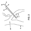
最初に、図2−4を参照すると、本発明の一形態では、閉塞器30が、提供される。閉塞器30は、弾性フィラメント35を備え、弾性フィラメント35は、非制約状態では、略非線形構成(例えば、コイル状塊)を備えているが、適切に拘束されると、線形構成(例えば、針45の細い管腔40内で、または、フィラメントが形状記憶材料から形成されている場合、その温度、ひいては、その形状を適切に制御することによって)を維持し得る。拘束が除去されると(例えば、弾性フィラメント35が針45の制約管腔40から押し出される、または形状記憶材料の温度が体温等によって上昇させられる)、弾性フィラメント35は、その略非線形構成に戻り、それによって、静脈を閉塞するための拡大塊を提供するであろう。
Initially, referring to FIGS. 2-4, in one form of the invention, an
本発明の一形態では、閉塞器は、形状記憶材料(例えば、ニチノール等の形状記憶合金または形状記憶ポリマー)から形成され、形状記憶材料は、超弾性、または温度誘発形状変化、または両方を提供するように構成される)。 In one form of the invention, the occluder is formed from a shape memory material (eg, a shape memory alloy or shape memory polymer such as Nitinol), which provides superelasticity, or temperature induced shape change, or both. To be configured).
一好ましい使用方法では、閉塞器30は、針45の細い管腔40(図2)内に据え付けられ、針は、経皮的に導入され、閉塞されるべき静脈(例えば、大伏在静脈15)を横断して前進させられ、閉塞器の第1の長さが、静脈の遠い側で針から押し出され、その結果、閉塞器の一部が静脈の遠い側でコイル状塊構成50(図3)に復元され、針が、静脈を横断して引き戻され、次いで、閉塞器の残りが静脈の近い側で押し出され(図4)、それに応じて、閉塞器の残りがコイル状塊構成55に復元され、閉塞器の部分57が静脈15の管腔60を横断して延び、静脈の遠い側および近い側の閉塞器の部分(すなわち、それぞれ、コイル状塊50および55)が、弾性フィラメントに固有のコイル力下、互に向かって引かれることにより、その間の静脈を圧迫し、その管腔60を閉塞し、それによって、静脈を通る血流を制限し、それによって、静脈瘤を治療する。
In one preferred method of use, the
前述のように、閉塞器30は、形状記憶材料(例えば、ニチノール等の形状記憶合金または形状記憶ポリマー等)から形成され得、形状記憶材料は、超弾性、または温度誘発形状変化、または両方を提供するように構成される。
As described above, the
図2−4に示される本発明の形態では、閉塞器30は、単一弾性フィラメント35から形成され、形状遷移(すなわち、略線形から一対の対向コイル状塊50、55に)が、標的血管の閉塞を生じさせるために使用される。この点では、前述のコイル状塊50、55は、略3次元構造に配列される弾性フィラメントの略無作為巻き(すなわち、若干、紐のボールに類似する)を備え得るか、またはコイル状塊50、55は、ループ、コイル等の高度に再現性の構造を備え得、これらのループ、コイル等は、略平面構造をとることも、とらないこともあることを理解されたい。例えば、コイル状塊50、55が高度に再現性のループおよびコイルを備えている、図5を参照されたい。
In the form of the invention shown in FIGS. 2-4, the
図6および7は、本発明の新規閉塞器を展開するために使用され得る、例示的注射器型挿入器65を示す。注射器型挿入器65は、1つの閉塞器30または複数の事前に装填された閉塞器30を含み得、例えば、注射器型挿入器65は、複数の閉塞器30を備え、閉塞器は、注射器型挿入器内に直列に配置され得るか、または、それは、注射器型挿入器内に互に平行に配置され得る(すなわち、「ガトリング砲」配置様式)。注射器型挿入器65が作動させられると、閉塞器30は、針45の遠位端から展開される。
6 and 7 show an exemplary syringe-
図2−4では、閉塞器30は、2つのコイル状塊50、55間の静脈を圧迫し、それによって、その管腔60を閉鎖することによって、静脈を閉塞するステップが示される。しかしながら、本発明の別の形態では、閉塞器30は、静脈を圧迫せずに、静脈を閉塞するために使用されることができる。これは、静脈の管腔内にコイル状塊を堆積し、それによって、静脈の管腔を通る血流を制限することによって行われる。より具体的には、ここで図8−10を参照すると、本発明の本形態では、針45は、静脈15の内部の中に通され、閉塞器30の1つのコイル状塊50が、静脈の管腔を閉塞するように、静脈の管腔60の中に押し出され(図8)、針45は、静脈の近い側に引き出され(図9)、次いで、別のコイル状塊55が、静脈の近い側に配置され(図10)、閉塞器の部分57は、閉塞器を静脈に対して安定化させるように(すなわち、閉塞器を静脈に取り付け、閉塞器が静脈に対して移動することを防止するように)、静脈の側壁を通して延びる。
In FIG. 2-4, the
図11−14は、別のアプローチを示し、閉塞器30のコイル状塊は、脈管を通る血流を妨害するように、血管の内部内に堆積される。より具体的には、本発明の本形態では、針45は、静脈を完全に貫通させられ(図11)、閉塞器のコイル状塊50は、静脈の遠い側に堆積され(図12)、針が、静脈の内部の中に引き戻され、閉塞器の別のコイル状塊55は、堆積され(図13)、次いで、針は、静脈の近い側まで引き出され、閉塞器30の別のコイル状塊70が、堆積される(図14)。本発明の本形態では、コイル状塊55は、静脈の管腔60内に常駐し、血流を妨害する一方、コイル状塊50および70は、静脈を内向きに圧迫し、管腔内コイル状塊55の配置を安定化させる。
FIGS. 11-14 illustrate another approach, where the coiled mass of the
図15および16は、弾性フィラメントの単一糸から形成される、閉塞器30を示す。図15では、閉塞器30は、比較的に整然としたコイルを備え、コイルの巻き72は、一方向性である。図16では、閉塞器30は、別の比較的に整然としたコイルを備えているが、巻きは、中間点75の異なる側で反対方向に回転する。当然ながら、閉塞器30は、比較的に整然としていないコイルを形成するようにも構築されることができ、すなわち、フィラメントの糸は、比較的に無作為パターンを辿る(例えば、図8−10に図示される整然としていないコイル参照)ことを理解されたい。実際、再形成されたコイル自体の塊が流動障害物を提供することが望ましい場合(例えば、再形成されたコイルが静脈を通る血流を阻止するように管腔内に配置される場合)、概して、弾性フィラメントは、より密度の高いフィラメント構成を提供するため、比較的に無作為配置を有する比較的に整然としていないコイルに再形成することが好ましい。
Figures 15 and 16 show an
図17は、弾性フィラメント35の複数の糸から形成される、閉塞器30を示す。本発明の一形態では、これらの複数の糸は、接合部80において一緒に接合される。再び、これらの複数の糸によって形成されるコイル(例えば、前述のコイル状塊50、55、70)は、比較的に整然としたまたは比較的に整然としていないものであることができる。図18および19は、図17の多糸閉塞器が、コイル状塊50、55を形成し、静脈を通る血流を制限するように、静脈の側壁を内向きに圧迫することによって、静脈を閉塞するために使用され得る方法を示す。図20は、図17の多糸閉塞器30が、コイル状塊55を静脈の管腔60内に堆積し、それによって、静脈の管腔を通る血流を制限することによって、静脈を閉塞するために使用され得る方法を示す。図20では、いくつかの弾性フィラメント35が、コイル状塊55を血管の管腔内の定位置に保持するように、静脈の側壁を穿刺するように示される。
FIG. 17 shows the
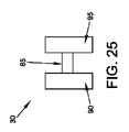
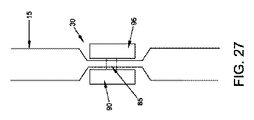
図21−24は、閉塞器30の別の形態を示し、閉塞器は、フィラメント以外の構造によって形成される。一例として、限定ではないが、閉塞器30は、経管腔区分85と、遠い側の側方突起90と、近い側の側方突起95とを備え得、遠い側の側方突起90および近い側の側方突起95は、静脈15の管腔60を閉鎖するように、互に反対に保持される。そのような配列は、例えば、図25−27に示される「二重Tバー」構造等の多くの異なるタイプの構造によって提供され得、閉塞器30の経管腔区分85は、脈管閉塞を提供するように、閉塞器の2つの対向Tバー90、95を一緒に引っ張る弾性材料から形成される。遠い側の側方突起90および近い側の側方突起95を一緒に接続し、引っ張るためのさらに他の配列は、本開示に照らして、当業者に明白であろう。さらなる実施例として、限定ではないが、遠い側の側方突起90および近い側の側方突起95は、縫合糸のループによって、一緒に接続され得、縫合糸のループは、スライド係止結びを用いて、縮小サイズ構成(すなわち、閉塞を維持するように)に係止可能である。
21-24 show another form of the
さらに、複数の閉塞器30が、単一血管または組織において使用され、血管をより完全に閉塞し、または複数の領域内の血管を閉塞し、または材料(例えば、薬物または細胞送達要素)を複数の場所において血管に取り付け得る。閉塞器は、薬物溶出化合物でコーティングされるか、または閉塞器は、電気的に帯電され、凝固を向上または防止し、あるいは所望の化合物または薬剤を血管等に送達し得る。所望に応じて、閉塞または取り付け要素の場所は、所望の化合物または薬剤を特定の解剖学的場所に送達するように精密に制御され得る。
In addition,
(腔内アプローチ)
腔内アプローチでは、閉塞器30は、カテーテルを使用して、閉塞器を静脈まで腔内で前進させ、次いで、閉塞器を静脈内で展開することによって、閉塞部位に送達され、閉塞器は、静脈を閉塞し、それによって、静脈瘤を治療するように作用する。本発明の本形態では、閉塞器は、好ましくは、閉塞器を静脈に対して安定化させるように、1つ以上の静脈の側壁を貫通させられる。本発明の一形態では、閉塞器は、静脈の管腔を通る血流を制限するように、塊を静脈の管腔内に堆積させることによって、静脈を閉塞するように構成され、本発明の別の形態では、閉塞器は、静脈を圧迫し、その管腔を閉鎖することによって、静脈を閉塞するように構成される。
(Intracavity approach)
In an intraluminal approach, the
より具体的には、ここで図28および29を参照すると、カテーテル100は、閉塞器30を静脈15の内部の展開部位まで腔内で前進させるために使用される。次いで、閉塞器の一端は、閉塞器30のコイル状塊50を静脈の外側に堆積させるように静脈の側壁を貫通させられ、閉塞器の残りは、静脈の管腔60内のコイル状塊55として堆積され、閉塞器の部分57は、閉塞器を静脈の側壁に取り付け、それによって、閉塞器を静脈に対して安定化させるように、静脈の側壁を通して延びる。したがって、本発明の本形態では、閉塞器のコイル状塊55は、静脈を通る血流を制限し、それによって、静脈瘤を治療するように、静脈の内部内に堆積される。
More specifically, referring now to FIGS. 28 and 29, the
図30および31は、各々が図28および29に示される様式で使用される2つの別個の閉塞器30が、静脈の管腔内に含まれる閉塞器のコイル状塊を増加させ、それによって、静脈の管腔の閉塞の範囲を増加させるために、どのように使用され得るかを示す。
FIGS. 30 and 31 show that two
図32および33は、閉塞器30が、どのように腔内で送達され、静脈の管腔を通る血流を閉塞するように、静脈の外壁を圧迫するために使用されるかを示す。より具体的には、本発明の本形態では、閉塞器30は、静脈を通して展開部位まで腔内を前進させられ、閉塞器の一端は、コイル状塊50を静脈の片側に堆積するように、静脈の片方の側壁を貫通させられ、閉塞器の他端は、別のコイル状塊55を静脈の他の側に堆積させるように、静脈の他方の側壁を貫通させられ、2つのコイル状塊は、閉塞器の中間部分57によって一緒に接続され、2つのコイル状塊は、圧迫対向力を静脈の両側に加え、それによって、静脈を圧迫し、その管腔を閉鎖するように、弾性フィラメントに固有のコイル力下、互に向かって引かれる。
FIGS. 32 and 33 show how the
(静脈切除と組み合わせた閉塞)
所望に応じて、本発明の新規閉塞器は、大静脈瘤の除去(すなわち、静脈切除)と併せて使用されることができる。静脈切除は、静脈の閉塞と同時に、または別の時に、行われることができる。本外科手術手技の場合、最小限の局所麻酔剤が、必要とされる。
(Occlusion combined with venectomy)
If desired, the novel occluder of the present invention can be used in conjunction with removal of a large varicose vein (ie, venectomy). Venectomy can be performed simultaneously with vein occlusion or at another time. For this surgical procedure, minimal local anesthetic is required.
(静脈瘤の治療以外の目的のための管状構造の閉塞)
本発明の新規閉塞器はまた、静脈瘤の治療以外の目的のために、管状構造を閉塞するために使用されることもできることを理解されるであろう。一例として、限定ではないが、本発明の新規閉塞器は、他の血管構造を閉塞する(例えば、出血を制御するように、動脈を閉塞する)、または身体内の他の管状構造(例えば、不妊症を誘発するように、卵管)を閉塞するため等に使用されることができる。
(Occlusion of tubular structure for purposes other than the treatment of varicose veins)
It will be appreciated that the novel occluder of the present invention can also be used to occlude tubular structures for purposes other than the treatment of varicose veins. By way of example, and not limitation, the novel occluder of the present invention occludes other vascular structures (eg, occludes an artery to control bleeding) or other tubular structures within the body (eg, Can be used to occlude the fallopian tube, etc., to induce infertility.
(薬物/細胞送達用途)
さらに、経皮的にまたは腔内のいずれかにおける、低侵襲的な中空管穿刺と脈管壁へのデバイスの取り付けおよび固定との前述の概念を使用して、閉塞器30は、血管系または他の中空身体構造内あるいはそれに隣接する固定点への薬物/細胞送達を可能にするように修正され得る。本発明の本形態では、デバイスは、薬物/細胞送達安定器として機能し、かつ閉塞器として機能することも、機能しないこともある。例えば、それに取り付けられた薬物/細胞送達体105を有する弾性フィラメント35が、針115を使用して、血管110を横断して前進させられ、弾性フィラメントの遠位端が、コイル状塊120を血管の遠い側に形成し、薬物/細胞送達体105が、血管の管腔125内に固定して配置される、図34および35を参照されたい。図36および37は、類似配列を示し、カテーテル130が、デバイスを腔内で送達するために使用される。図38および39は、別の配列を示し、デバイスは、コイル状塊が血管の管腔125の内側に配置され、薬物/細胞送達体105が血管の外側に配置されるように、経皮的に送達され、図40および41は、デバイスが、コイル状塊が、内側血管の管腔125の内側に配置され、薬物/細胞送達体105が、血管の外側に配置されるように、どのように腔内で送達されるかを示す。これらの薬物/細胞送達デバイスは、不活性または活性のポリマーまたはシリコンベース、あるいはマイクロおよびナノ技術のデバイス、もしくは材料のマトリクス等であり得る。
(Drug / cell delivery applications)
Furthermore, using the aforementioned concepts of minimally invasive hollow tube puncture and attachment and fixation of the device to the vessel wall, either percutaneously or intracavitary, the
(2部構成閉塞器)
次に図42を参照すると、本発明に従って形成される2部構成閉塞器200が、示される。2部構成閉塞器200は、概して、遠位インプラント205と、近位インプラント210とを備えている。
(2-part occluder)
Referring now to FIG. 42, a two-
遠位インプラント205は、図43−46にさらに詳細に示される。遠位インプラント205は、遠位インプラント本体215と、遠位インプラント係止管220とを備えている。遠位インプラント本体215は、遠位端226と、近位端227と、その間に延びる管腔230とを有する管225を備えている。管225は、複数の脚部235を画定するように、その長さの中間にスリットが入れられている。一組の内向きに突出するつまみ240が、脚部235と近位端227との間の管225内に形成される。一組の窓245が、内向きに突出するつまみ240と近位端227との間の管225内に形成される。遠位インプラント本体215は、好ましくは、弾性材料(例えば、超弾性プラスチックを含む、ニチノールまたは超弾性ポリマー等の超弾性特性を有する形状記憶材料)から形成され、その脚部235が、通常、管225の縦軸から側方に突出するように構築される(例えば、図43および44に示される様式において)が、しかしながら、遠位インプラント本体215を形成するために使用される材料の弾性性質に起因して、脚部235は、遠位インプラント本体215が略線形配置をとり得るように、内向きに制約されることができる(例えば、以下に論じられるように、送達針の管腔内に)。例えば、図43および44に示される位置に対して内向きに移動させられた脚部235を示す、図46を参照されたい。しかしながら、任意のそのような制約が除去されると、遠位インプラント本体215を形成するために使用される材料の弾性性質は、脚部235に図43および44に示される位置に戻らせる。
The
遠位インプラント係止管220(図45)は、遠位端250と、近位端260と、その間に延びる管腔262とを有する、略管状構造を備えている。一組の窓265が、遠位インプラント係止管220内に形成され、窓265は、近位端260の遠位に配置される。
The distal implant locking tube 220 (FIG. 45) comprises a generally tubular structure having a
遠位インプラント係止管220は、遠位インプラント本体215の管腔230内に配置される。遠位インプラント205がその前述の略線形状態(すなわち、脚部235が列に並んだ状態に拘束される)にあるとき、遠位インプラント係止管220は、遠位インプラント本体215の近位端227が、遠位インプラント本体215の遠位端226に対して縦方向に移動し得るように、遠位インプラント本体215のつまみ240のずっと手前で終了する。しかしながら、遠位インプラント本体215の近位端227が、脚部235の完全半径方向拡張(図42参照)を可能にするために十分な距離に遠位に移動させられると、遠位インプラント本体215の係止つまみ240は、遠位インプラント係止管220の窓265内に受け取られ、それによって、遠位インプラント205をその半径方向に広げられた状態(すなわち、脚部235が、例えば、図43および44に示される様式において、管225の縦軸から離れるように側方に突出する)に係止するであろう。遠位インプラント本体215の遠位端226内に形成される開口部270を介して適用されるスポット溶接は、遠位インプラント係止管220を遠位インプラント本体215に係止し、それによって、一体型構造(図43および46参照)を形成する役割を果たす。
Distal
次に図47および48を参照すると、近位インプラント210は、遠位端280と、近位端285と、その間に延びる管腔290とを有する、管275を備えている。管275は、複数の脚部295を画定するように、その遠位端にスリットが入れられる。一組の内向きに突出するつまみ300が、脚部295と近位端285との間の管275内に形成される。近位インプラント210は、好ましくは、弾性材料(例えば、ニチノール等の超弾性特性を有する形状記憶材料)から形成され、その脚部295が、通常、管275の縦軸から離れるように側方に突出する(例えば、図47に示される様式において)が、しかしながら、脚部295は、近位インプラント210が略線形配置をとり得るように、内向きに制約されることができる(例えば、以下に論じられるように、送達管の管腔内に)。例えば、図47に示される位置に対して内向きに移動させられた脚部295を示す、図48を参照されたい。しかしながら、任意のそのような制約が除去されると、近位インプラント210を形成するために使用される材料の弾性性質は、脚部295に図47に示される位置に戻らせる。
47 and 48, the
以下に論じられるように、遠位インプラント205および近位インプラント210は、遠位インプラント本体215の管225が、近位インプラント210の管腔290内に受け取られ、遠位インプラント205の広げられた脚部235が近位インプラント210の広げられた脚部295に対向し(例えば、図82参照)、それによって、以下にさらに詳細に論じられるように、その間に配置される血管(例えば、静脈)の側壁に締め付け作用を与え、それによって、血管を閉塞し得る(または、代替として、近位および遠位インプラントの対向する広げられた脚部が、締め付け作用を与えるように互いに組み合わさり得る)ように構成され、かつサイズ決定される。さらに、遠位インプラント205および近位インプラント210は、この位置に係止され、したがって、近位インプラント210の内向きに突出するつまみ300が、遠位インプラント205の窓245の中に突出し得るように構成され、かつサイズ決定される。
As discussed below,
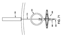
2部構成閉塞器200は、関連付けられた据え付け装置を使用して展開されることが意図される。この関連付けられた据え付け装置は、好ましくは、以下に論じられるように、組織を穿刺するための中空針305(図49)と、遠位インプラント205を中空針305を通して閉塞されるべき血管の遠い側に送達するための遠位インプラント送達管310(図50)と、送達および展開の間、種々の構成要素に支持を供給するための複合ガイドワイヤ315(図51−56)と、複合ガイドワイヤ315の上で種々の構成要素を送達するためのプッシュロッド320(図57)と、遠位インプラント205と嵌合するための近位インプラント210を送達するための近位インプラント送達管330(図58)とを備えている。
The two-
中空針305(図49)は、遠位端335と、近位端340と、その間に延びる管腔345とを備えている。遠位端335は、鋭くとがった先350で終了する。本発明の一つの好ましい形態では、中空針305は、管腔345と連通する側面ポート355を備えている。
The hollow needle 305 (FIG. 49) includes a
遠位インプラント送達管310(図50)は、遠位端360と、近位端365と、その間に延びる管腔370とを備えている。
The distal implant delivery tube 310 (FIG. 50) includes a
複合ガイドワイヤ315(図51−56)は、ガイドワイヤロッド370と、ガイドワイヤシース380とを備えている。ガイドワイヤロッド370は、遠位端385と、近位端390とを備えている。遠位端385は、拡大部395で終了する。ガイドワイヤシース380は、遠位端400と、近位端405と、その間に延びる管腔410とを備えている。ガイドワイヤシース380の遠位端400は、少なくとも1つ、好ましくは、複数の近位に延びるスリット415を備えている。近位に延びるスリット415は、ガイドワイヤシース380の遠位端で開放し、ガイドワイヤシース380の遠位端が、若干、半径方向に広がることを可能にする。以下に論じられるように、ガイドワイヤロッド370およびガイドワイヤシース380は、ガイドワイヤロッド370がガイドワイヤシース380の管腔410内に受け取られ得るように構成され、かつサイズ決定される。さらに、ガイドワイヤロッド370が、ガイドワイヤシース380に対して近位に押されると、ガイドワイヤシース380内の近位に延びるスリット415は、ガイドワイヤシース380の遠位端が、ガイドワイヤロッド370の遠位端に形成される拡大部395の少なくとも一部を受け取るように、若干、広がることを可能にする。これが生じるにつれて、ガイドワイヤシース380の遠位端は、半径方向に広がるであろう。
The composite guide wire 315 (FIGS. 51-56) includes a
プッシュロッド320(図57)は、遠位端420と、近位端425と、その間に延びる管腔430とを備えている。
Push rod 320 (FIG. 57) includes a
近位インプラント送達管330(図58)は、遠位端435と、近位端440と、その間に延びる管腔445とを備えている。
Proximal implant delivery tube 330 (FIG. 58) includes a
2部構成閉塞器200およびその関連付けられた据え付け装置は、好ましくは、以下のように使用される。
The two-
最初に、中空針305(遠位インプラント送達管310をその中に支持し、そして、遠位インプラント送達管310は、複合ガイドワイヤ315をその中に含み、複合ガイドワイヤ315の上に遠位インプラント205が搭載される)が、患者の皮膚を通して、介在組織を通して、閉塞されるべき血管(例えば、静脈450)を横断して貫通させられる。図59−61を参照されたい。これが行われると、側面ポート355から流出する任意の血液が監視されることができる(過剰または脈動血流は、中空針が偶発的に動脈に衝打したことを示し得る)。
Initially, the hollow needle 305 (supports a distal
次に、中空針305は、後退させられ、遠位インプラント送達管310が血管を横断して延びたままにしておく。図62を参照されたい。
The
次いで、遠位インプラント送達管310は、複合ガイドワイヤ、すなわちロッド315の遠位端と、遠位インプラント205の遠位端とを露出させるように、若干、後退させられる。図63を参照されたい。
The distal
次に、複合ガイドワイヤ315、プッシュロッド320、および遠位インプラント205は全て、複合ガイドワイヤ315および遠位インプラント205の遠位端を遠位インプラント送達管310の遠位端の外へ前進させるように、遠位に移動させられる。これが生じると、遠位インプラント205の脚部235は、遠位インプラント送達管310の制約から解放され、半径方向に広がる。図64および65を参照されたい。
Next,
次いで、プッシュロッド320が遠位インプラント205の近位端に対して定位置に保持された状態で、複合ガイドワイヤ315が、遠位インプラント205の遠位端を遠位インプラント205の近位端の方に引き寄せるように近位に引かれ、それによって、遠位インプラント本体215の係止つまみ240に遠位インプラント係止管220の窓265に入らせ、それによって、脚部235をその半径方向に広げられた状態(図66参照)に係止する。
The
この時点において、中空針305、遠位インプラント送達管310、およびプッシュロッド320は、除去され(図67)、遠位インプラント205を複合ガイドワイヤ315上に搭載されたままにし、脚部235が血管の遠い側において完全に展開され、遠位インプラント205の近位端が血管の内部の中に延び得る(図68)。
At this point, the
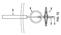
次に、近位インプラント送達管330(近位インプラント210をその中に支持する)が、近位インプラント送達管330の遠位端が血管のすぐ近位に位置するまで、複合ガイドワイヤ315に沿って前進させられる(図69−72)。
Next, the proximal implant delivery tube 330 (supporting the
次いで、プッシュロッド320が、近位インプラント210の遠位端を近位インプラント送達管330の遠位端から前進させるために使用される。これが生じると、脚部295は、近位インプラント送達管330の制約から解放され、半径方向に開放する。図73−76を参照されたい。
Push
次に、プッシュロッド320を使用して、近位インプラント210が、遠位インプラント205が複合ガイドワイヤ315を使用して近位に引かれると、遠位に押される。より具体的には、ガイドワイヤロッド370が近位に引かれ、ガイドワイヤロッド370は、ガイドワイヤロッド370の遠位端の拡大部395にガイドワイヤシース380を遠位インプラント係止管220内の管腔262より大きいサイズまで広げさせ、これは、ガイドワイヤシース380に近位に移動させ、これは、遠位インプラント205の近位移動を生じさせる。遠位インプラント205と近位インプラント210とが一緒に移動するにつれて、それらの脚部235、295は、血管を圧迫し、それによって、血管を閉塞する。遠位インプラント205および近位インプラント210は、近位インプラント210の内向きに突出するつまみ300が遠位インプラント205の窓245に入り、それによって、2つの部材を互に対して定位置に係止するまで、一緒に移動し続ける。図77を参照されたい。
Next, using
この時点において、プッシュロッド320および近位インプラント送達管330は、除去される。図78を参照されたい。
At this point, push
次に、複合ガイドワイヤ315が、除去される。これは、最初に、ガイドワイヤロッド370を遠位に前進させることによって行われ(図79)、ガイドワイヤシース380の遠位端が、内向きに弛緩し、それによって、その外径を遠位インプラント係止管220内の管腔262より小さいサイズまで縮小することを可能にする。その結果、ガイドワイヤシース380は、次いで、2部構成閉塞器200の内部を通して近位に引き出されることができる。図80を参照されたい。次いで、ガイドワイヤロッド370は、2部構成閉塞器200の内部を通して近位に引き出されることができる。図81を参照されたい。
Next, the
前述の手技は、2部構成閉塞器200を血管を横断して定位置に係止されたままにし、対向脚部235、295は、血管を圧迫し、それによって、血管を閉塞する。
The foregoing procedure leaves the two-
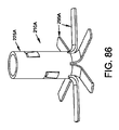
図83−86は、遠位インプラント205Aと、近位インプラント210Aとを有する、別の2部構成閉塞器200Aを図示する。2部構成閉塞器200Aは、概して、前述の2部構成閉塞器200に類似するが、遠位インプラント205Aは、一体型構造を利用する。
FIGS. 83-86 illustrate another two-
図87−90は、別の2部構成閉塞器200Bを図示する。2部構成閉塞器200Bは、概して、前述の2部構成閉塞器200Aに類似するが、遠位インプラント205Bは、摩擦嵌めを利用し、遠位インプラント205Bを近位インプラント210Bに係止させる。
87-90 illustrate another two-
図91−94は、遠位インプラント205Cおよび近位インプラント210Cを有する、別の2部構成閉塞器200Cを図示する。2部構成閉塞器200Cは、概して、前述の2部構成閉塞器200に類似するが、遠位インプラント205Cは、脚部235Cの近位端を受け取り、固定する管225Cを備えている。脚部235Cは、好ましくは、所望の弾性率を伴う脚部235Cを提供するように、超弾性形状記憶材料から形成される細長い要素(例えば、曲げられたワイヤ)である。
FIGS. 91-94 illustrate another two-
図95−100は、遠位インプラント205Dと、近位インプラント210Dとを有する、別の2部構成閉塞器200Dを図示する。2部構成閉塞器200Dは、概して、前述の2部構成閉塞器200に類似するが、遠位インプラント205Dは、脚部235Dの近位端を受け取り、固定する、管またはロッド225Dを備えている。脚部235Dは、好ましくは、所望の弾性率を伴う脚部235Dを提供するように、超弾性形状記憶材料から形成されるコイルである。
FIGS. 95-100 illustrate another two-
前述の開示では、遠位インプラント205および近位インプラント210を生体構造に送達する際に使用するための複合ガイドワイヤ315が、開示される。前述のように、複合ガイドワイヤ315は、2つの部品、すなわち、ガイドワイヤロッド370と、ガイドワイヤシース380とから形成される。複合ガイドワイヤ315にこの2部構成構造を提供することによって、複合ガイドワイヤ315は、複合ガイドワイヤ315が、遠位インプラント205に結合するか、または遠位インプラント205から分離可能であることを可能であるように、それぞれ、所望に応じて拡大または縮小されるその遠位直径を有することができる。しかしながら、所望に応じて、複合ガイドワイヤ315は、代替ガイドワイヤを遠位インプラント205に取り外し可能に結合するための機構を含む、代替ガイドワイヤによって置換されることができる。一例として、限定ではないが、そのような代替ガイドワイヤは、ねじ山を含み得、遠位インプラント205は、代替ガイドワイヤが、選択的に、すなわち、ねじ作用によって、遠位インプラント205に固定される、またはそこから解放され得るように、ねじ陥凹を含み得る。
In the foregoing disclosure, a
次に図101−104を参照すると、本発明に従って形成される2部構成閉塞器200Eが、示される。2部構成閉塞器200Eは、概して、遠位インプラント205Eと、近位インプラント210Eとを備えている。
Referring now to FIGS. 101-104, a two-
遠位インプラント205Eは、遠位インプラント本体215Eと、遠位インプラント係止管220Eとを備えている。遠位インプラント本体215Eは、遠位端226Eと、対向近位端とを有する、管225Eを備えている。好ましくは、遠位インプラント205Eは、その近位端から遠位に延びる管腔230Eを有する。管腔230Eが、遠位インプラント本体215Eの全長に沿って延び得るか、または遠位インプラント本体215Eの遠位端の手前で終了し得る。一例として、限定ではないが、2部構成閉塞器200Eが、ガイドワイヤの上に設定されるべき場合、管腔230Eは、遠位インプラント本体215Eの全長に沿って延びる。管225Eは、複数の脚部235Eを画定するように、その長さの中間にスリットが入れられる。遠位インプラント本体215Eは、好ましくは、少なくとも部分的に、弾性材料(例えば、超弾性プラスチックを含む、ニチノールまたは超弾性ポリマー等の超弾性特性を有する、形状記憶材料)から形成され、その脚部235Eが、通常、管225Eの縦軸から離れるように側方に突出するように構成される(例えば、図101−104に示される様式において)が、しかしながら、遠位インプラント本体215Eの少なくとも脚部235Eを形成するために使用される材料の弾性性質に起因して、脚部235Eは、遠位インプラント本体215Eが略線形配置をとり得る(その場合、脚部235Eの最遠位先端が収束し、遠位インプラント本体215Eの前述の近位端を形成する)ように、内向きに制約されることができる(例えば、以下に論じられるように、送達針の管腔内に)。しかしながら、任意のそのような制約が除去されると(例えば、遠位インプラント本体215が、もはや送達針内に制約されなくなると)、遠位インプラント本体215Eの少なくとも脚部235Eを形成するために使用される材料の弾性性質は、脚部235Eに図101−104に示される位置をとらせる。
The
本発明の一好ましい形態では、図101−103に見られるように、遠位インプラント205Eの脚部235Eは、脚部235Eが、集合的に、凹面領域236Eを画定するように、遠位インプラント205Eの縦軸に鋭角に延びる。
In one preferred form of the invention, as seen in FIGS. 101-103, the
遠位インプラント係止管220E(図101−104)は、遠位端250Eと、近位端260Eとを有する略管状構造を備えている。好ましくは、遠位インプラント係止管220Eは、近位端260Eから遠位に延びる管腔262Eを有する。管腔262Eは、遠位インプラント係止管220Eの全長に沿って延び得るか、または遠位インプラント係止管220Eの遠位端の手前で終了し得る。一例として、限定ではないが、2部構成閉塞器200Eが、ガイドワイヤの上に設定されるべき場合、遠位インプラント係止管220Eの管腔262Eは、遠位インプラント係止管220Eの全長に沿って延びる。一組の円周方向溝または陥凹265Eが、遠位インプラント係止管220E内に形成され、溝または陥凹265Eは、遠位端250Eと近位端260Eの中間に配置される。遠位インプラント係止管220Eはまた、遠位インプラント係止管220E(ひいては、遠位インプラント205E)を遠位インプラント送達管310Eに取り外し可能に固定するための第1の機械的相互係止半体266Eを備えている(以下参照)。遠位インプラント係止管220Eは、好ましくは、その長さに沿って比較的に非弾性であり、それによって、長さ方向の伸びを最小限にするが、若干、可撓性であり、それによって、湾曲経路を介して送達されることを可能にし得る、生体適合性材料から形成される。一例として、限定ではないが、遠位インプラント係止管220Eは、Ti5AL−4V等のチタン合金から形成され得る。
The distal
遠位インプラント係止管220Eは、遠位インプラント本体215Eの管腔230E内に配置され、そこから近位に延びる。遠位インプラント係止管220Eは、当技術分野において周知の方法において(例えば、スポット溶接、接着剤、機械的相互係止等によって)、遠位インプラント本体215Eに固定され、それによって、集合的に、一体型構造を形成する(図101−104参照)。遠位インプラント本体215Eを弾性材料から形成することによって、および遠位インプラント係止管220Eをその長さに沿って比較的に非弾性である材料から形成することによって、遠位インプラント本体215Eは、容易に変形可能である(例えば、その脚部235Eが、送達針内に制約され得るように)一方、遠位インプラント係止管220Eは、構成が固定される(例えば、以下に論じられるように、近位インプラント210Eを遠位インプラント205Eに保持する役割を果たし得るように)ことに留意されたい。
Distal
さらに、ここで図101−104を参照すると、近位インプラント210Eは、遠位端と、近位端285Eと、その間に延びる管腔290Eとを有する管275Eを備えている。管275Eは、複数の脚部295Eを画定するように、その遠位端にスリットが入れられる。一組の内向きに突出するつまみ300Eが、以下に論じられるように、前述の溝または陥凹265Eを遠位インプラント係止管220E内に係合するために、脚部295Eと近位端285Eとの間の管275E内に形成される(所望に応じて、溝または陥凹265Eおよびつまみ300Eの場所および構成は、反転されることができ、すなわち、外向きに突出するつまみ300Eが、遠位インプラント係止管220E上に提供されることができ、溝または陥凹265Eが、管275Eの内側側壁上に提供されることができるか、または他の手段が、近位インプラント210Eの管275Eを遠位インプラント205Eの遠位インプラント係止管220Eに接続するために提供されることができることに留意されたい)。近位インプラント210Eは、好ましくは、少なくとも部分的に、弾性材料(例えば、ニチノール等の超弾性特性を有する形状記憶材料)から形成され、その脚部295Eが、通常、管275Eの縦軸から離れるように側方に突出するように構築される(例えば、図101−104に示される様式において)が、しかしながら、脚部295Eは、近位インプラント210Eが略線形配置をとり得るように(脚部295Eの遠位端は、集合的に、近位インプラント210Eの遠位端を形成する)、内向きに制約されることができる(例えば、以下に論じられるように、送達針の管腔内に)。しかしながら、任意のそのような制約が除去されると、近位インプラント210Eの少なくとも脚部295Eを形成するために使用される材料の弾性性質は、脚部295E図101−104に示される位置に戻らせる。
Further, referring now to FIGS. 101-104, the
本発明の一好ましい形態では、図101−104に見られるように、近位インプラント210Eの脚部295Eは、脚部295Eが、集合的に、凹面領域301Eを画定するように、近位インプラント210Eの縦軸に鈍角で延びる。
In one preferred form of the invention, as seen in FIGS. 101-104,
遠位インプラント205Eの凹面領域236Eの凹面は、近位インプラント210Eの凹面領域301Eの凹面の逆であることに留意されたい(言い換えると、図101−104に見られるように、遠位インプラント205Eの凹面領域236Eの凹面は、近位インプラント210Eの凹面領域301Eの凹面に面する)。
Note that the concave surface of the
以下に論じられるように、遠位インプラント205Eおよび近位インプラント210Eは、さらに詳細に以下に論じられるように、遠位インプラント205Eの遠位インプラント係止管220Eが、近位インプラント210Eの管腔290E内に受け取られ、遠位インプラント205Eの広げられた脚部235Eが、近位インプラント210Eの広げられた脚部295Eに対向し(例えば、図103および104参照)、それによって、締め付け作用をその間に配置される血管(例えば、静脈)の側壁に与え、それによって、血管を閉塞し得る(または、代替として、近位および遠位インプラントの対向する広げられた脚部は、締め付け作用をさらに向上させるように、互いに入り込み合い得る)ように構成され、かつサイズ決定される。さらに、遠位インプラント205Eおよび近位インプラント210Eは、この位置に係止され、したがって、近位インプラント210Eの内向きに突出するつまみ300Eが、遠位インプラント205Eの遠位インプラント係止管220Eの円周方向溝または陥凹265Eの中に突出し、それによって、近位インプラント210Eを遠位インプラント205Eに固定するように構成され、かつサイズ決定される。遠位インプラント係止管220Eの円周方向溝または陥凹265Eおよび近位インプラント210Eの内向きに突出するつまみ300Eの位置は、近位インプラント210Eの内向きに突出するつまみ300Eが、遠位インプラント係止管220Eの円周方向溝または陥凹265E内に配置されると、遠位インプラント205Eの脚部235Eおよび近位インプラント210Eの脚部295Eが、その間に配置される血管(または、他の管状構造)の適正な締め付けを確実にするために十分に近接するように調整されることに留意されたい。
As discussed below,
2部構成閉塞器200Eは、関連付けられた据え付け装置を使用して展開されるように意図される。本発明の一好ましい形態では、そのような関連付けられた据え付け装置は、好ましくは、以下に論じられるように、組織を穿刺するための中空針305E(図109)と、遠位インプラント205Eを中空針305Eを通して閉塞されるべき血管(または、他の管状構造)の遠い側に送達するための遠位インプラント送達管310E(図110)と、遠位インプラント205Eと嵌合するための近位インプラント210Eを送達するための近位インプラント送達管330E(図110)とを備えている。
Two-
所望に応じて、関連付けられた据え付け装置は、図105−113に示されるように、腹腔鏡下デバイス331Eの形態で提供され得る。腹腔鏡下デバイス331Eは、以下に説明される機能性を伴う、ハンドル332Eと、外側シース333Eと、ノブ334Eと、第1のトリガ336Eと、第2のトリガ337Eと、解除レバー338Eとを備えている。
If desired, the associated mounting device can be provided in the form of a
より具体的には、中空針305E(図109)は、遠位端335Eと、近位端(図示されないが、腹腔鏡下デバイス331E内に含まれる)と、その間に延びる管腔345Eとを備えている。中空針305Eの遠位端335Eは、鋭くとがった先350Eで終了する。
More specifically, the
遠位インプラント送達管310E(図110)は、遠位端360Eと、近位端(図示されないが、腹腔鏡下デバイス331E内に含まれる)とを備えている。遠位インプラント送達管310Eの遠位端360Eはまた、遠位インプラント送達管310Eの遠位端を遠位インプラント205Eの近位端に取り外し可能に固定するために、すなわち、第1の機械的相互係止半体266E(遠位インプラント係止管220Eの近位端によって支持される)と第2の機械的相互係止半体361E(遠位インプラント送達管310Eの遠位端によって支持される)の解放可能相互接続によって固定するために、第2の機械的相互係止半体361Eを備えている。
Distal
近位インプラント送達管330E(図110)は、遠位端435Eと、近位端(図示されないが、腹腔鏡下デバイス331E内に含まれる)と、その間に延びる管腔445Eとを備えている。
Proximal implant delivery tube 330E (FIG. 110) includes a
2部構成閉塞器200Eおよびその関連付けられた据え付け装置(例えば、腹腔鏡下デバイス331E)は、好ましくは、以下のように使用される。
The two-
最初に、中空針305Eが、好ましくは、針305Eが、腹腔鏡下デバイス331E(図107)のシース333E内に含まれている間に閉塞部位に通される。次いで、シース333Eは、例えば、ノブ334E(図108)を旋回させることによって、後退させられ、中空針305Eは、閉塞されるべき血管(例えば、静脈)を横断して通される(または、閉塞されるべき別の管状構造を横断して通されるか、あるいは固形臓器または組織の層等、互に固定されるべき組織または物体を貫通させられる)。
Initially, the
次に、中空針305Eは、例えば、第1のトリガ336E(図109)を介して、血管を横断して後方に近位に後退させられる。この作用は、遠位インプラント205Eの脚部235Eが、血管の遠い側で半径方向に広がることを可能にする。この時点において、遠位インプラント係止管220Eは、血管を通して近位に延びる。
The
次いで、遠位インプラント送達管310Eが、遠位インプラント送達管310Eと、遠位インプラント係止管220E(ひいては、遠位インプラント205)とのその相互係止とを介して、定位置に保持された状態で、中空針305Eは、近位インプラント210Eが、もはや中空針305E内に制約されなくなるまで(図110)、さらに近位に引き出される(例えば、第1のトリガ336Eを介して)。これが生じると、近位インプラント210Eの脚部295Eは、中空針305Eの制約から解放され、半径方向に開放する。
The distal
近位インプラント送達管330Eは、次いで、例えば、近位インプラント210Eと遠位インプラント205Eとが合体するまで、第2のトリガ337Eを使用して、遠位に前進させられる(図111)。遠位インプラント205Eおよび近位インプラント210Eが一緒に移動するにつれて、その脚部235E、295Eは、その間の血管を圧迫し、それによって、血管を閉塞する。遠位インプラント205Eおよび近位インプラント210Eは、近位インプラント210Eの内向きに突出するつまみ300Eが、遠位インプラント205Eの円周方向溝または陥凹245Eに入り、それによって、2つの部材を互に対して定位置に係止するまで、一緒に移動し続ける。
Proximal implant delivery tube 330E is then advanced distally using
この時点において、近位インプラント送達管330Eは、引き出され(図112)、遠位インプラント送達管310Eは、遠位インプラント205Eから解放され(すなわち、レバー338Eを使用して、第2の機械的相互係止半体361E(遠位インプラント送達管310Eの遠位端によって支持される)を機械的相互係止の第1の半体266E(遠位インプラント係止管220Eの近位端によって支持される)から係止解除することによって)、次いで、据え付けデバイスが、引き出される(図113)。
At this point, the proximal implant delivery tube 330E is withdrawn (FIG. 112) and the distal
前述の手技は、2部構成閉塞器200Eを血管を横断して定位置に係止されるように残し、対向脚部235E、295Eは、その間の血管を圧迫し、それによって、血管を閉塞する。
The foregoing procedure leaves the two-
前述の開示では、2部構成閉塞器200Eは、その脚部235E、295Eの弾性を使用して、その脚部235E、295Eを直径方向に縮小された構成(例えば、送達針内に制約されるとき)から直径方向に広げられた構成(例えば、送達針の制約から解放されるとき)に再構成させる観点から論じられる。しかしながら、また、脚部235E、295Eが形状記憶材料(例えば、ニチノール)から形成される場合、温度変化が、脚部235E、295Eを直径方向に縮小された構成から直径方向に広げられた構成に再構成するために使用され得ることを理解されたい。一例として、限定ではないが、本発明の本形態では、脚部235E、295Eは、体温を下回る温度に維持されるとき、直径方向に縮小された構成を有するように構築され得、脚部235E、295Eは、体温に維持されるとき、直径方向に広げられた構成を有するように構築され得る。その結果、2部構成閉塞器200Eを体温を下回る温度まで冷却し、2部構成閉塞器を身体の中に挿入し、次いで、2部構成閉塞器を体温まで加熱させることによって、脚部235E、295Eは、その直径方向に縮小された構成から直径方向に広げられた構成に再構成させられ得る。
In the foregoing disclosure, the two-
図114−120は、据え付けデバイスの代替形態を示す。より具体的には、図114−120は、別の腹腔鏡下デバイス331Eを示す。図114−120に示される腹腔鏡下デバイス331Eは、概して、図105−113に示される腹腔鏡下デバイス331Eに類似するが、第2のトリガ337Eは、省略され、レバー338Eが、(i)近位インプラント210Eと遠位インプラント205Eとが合体するまで、近位インプラント送達管330Eを前進させること(図119)と、(ii)遠位インプラント205Eを遠位インプラント係止管220Eから解放する(すなわち、第2の機械的相互係止半体361Eを(遠位インプラント送達管310Eの遠位端によって支持される)第1の機械的相互係止半体266E(遠位インプラント係止管220Eの近位端によって支持される)から係止解除することによって)こと(図120)との両方を行うために使用される。
114-120 illustrate an alternative form of installation device. More specifically, FIGS. 114-120 illustrate another
図121−123は、同様に本発明に従って形成される別の2部構成閉塞器200Eを示す。図121−123に示される閉塞器200Eは、図101−120に示される閉塞器200Eと実質的に同一であるが、遠位インプラント205Eの脚部235Eおよび近位インプラント210Eの脚部295Eは、脚部235E、295Eが、互に向かい合うのではなく、互に入れ子になるように、同一方向に施行されたその凹面を有する。加えて、図121−123に見られるように、遠位インプラント205Eの管225Eは、部分的に、近位インプラント210Eの管腔290E内に受け取られる。
FIGS. 121-123 illustrate another two-
図124−126は、図121−123の2部構成閉塞器200Eの遠位インプラント205Eを遠位インプラント送達管310Eに取り外し可能に固定するための一好ましい構造を示す。より具体的には、本発明の本形態では、ここで図124−126を参照すると、第1の機械的相互係止半体266E(遠位インプラント係止管220Eの近位端によって支持される)は、段付き構成433Eを備え、第2の機械的相互係止半体361E(遠位インプラント送達管360Eの遠位端によって支持される)は、別の段付き構成434Eを備え、段付き構成433Eと段付き構成434Eとは、一緒に嵌合するように、相互の逆である。第2の機械的相互係止半体361E(遠位インプラント送達管310Eの遠位端によって支持される)が、第1の機械的相互係止半体266E(遠位インプラント係止管220Eの近位端によって支持される)に固定された後、遠位インプラント送達管310Eと遠位インプラント205Eとの間の接続は、例えば、係止ロッド436Eを遠位インプラント送達管310Eの中心管腔437Eからインプラント係止管220Eの管腔262Eの中に入れ子式で突出させることによって強化されることができる。本発明の本形態では、据え付けデバイス(例えば、図105−113の腹腔鏡下デバイス331Eまたは図114−120の腹腔鏡下デバイス331E)は、係止ロッド436Eを遠位インプラント送達管310Eの中心管腔437Eからインプラント係止管220Eの管腔262Eの中に入れ子式で移動させるための適切な制御手段(例えば、解除レバー338E)を含む。代替として、本発明の別の形態では、内部係止ロッド436Eは、遠位インプラント205Eの遠位インプラント送達管310Eおよび遠位インプラント係止管220Eの上で入れ子式で突出し、それによって、部材間の接続を向上させる、オーバーチューブ(図示せず)によって置換され得る。
FIGS. 124-126 illustrate one preferred structure for releasably securing the
また、図121−123の2部構成閉塞器200Eの遠位インプラント205Eを遠位インプラント送達管310Eに取り外し可能に固定するための他の形態の機械的相互係止も、使用され得ることを理解されたい。一例として、限定ではないが、ねじ相互係止が、使用され得、例えば、第1の機械的相互係止半体266E(遠位インプラント係止管220Eの近位端によって支持される)は、ねじ山付きボアを備え得、第2の機械的相互係止半体361E(遠位インプラント送達管360Eの遠位端によって支持される)は、ねじ山付き支柱を備え得、遠位インプラント送達管360Eの遠位端によって支持されるねじ山付き支柱は、遠位インプラント係止管220Eのねじ山付きボア内に受け取られ得る。代替として、ねじ相互係止の他の構成が、使用され得るか、または他の形態の機械的相互係止が、使用され得る。
It will also be appreciated that other forms of mechanical interlocks for removably securing the
図101−143に示される構造では、機械的相互係止(例えば、遠位インプラント係止管220Eの近位端によって支持される第1の半体266Eおよび遠位インプラント送達管310Eの遠位端によって支持される第2の半体361E)が、遠位インプラント係止管220E(ひいては、遠位インプラント205E)を遠位インプラント送達管310Eに接続するために使用される。代替として、所望に応じて、遠位インプラント係止管220Eは、遠位インプラント送達管310Eと一体型に形成されることができ、脆弱区分が、その交差部に配置され、2つの部材は、機械的破壊作用によって分離される。
In the structure shown in FIGS. 101-143, mechanical interlocks (eg, the
ある状況では、組織に加えられる負荷をより良好に分散させるために、管状構造に接触する閉塞器のそれらの部分の表面積を増加させることが望ましくあり得ることを理解されるであろう。この状況では、脚部(例えば、2部構成閉塞器205Eの脚部235Eおよび/または脚部295E等)の幅を増加させること、および/または可撓性材料もまた負荷を支持し得るように、隣接する脚部の間の区域内に可撓性材料を提供すること(例えば、傘の様式において)(すなわち、本質的に、脚部235Eおよび/または脚部295Eの有効幅を増加させる)が有用であり得る。例えば、脚部235Eの間、および脚部295Eの間に延びる可撓性材料438Eを示す、図127を参照されたい。
It will be appreciated that in certain situations it may be desirable to increase the surface area of those portions of the occluder that contact the tubular structure in order to better distribute the load applied to the tissue. In this situation, increasing the width of the leg (eg,
図128−133は、血管(または、他の中空管状体)を閉塞するように、閉塞器(例えば、2部構成閉塞器200E)の適切な配置を促進するための配置デバイス500を示す。配置デバイス500は、概して、比較的に小径(例えば、21ゲージまたはより小さい)の針である、血管探知器針505と、誘導構成要素510(プラスチック等の安価な材料から製造され得る)とを備えている。誘導構成要素510は、血管探知器針505を受け取るための座部515と、閉塞器(例えば、図105−113の腹腔鏡下デバイス331Eまたは図114−120の腹腔鏡下デバイス331E等)を設定するための据え付けデバイスのシャフトをスライド可能に収容するための開口部520とを含む。
128-133 illustrate a
使用時、血管探知器針505は、誘導構成要素510の座部515内に位置付けられ、次いで、血管探知器針505は、標的血管を貫いて前進させられる(例えば、超音波誘導下)。図129を参照されたい。血管探知器針505の適切な配置は、血液が血管探知器針505の近位端から流出し始めると確認される。次に、閉塞器(例えば、図105−113の腹腔鏡下デバイス331Eまたは図114−120の腹腔鏡下デバイス331E等)を設定するための据え付けデバイスのシャフトが、誘導構成要素510の開口部520を通して前進させられる。図130を参照されたい。前進は、据え付けデバイスのシャフト上の停止部525が、誘導構成要素510の近位端を係合するまで生じる。図131を参照されたい。据え付けデバイスのシャフト上の停止部525が、誘導構成要素510の近位端を係合している場合、据え付けデバイスのシャフトの遠位端は、標的血管を貫通しているであろう。図132を参照されたい。この時点において、血管探知器針505は、引き出され(図133参照)、閉塞器の展開が、前述のように進む。
In use, the
ある状況では、閉塞されるべき血管(または、他の管状構造)は、下にある層解剖学的構造、例えば、器官、神経、別の管状構造等に近接して位置付けられ得ることを理解されるであろう。この状況では、下にある層解剖学的構造との任意のそのような係合が、外傷を下にある層解剖学的構造に生じさせ得るため、展開針の鋭くとがった遠位先端が、下にある層解剖学的構造を係合しないように、および閉塞器の遠位端(例えば、2部構成閉塞器200Eの遠位インプラント205E)が、下にある層解剖学的構造を係合しないように、血管(または、他の管状構造)を上向きに下にある層解剖学的構造から離れるように持ち上げることが有用であり得る。そのために、ここで図134を参照すると、クランプ鉗子530(または、二またの遠位端を有する他のツール)が、血管(または、他の管状構造)と下にある層解剖学的構造との間に配置され、次いで、標的血管(または、他の管状構造または組織)を下にある層解剖学的構造から分離するように、下にある層解剖学的構造から離れるように上向きに引かれ得る。閉塞器(例えば、2部構成閉塞器200E)は、次いで、標的血管(または、他の管状構造)を安全に貫通させられ、ツールの二またの遠位端間を通り、前述のように展開され得る。
It will be appreciated that in certain situations, the blood vessel (or other tubular structure) to be occluded may be located in proximity to the underlying layer anatomy, eg, organ, nerve, another tubular structure, etc. It will be. In this situation, any such engagement with the underlying layer anatomy can cause trauma to the underlying layer anatomy so that the sharpened distal tip of the deployment needle is Disengage the underlying layer anatomy and the distal end of the occluder (eg, the
(血管以外の管状構造を閉塞するための閉塞器の使用)
本発明の閉塞器はまた、血管以外の管状構造を閉塞するために使用されることができることを理解されるであろう。一例として、限定ではないが、本発明の一時的閉塞器は、身体内の他の構造(例えば、一時的または恒久的滅菌のための卵管および/または輸精管等の管、胸管、瘻管等を含む、胆嚢摘出術のための胆管および胆嚢管、リンパ管等の導管)を閉鎖するために使用されることができる。
(Use of an occluder to occlude tubular structures other than blood vessels)
It will be appreciated that the occluder of the present invention can also be used to occlude tubular structures other than blood vessels. By way of example, and not limitation, the temporary occluder of the present invention may be used for other structures in the body (eg, tubes such as the fallopian tube and / or vas deferens for temporary or permanent sterilization, thoracic tubes, fistulas, etc. And bile ducts for cholecystectomy and gallbladder ducts, lymphatic ducts, etc.).
(構造内の開口部を閉鎖するため、および/または少なくとも2つの物体を一緒に固定するための閉塞器の使用)
前述の開示では、閉塞器は、管状構造を閉塞するために、管状構造の対向側壁を一緒に締め付けることによって、管状構造(例えば、血管、卵管、リンパ管等)を閉塞する観点から論じられる。そのような用途では、閉塞器は、管状構造の片側壁を管状構造の対向側壁に効果的に固定させる。しかしながら、本発明の閉塞器は、構造内の開口部を閉鎖し、および/または他の用途のために、2つ以上の物体を一緒に固定するためにも使用され得ることを理解されたい。
(Use of an occluder to close an opening in the structure and / or to fix at least two objects together)
In the foregoing disclosure, the occluder is discussed in terms of occluding tubular structures (eg, blood vessels, fallopian tubes, lymphatic vessels, etc.) by clamping together opposing sidewalls of the tubular structure to occlude the tubular structure. . In such applications, the occluder effectively secures one side wall of the tubular structure to the opposite side wall of the tubular structure. However, it should be understood that the occluder of the present invention can also be used to close an opening in a structure and / or to secure two or more objects together for other applications.
一例として、限定ではないが、本発明の閉塞器は、切開を閉鎖するように、組織の2つ以上の部分を一緒に固定するために使用され得る。 By way of example and not limitation, the occluder of the present invention can be used to secure two or more portions of tissue together to close the incision.
さらなる実施例として、限定ではないが、本発明の閉塞器は、胃のサイズを縮小するために、胃の対向側壁を合体させるための「胃ステープル留め」手技において使用され得る。 By way of further example, but not by way of limitation, the occluder of the present invention can be used in a “gastric stapling” procedure to coalesce the opposite side walls of the stomach to reduce the size of the stomach.
さらに、本発明の閉塞器は、切除された器官の周縁を密閉するように、器官切除(例えば、肝臓切除)において使用され得る。 Further, the occluder of the present invention can be used in organ resection (eg, liver resection) to seal the periphery of the resected organ.
さらなる実施例として、限定ではないが、ここで図135−137を参照すると、本発明の閉塞器は、組織圧迫を通して種々の領域内の血流または血液損失を選択的に停止させるように、固形臓器の領域を選択的に締め付けまたは閉塞するために使用されることができる。閉塞器は、腎臓または肝臓または他の器官の固形臓器切除において使用され得る。血液損失および分泌物漏出(例えば、胆汁、尿等)は、既存の固形臓器切除手技において問題となり得る。肝臓切除のための平均血液損失は、700〜1200mlである。本発明の閉塞器を用いて固形臓器の所望の領域を締め付けることによって、望ましくない流体損失(血液損失、分泌物漏出等)の量を有意に低下させることが可能となる。本発明の閉塞器は、圧力を広面積の器官に選択的に加えるために使用されることができ、加えて、器官と身体の他の領域を接続する選択的管状構造および脈管を閉鎖するためにも使用され得る。一実施形態および方法では、複数の個別の閉塞器要素が、器官の領域を横断して個々に選択的に展開され得る。例えば、切除される肝臓を閉鎖するための閉塞器の複数の単一の別個の穿刺場所を示す、図135を参照されたい。閉塞器の複数の単一の別個の穿刺配置が使用される場合、固形臓器の異なる領域が、異なる制御可能程度まで圧迫され得ることに留意されたい。 By way of further example and not limitation, referring now to FIGS. 135-137, the occluder of the present invention can be used to selectively stop blood flow or blood loss in various regions through tissue compression. It can be used to selectively tighten or occlude areas of the organ. The occluder can be used in solid organ resection of the kidney or liver or other organs. Blood loss and secretion leakage (eg, bile, urine, etc.) can be a problem in existing solid organ resection procedures. The average blood loss for liver resection is 700-1200 ml. By tightening the desired area of the solid organ using the occluder of the present invention, the amount of undesirable fluid loss (blood loss, secretion leakage, etc.) can be significantly reduced. The occluder of the present invention can be used to selectively apply pressure to a large area organ, in addition to closing selective tubular structures and vessels connecting the organ and other areas of the body. Can also be used for. In one embodiment and method, a plurality of individual occluder elements can be selectively deployed individually across the region of the organ. See, for example, FIG. 135, which shows a plurality of single separate puncture locations of an occluder for closing the resected liver. Note that if multiple single separate puncture arrangements of the occluder are used, different regions of the solid organ can be compressed to different controllable degrees.
本発明の新規実施形態では、身体内に残る(遠位インプラント205Eの)遠位インプラント係止管220Eの長さは、遠位インプラント係止管220Eおよび/または遠位インプラント送達管310Eに脆弱(例えば、壊れやすい)区分を提供することによって、かつ遠位インプラント係止管220Eを近位インプラント210Eの上方の領域において遠位インプラント送達管310Eから切り離すことによって、閉塞器の締め付けがもたらされた時点で決定されることができる。この切り離しは、選択的脆弱領域が、ねじり、またはトルク、または曲げ、または引っ張り、または選択的な他の歪みまたは応力等を受けると、遠位インプラント係止管220Eが、近位インプラント210Eの近位の場所において、遠位インプラント送達管310Eから分離するであろうように、選択的脆弱領域を遠位インプラント係止管220Eおよび/または遠位インプラント送達管310Eの中に組み込むことによって達成されることができる。締め付けが、組織を横断してもたらされるため、遠位インプラント送達管310Eは、遠位インプラント係止管220Eから離れるであろうが、遠位インプラント本体215および近位インプラント210Eを接続する遠位インプラント係止管220Eは、移動しないであろう。遠位インプラント本体215Eおよび近位インプラント210Eを接続する、遠位インプラント係止管220Eは、中実または可撓性であり得る。
In the novel embodiment of the invention, the length of the distal
本発明の他の実施形態では、遠位インプラント係止管220Eは、複数の相互係止区分から成り、封入シースによって制約され得るか、または展開されると、周囲組織によって制約され得る。組織の締め付けが達成されると、シースは、近位インプラントを越えて後退させられることができ、遠位インプラント係止管220E区分間の相互係止領域を露出させ、次いで、ねじりまたは適切な係止解除機構を用いて、閉塞器が遠位インプラント送達管310Eから切り離されることを可能にする。
In other embodiments of the present invention, the distal
本構造は、遠位インプラント205Eと近位インプラント210Eとの間の締め付け距離が制御可能となることを可能にし、有意な組織厚が締め付けられることを可能にする。
This structure allows the clamping distance between the
図136に示される実施形態では、閉塞器は、ポリマーまたは他の組織材料または金属または当技術分野において公知の他の一般に使用される材料であり得る、単一または複数の圧迫バンド550と併せて送達される。圧迫バンド550は、送達針またはシースの中に巻いて入れられ、閉塞器の送達に先立って展開され得る。圧迫バンド550は、本発明の閉塞器を受け取る、器官または組織のより広い領域を横断して圧力を広げる。 In the embodiment shown in FIG. 136, the occluder is in conjunction with single or multiple compression bands 550, which can be a polymer or other tissue material or metal or other commonly used material known in the art. Delivered. The compression band 550 can be wrapped into a delivery needle or sheath and deployed prior to delivery of the occluder. The compression band 550 spreads pressure across a larger area of the organ or tissue that receives the occluder of the present invention.
他の実施形態では、閉塞器の脚部は、可撓性であるが、フィンガ間に接続し、締め付けられる組織、脈管、器官等にかかる圧力のさらなる分散を可能にする、薄い金属またはポリマーメッシュまたはフィルムを有し得る。 In other embodiments, the occluder leg is flexible, but is a thin metal or polymer that connects between the fingers and allows further distribution of pressure on the tissue, vessel, organ, etc. to be clamped Can have a mesh or film.
図137の実施形態では、複数の閉塞器が、器官、組織、管状構造等に並列に送達されることができる。本発明の本形態では、そこから延びる複数の展開針565を有する本体560を備えている、据え付けデバイス555が、複数の閉塞器を設定するために使用されることができる。据え付けデバイス555は、1度に1つずつであるが、閉塞器間に事前に定義された間隔(展開針565間の所定の間隔によって決定される)を伴う空間的に制約された方法で展開される、単一閉塞器を送達することができるか、または単一作動制御を用いて、複数の閉塞器を全て同時に送達することができるかのいずれかである。本構造は、同時閉塞器展開を提供することによって、切除等の手技のために要求される時間量を短縮することができる。
In the embodiment of FIG. 137, multiple occluders can be delivered in parallel to organs, tissues, tubular structures, and the like. In this form of the invention, an installation device 555 comprising a
本発明の他の実施形態では、閉塞器は、複数の組織あるいは同一組織、器官、または管状構造の複数の襞を横断して展開されることができる。本発明のある実施形態では、遠位インプラント係止管220Eは、弾性であり、締め付けられた組織のある程度の移動を可能にするが、依然として、組織にかかる所望の締め付け力または圧力を維持し得る。
In other embodiments of the invention, the occluder can be deployed across multiple tissues or multiple folds of the same tissue, organ, or tubular structure. In certain embodiments of the present invention, the distal
前述の発明の閉塞器はまた、肥満外科手術のために、あるいは胃を縮小または折り重ねるために、もしくは胃瘻造設スリーブを作成するために使用され得る。 The occluder of the foregoing invention can also be used for bariatric surgery, or to reduce or fold the stomach, or to create a gastrostomy sleeve.
本発明の別の実施形態では、無解放遠位インプラント205Eは、開創器として使用され、組織を任意の器官または組織またはその下の主要血管から離れるように開創し、他の閉塞器の後続展開が、器官のサイズの縮小を可能にし、器官を一緒に接合し、腸等内の裂開を閉鎖し得る様式において行われることを可能にすることができる。他の所望の閉塞器が展開されると、第1の閉塞器の展開(すなわち、近位インプラント210Eと遠位閉塞器205Eの統合)は、完了することができる。
In another embodiment of the present invention, the unreleased
(直接可視化および/または間接可視化下における本発明の使用)
有意には、本発明は、直接可視化(例えば、「観血」外科手術の間)または間接可視化(例えば、可視化が顕微鏡の使用を通して提供される腹腔鏡下外科手術の間、または可視化が超音波撮像機、X線撮像機等の撮像装置の使用を通して提供される経皮的外科手術の間)下において実践され得る。
(Use of the invention under direct and / or indirect visualization)
Significantly, the present invention provides direct visualization (eg, during “open” surgery) or indirect visualization (eg, during laparoscopic surgery where visualization is provided through the use of a microscope, or visualization is ultrasound). Under percutaneous surgery provided through the use of an imaging device such as an imager, x-ray imager, etc.
(好ましい実施形態の修正)
本発明の本質を説明するために本明細書に説明および図示される、詳細、材料(例えば、恒久的または経時的に溶解する形状記憶ポリマー、あるいはカーボンナノチューブベース)、ステップ、および部品の配列の多くの追加の変更が、当業者によって行われ得、これは、依然として、本発明の原理および範囲内にあることを理解されたい。
(Modification of Preferred Embodiment)
Details, materials (eg, shape memory polymers that dissolve permanently or over time, or carbon nanotube-based), steps, and arrangements of components described and illustrated herein to illustrate the nature of the invention It should be understood that many additional modifications can be made by those skilled in the art and still remain within the principles and scope of the present invention.
Claims (27)
前記遠位インプラントは、本体および前記本体に搭載されている係止シャフトを備え、前記遠位インプラントの本体は、(i)管の管腔内での配置のための直径方向に縮小された構成、および(ii)前記第1の構造に対する配置のための直径方向に広げられた構成をとり得る複数の脚部を備え、さらに、前記係止シャフトは、前記近位インプラントへの選択的接続のための第1の係止要素と、前記閉塞器を展開するための挿入器への選択的接続のための第2の係止要素とを備え、
前記近位インプラントは、開口部を有する本体を備え、前記近位インプラントの本体は、(i)管の管腔内での配置のための直径方向に縮小された構成、および(ii)前記第2の構造に対する配置のための直径方向に広げられた構成をとり得る複数の脚部を備え、さらに、前記近位インプラントの本体は、前記遠位インプラントの前記第1の係止要素への選択的接続のための第3の係止要素を備え、
前記遠位インプラントの前記係止シャフトは、前記近位インプラントの本体内の前記開口部内にスライド可能に受け取り可能であり、さらに、前記遠位インプラントの前記第1の係止要素および前記近位インプラントの前記第3の係止要素は、前記遠位インプラントおよび前記近位インプラントを互に対して固定された位置に保持するように、互に選択的に係合可能である、装置。 An apparatus for occluding a space between a first structure and a second structure, the apparatus comprising an occluder, the occluder comprising a distal implant and a proximal implant;
The distal implant comprises a body and a locking shaft mounted on the body, the body of the distal implant being (i) a diametrically reduced configuration for placement within a lumen of a tube And (ii) a plurality of legs that can take a diametrically expanded configuration for placement relative to the first structure, and wherein the locking shaft is for selective connection to the proximal implant A first locking element for, and a second locking element for selective connection to an inserter for deploying the occluder,
The proximal implant comprises a body having an opening, wherein the body of the proximal implant is (i) a diametrically reduced configuration for placement within the lumen of a tube, and (ii) the first A plurality of legs that can take a diametrically expanded configuration for placement relative to the two structures, and wherein the body of the proximal implant is a choice for the first locking element of the distal implant A third locking element for mechanical connection,
The locking shaft of the distal implant is slidably receivable within the opening in the body of the proximal implant, and further the first locking element of the distal implant and the proximal implant Wherein the third locking element is selectively engageable with each other to hold the distal implant and the proximal implant in a fixed position relative to each other.
装置を提供することであって、前記装置は、閉塞器を備え、前記閉塞器は、遠位インプラントおよび近位インプラントを備え、
前記遠位インプラントは、本体および前記本体に搭載されている係止シャフトを備え、前記遠位インプラントの本体は、(i)管の管腔内での配置のための直径方向に縮小された構成、および(ii)前記第1の構造に対する配置のための直径方向に広げられた構成をとり得る複数の脚部を備え、さらに、前記係止シャフトは、前記近位インプラントへの選択的接続のための第1の係止要素と、前記閉塞器を展開するための挿入器への選択的接続のための第2の係止要素とを備え、
前記近位インプラントは、開口部を有する本体を備え、前記近位インプラントの本体は、(i)管の管腔内での配置のための直径方向に縮小された構成、および(ii)前記第2の構造に対する配置のための直径方向に広げられた構成をとり得る複数の脚部を備え、さらに、前記近位インプラントの本体は、前記遠位インプラントの前記第1の係止要素への選択的接続のための第3の係止要素を備え、
前記遠位インプラントの前記係止シャフトは、前記近位インプラントの本体内の前記開口部内にスライド可能に受け取り可能であり、さらに、前記遠位インプラントの前記第1の係止要素および前記近位インプラントの前記第3の係止要素は、前記遠位インプラントおよび前記近位インプラントを互に対して固定された位置に保持するように、互に選択的に係合可能である、ことと、
前記遠位インプラントの複数の脚部が前記第1の構造に対して配置され、前記近位インプラントの複数の脚部が前記第2の構造に対して配置され、前記係止シャフトが前記第1の構造と前記第2の構造との間の空間を横断して延びるように、前記閉塞器を位置付けることと
を含む、方法。 A method of closing a space between a first structure and a second structure, the method comprising:
Providing a device, the device comprising an occluder, the occluder comprising a distal implant and a proximal implant;
The distal implant comprises a body and a locking shaft mounted on the body, the body of the distal implant being (i) a diametrically reduced configuration for placement within a lumen of a tube And (ii) a plurality of legs that can take a diametrically expanded configuration for placement relative to the first structure, and wherein the locking shaft is for selective connection to the proximal implant A first locking element for, and a second locking element for selective connection to an inserter for deploying the occluder,
The proximal implant comprises a body having an opening, wherein the body of the proximal implant is (i) a diametrically reduced configuration for placement within the lumen of a tube, and (ii) the first A plurality of legs that can take a diametrically expanded configuration for placement relative to the two structures, and wherein the body of the proximal implant is a choice for the first locking element of the distal implant A third locking element for mechanical connection,
The locking shaft of the distal implant is slidably receivable within the opening in the body of the proximal implant, and further the first locking element of the distal implant and the proximal implant The third locking elements are selectively engageable with each other to hold the distal and proximal implants in a fixed position relative to each other;
A plurality of legs of the distal implant are disposed with respect to the first structure, a plurality of legs of the proximal implant are disposed with respect to the second structure, and the locking shaft is disposed on the first structure. Positioning the occluder so as to extend across the space between the structure and the second structure.
前記第2の構造を通し、かつ前記第1の構造を通して前記遠位インプラントを前進させることと、
前記近位インプラントおよび前記遠位インプラントのうちの少なくとも一方を、前記第1の構造および前記第2の構造が一緒に押し付けられるように、前記近位インプラントおよび前記遠位インプラントのうちの他方に向かって移動させることと
を含む、請求項21に記載の方法。 Positioning the occluder
Advancing the distal implant through the second structure and through the first structure;
At least one of the proximal implant and the distal implant is directed toward the other of the proximal implant and the distal implant such that the first structure and the second structure are pressed together. The method of claim 21, comprising: moving.
Applications Claiming Priority (3)
| Application Number | Priority Date | Filing Date | Title |
|---|---|---|---|
| US201361820589P | 2013-05-07 | 2013-05-07 | |
| US61/820,589 | 2013-05-07 | ||
| PCT/US2014/037199 WO2014182849A1 (en) | 2013-05-07 | 2014-05-07 | Method and apparatus for occluding a blood vessel and/or securing two objects together |
Related Child Applications (1)
| Application Number | Title | Priority Date | Filing Date |
|---|---|---|---|
| JP2018206410A Division JP6689346B2 (en) | 2013-05-07 | 2018-11-01 | Methods and devices for clamping layers of tissue and occluding tubular body cavities |
Publications (3)
| Publication Number | Publication Date |
|---|---|
| JP2016517776A true JP2016517776A (en) | 2016-06-20 |
| JP2016517776A5 JP2016517776A5 (en) | 2017-06-22 |
| JP6473140B2 JP6473140B2 (en) | 2019-02-20 |
Family
ID=51867728
Family Applications (2)
| Application Number | Title | Priority Date | Filing Date |
|---|---|---|---|
| JP2016513053A Expired - Fee Related JP6473140B2 (en) | 2013-05-07 | 2014-05-07 | Method and apparatus for clamping a layer of tissue and occluding a tubular body cavity |
| JP2018206410A Active JP6689346B2 (en) | 2013-05-07 | 2018-11-01 | Methods and devices for clamping layers of tissue and occluding tubular body cavities |
Family Applications After (1)
| Application Number | Title | Priority Date | Filing Date |
|---|---|---|---|
| JP2018206410A Active JP6689346B2 (en) | 2013-05-07 | 2018-11-01 | Methods and devices for clamping layers of tissue and occluding tubular body cavities |
Country Status (5)
| Country | Link |
|---|---|
| US (2) | US10076339B2 (en) |
| EP (1) | EP2994059A4 (en) |
| JP (2) | JP6473140B2 (en) |
| CN (2) | CN110101422A (en) |
| WO (1) | WO2014182849A1 (en) |
Families Citing this family (20)
| Publication number | Priority date | Publication date | Assignee | Title |
|---|---|---|---|---|
| US10820895B2 (en) * | 2011-01-11 | 2020-11-03 | Amsel Medical Corporation | Methods and apparatus for fastening and clamping tissue |
| US12070222B2 (en) | 2011-01-11 | 2024-08-27 | Amsel Medical Corporation | Apparatus and method for temporary occlusion of blood vessels |
| CA2919480C (en) * | 2012-07-27 | 2020-08-18 | Venovation Inc. | Apparatus and methods for closing vessels |
| CN110101422A (en) * | 2013-05-07 | 2019-08-09 | 阿姆泽尔医药公司 | For occluding vascular and/or the method and apparatus that two objects are fixed together |
| EP3884906A1 (en) | 2015-02-05 | 2021-09-29 | Tendyne Holdings, Inc. | Expandable epicardial pads and devices and methods for delivery of same |
| EP3386435B1 (en) * | 2015-12-07 | 2024-02-28 | Sano V Pte Ltd | System for controlling blood flow through a vein |
| US10058426B2 (en) * | 2016-07-20 | 2018-08-28 | Abbott Cardiovascular Systems Inc. | System for tricuspid valve repair |
| CN106175986B (en) * | 2016-07-26 | 2017-12-01 | 复旦大学附属中山医院 | A kind of valve clamping machine |
| WO2018031539A1 (en) * | 2016-08-12 | 2018-02-15 | Essential Medical, Inc. | Vascular closure device with locking assembly for a tamper |
| US10420565B2 (en) | 2016-11-29 | 2019-09-24 | Abbott Cardiovascular Systems Inc. | Cinch and post for tricuspid valve repair |
| US10548614B2 (en) | 2016-11-29 | 2020-02-04 | Evalve, Inc. | Tricuspid valve repair system |
| US10952852B2 (en) | 2017-02-24 | 2021-03-23 | Abbott Cardiovascular Systems Inc. | Double basket assembly for valve repair |
| US10667934B2 (en) * | 2017-04-04 | 2020-06-02 | Medtronic Vascular, Inc. | System for loading a transcatheter valve prosthesis into a delivery catheter |
| WO2019075494A1 (en) * | 2017-10-13 | 2019-04-18 | Amsel Medical Corporation | Apparatus and methods for clamping tissue layers and occluding hollow vessels |
| WO2019213455A1 (en) * | 2018-05-02 | 2019-11-07 | Femasys, Inc. | Methods and devices for controlled delivery |
| US11191548B2 (en) * | 2018-09-18 | 2021-12-07 | Amsel Medical Corporation | Method and apparatus for intraluminally occluding hollow or tubular body structures |
| US11534303B2 (en) | 2020-04-09 | 2022-12-27 | Evalve, Inc. | Devices and systems for accessing and repairing a heart valve |
| EP4199860A1 (en) | 2020-08-19 | 2023-06-28 | Tendyne Holdings, Inc. | Fully-transseptal apical pad with pulley for tensioning |
| US20220361884A1 (en) * | 2021-05-17 | 2022-11-17 | Justin D'Addario | Venous valve apparatuses and methods for controlling blood flow |
| CN115969452B (en) * | 2023-03-21 | 2023-07-18 | 北京普益盛济科技有限公司 | Lumen access closure and lumen access closure delivery system |
Citations (9)
| Publication number | Priority date | Publication date | Assignee | Title |
|---|---|---|---|---|
| US3874388A (en) * | 1973-02-12 | 1975-04-01 | Ochsner Med Found Alton | Shunt defect closure system |
| US6960220B2 (en) * | 2003-01-22 | 2005-11-01 | Cardia, Inc. | Hoop design for occlusion device |
| US20070135826A1 (en) * | 2005-12-01 | 2007-06-14 | Steve Zaver | Method and apparatus for delivering an implant without bias to a left atrial appendage |
| WO2007092274A1 (en) * | 2006-02-02 | 2007-08-16 | Boston Scientific Limited | Occlusion apparatus, system, and method |
| JP2007535997A (en) * | 2004-05-07 | 2007-12-13 | エヌエムティー メディカル, インコーポレイティッド | Capturing mechanism of tubular septal occluder |
| JP2008512139A (en) * | 2004-09-07 | 2008-04-24 | セント ジュード メディカル カーディオロジー ディビジョン インコーポレイテッド | Closing device, method of supplying the closing device, and method of using the closing device |
| JP2008541952A (en) * | 2005-06-02 | 2008-11-27 | コーディス・コーポレイション | Device for closing the patent foramen ovale |
| US8133242B1 (en) * | 2007-04-27 | 2012-03-13 | Q-Tech Medical Incorporated | Image-guided extraluminal occlusion |
| US20120283758A1 (en) * | 2011-01-11 | 2012-11-08 | Arnold Miller | Method and apparatus for treating varicose veins |
Family Cites Families (108)
| Publication number | Priority date | Publication date | Assignee | Title |
|---|---|---|---|---|
| US3971284A (en) | 1974-03-04 | 1976-07-27 | Hammond Corporation | Plural mode envelope generator for voltage controlled amplifier |
| US4007743A (en) | 1975-10-20 | 1977-02-15 | American Hospital Supply Corporation | Opening mechanism for umbrella-like intravascular shunt defect closure device |
| US4235238A (en) | 1978-05-11 | 1980-11-25 | Olympus Optical Co., Ltd. | Apparatus for suturing coeliac tissues |
| JPS5542626A (en) | 1978-09-20 | 1980-03-26 | Olympus Optical Co | Blocking device of coelom inside tubular portion |
| US5601557A (en) | 1982-05-20 | 1997-02-11 | Hayhurst; John O. | Anchoring and manipulating tissue |
| US4573469A (en) | 1983-06-20 | 1986-03-04 | Ethicon, Inc. | Two-piece tissue fastener with coinable leg staple and retaining receiver and method and instrument for applying same |
| US4548202A (en) | 1983-06-20 | 1985-10-22 | Ethicon, Inc. | Mesh tissue fasteners |
| US4532926A (en) | 1983-06-20 | 1985-08-06 | Ethicon, Inc. | Two-piece tissue fastener with ratchet leg staple and sealable latching receiver |
| US4800879A (en) | 1987-07-09 | 1989-01-31 | Vladimir Golyakhovsky | Disposable vascular occluder |
| US5026379A (en) | 1989-12-05 | 1991-06-25 | Inbae Yoon | Multi-functional instruments and stretchable ligating and occluding devices |
| DE4104702C2 (en) | 1991-02-15 | 1996-01-18 | Malte Neuss | Implants for organ pathways in spiral form |
| US5217484A (en) | 1991-06-07 | 1993-06-08 | Marks Michael P | Retractable-wire catheter device and method |
| US5290299A (en) | 1991-12-11 | 1994-03-01 | Ventritex, Inc. | Double jaw apparatus for attaching implanted materials to body tissue |
| EP0623003B1 (en) | 1992-01-21 | 1999-03-31 | Regents Of The University Of Minnesota | Septal defect closure device |
| US5282811A (en) | 1992-04-16 | 1994-02-01 | Cook Pacemaker Corporation | Two part surgical ligating clip, applicator and method of use |
| US6776754B1 (en) | 2000-10-04 | 2004-08-17 | Wilk Patent Development Corporation | Method for closing off lower portion of heart ventricle |
| US5536275A (en) | 1994-04-12 | 1996-07-16 | Thies Gmbh & Co. | Method for the pretreatment of cotton-containing fabric |
| US5879366A (en) | 1996-12-20 | 1999-03-09 | W.L. Gore & Associates, Inc. | Self-expanding defect closure device and method of making and using |
| US5709224A (en) | 1995-06-07 | 1998-01-20 | Radiotherapeutics Corporation | Method and device for permanent vessel occlusion |
| US5707389A (en) | 1995-06-07 | 1998-01-13 | Baxter International Inc. | Side branch occlusion catheter device having integrated endoscope for performing endoscopically visualized occlusion of the side branches of an anatomical passageway |
| US6132438A (en) | 1995-06-07 | 2000-10-17 | Ep Technologies, Inc. | Devices for installing stasis reducing means in body tissue |
| US5853422A (en) * | 1996-03-22 | 1998-12-29 | Scimed Life Systems, Inc. | Apparatus and method for closing a septal defect |
| US6565581B1 (en) | 1996-09-16 | 2003-05-20 | Origin Medsystems, Inc. | Apparatus and method for performing an anastomosis |
| CA2224366C (en) | 1996-12-11 | 2006-10-31 | Ethicon, Inc. | Meniscal repair device |
| US6071292A (en) | 1997-06-28 | 2000-06-06 | Transvascular, Inc. | Transluminal methods and devices for closing, forming attachments to, and/or forming anastomotic junctions in, luminal anatomical structures |
| US5954766A (en) | 1997-09-16 | 1999-09-21 | Zadno-Azizi; Gholam-Reza | Body fluid flow control device |
| US5976127A (en) | 1998-01-14 | 1999-11-02 | Lax; Ronald | Soft tissue fixation devices |
| US6113611A (en) | 1998-05-28 | 2000-09-05 | Advanced Vascular Technologies, Llc | Surgical fastener and delivery system |
| US7128073B1 (en) | 1998-11-06 | 2006-10-31 | Ev3 Endovascular, Inc. | Method and device for left atrial appendage occlusion |
| US7416554B2 (en) | 2002-12-11 | 2008-08-26 | Usgi Medical Inc | Apparatus and methods for forming and securing gastrointestinal tissue folds |
| US6387104B1 (en) | 1999-11-12 | 2002-05-14 | Scimed Life Systems, Inc. | Method and apparatus for endoscopic repair of the lower esophageal sphincter |
| US8016877B2 (en) | 1999-11-17 | 2011-09-13 | Medtronic Corevalve Llc | Prosthetic valve for transluminal delivery |
| US6458153B1 (en) | 1999-12-31 | 2002-10-01 | Abps Venture One, Ltd. | Endoluminal cardiac and venous valve prostheses and methods of manufacture and delivery thereof |
| US6790218B2 (en) | 1999-12-23 | 2004-09-14 | Swaminathan Jayaraman | Occlusive coil manufacture and delivery |
| US6540782B1 (en) | 2000-02-02 | 2003-04-01 | Robert V. Snyders | Artificial heart valve |
| US7101366B2 (en) | 2000-02-15 | 2006-09-05 | Eva Corporation | Apparatus and method for performing a surgical procedure |
| US6319278B1 (en) | 2000-03-03 | 2001-11-20 | Stephen F. Quinn | Low profile device for the treatment of vascular abnormalities |
| US6485504B1 (en) | 2000-06-22 | 2002-11-26 | James A. Magovern | Hard or soft tissue closure |
| AU2001292563A1 (en) | 2000-09-01 | 2002-03-13 | Advanced Vascular Technologies, Llc | Multi-fastener surgical apparatus and method |
| US6616684B1 (en) | 2000-10-06 | 2003-09-09 | Myocor, Inc. | Endovascular splinting devices and methods |
| US6551533B1 (en) | 2000-11-28 | 2003-04-22 | Chemat Technology, Inc. | Method of forming fibrous materials and articles therefrom |
| US6645242B1 (en) | 2000-12-11 | 2003-11-11 | Stephen F. Quinn | Bifurcated side-access intravascular stent graft |
| US20130274857A1 (en) | 2000-12-11 | 2013-10-17 | W. L. Gore & Associates, Inc. | Bifurcated side-access intravascular stent graft |
| US8870946B1 (en) | 2000-12-11 | 2014-10-28 | W.L. Gore & Associates, Inc. | Method of deploying a bifurcated side-access intravascular stent graft |
| FR2822370B1 (en) | 2001-03-23 | 2004-03-05 | Perouse Lab | TUBULAR ENDOPROSTHESIS COMPRISING A DEFORMABLE RING AND REQUIRED OF INTERVENTION FOR ITS IMPLANTATION |
| US20030120286A1 (en) | 2001-03-28 | 2003-06-26 | Vascular Control System | Luminal clip applicator with sensor |
| US20020173803A1 (en) | 2001-05-01 | 2002-11-21 | Stephen Ainsworth | Self-closing surgical clip for tissue |
| AU2002340749A1 (en) | 2001-05-04 | 2002-11-18 | Concentric Medical | Coated combination vaso-occlusive device |
| US20060052821A1 (en) | 2001-09-06 | 2006-03-09 | Ovalis, Inc. | Systems and methods for treating septal defects |
| US20070129755A1 (en) | 2005-12-05 | 2007-06-07 | Ovalis, Inc. | Clip-based systems and methods for treating septal defects |
| US20030139819A1 (en) | 2002-01-18 | 2003-07-24 | Beer Nicholas De | Method and apparatus for closing septal defects |
| US7192434B2 (en) | 2002-03-08 | 2007-03-20 | Ev3 Inc. | Vascular protection devices and methods of use |
| US8721713B2 (en) | 2002-04-23 | 2014-05-13 | Medtronic, Inc. | System for implanting a replacement valve |
| CN2543502Y (en) * | 2002-05-29 | 2003-04-09 | 郑宏 | Block for cardiac septal defect and vascular permeability abnormity |
| WO2004012603A2 (en) * | 2002-07-31 | 2004-02-12 | Abbott Laboratories Vascular Enterprises, Limited | Apparatus for sealing surgical punctures |
| US7041132B2 (en) | 2002-08-16 | 2006-05-09 | 3F Therapeutics, Inc, | Percutaneously delivered heart valve and delivery means thereof |
| US20040044364A1 (en) | 2002-08-29 | 2004-03-04 | Devries Robert | Tissue fasteners and related deployment systems and methods |
| ATE536201T1 (en) * | 2002-09-26 | 2011-12-15 | Pacesetter Inc | CARDIOVASCULAR ANCHORING DEVICE |
| US7416557B2 (en) | 2002-10-24 | 2008-08-26 | Boston Scientific Scimed, Inc. | Venous valve apparatus and method |
| US7942884B2 (en) | 2002-12-11 | 2011-05-17 | Usgi Medical, Inc. | Methods for reduction of a gastric lumen |
| TW200501913A (en) | 2003-01-15 | 2005-01-16 | Univ Miami | Venous anti-reflux implant |
| FR2851451B1 (en) | 2003-02-24 | 2005-05-20 | Sofradim Production | DEVICE FOR LIGATURING ANATOMICAL STRUCTURE |
| US7220274B1 (en) | 2003-03-21 | 2007-05-22 | Quinn Stephen F | Intravascular stent grafts and methods for deploying the same |
| WO2004112615A2 (en) * | 2003-06-16 | 2004-12-29 | Galdonik Jason A | Temporary hemostatic plug apparatus and method of use |
| US7678123B2 (en) | 2003-07-14 | 2010-03-16 | Nmt Medical, Inc. | Tubular patent foramen ovale (PFO) closure device with catch system |
| US7261732B2 (en) | 2003-12-22 | 2007-08-28 | Henri Justino | Stent mounted valve |
| US20050267524A1 (en) | 2004-04-09 | 2005-12-01 | Nmt Medical, Inc. | Split ends closure device |
| US7704268B2 (en) | 2004-05-07 | 2010-04-27 | Nmt Medical, Inc. | Closure device with hinges |
| US20050267529A1 (en) | 2004-05-13 | 2005-12-01 | Heber Crockett | Devices, systems and methods for tissue repair |
| US7736379B2 (en) | 2004-06-09 | 2010-06-15 | Usgi Medical, Inc. | Compressible tissue anchor assemblies |
| US7462183B2 (en) | 2004-07-07 | 2008-12-09 | Percutaneous Systems, Inc. | Methods for deploying conformed structures in body lumens |
| US6951571B1 (en) | 2004-09-30 | 2005-10-04 | Rohit Srivastava | Valve implanting device |
| EP1841383A1 (en) | 2004-12-15 | 2007-10-10 | Mednua Limited | A medical device suitable for use in treatment of a valve |
| DE202005009061U1 (en) | 2005-05-31 | 2006-10-12 | Karl Storz Gmbh & Co. Kg | Clip and clip setter for closing blood vessels |
| US7766816B2 (en) | 2005-06-09 | 2010-08-03 | Chf Technologies, Inc. | Method and apparatus for closing off a portion of a heart ventricle |
| DE102005032308A1 (en) | 2005-07-11 | 2007-01-18 | Campus Gmbh & Co. Kg | Endovascular implant for the occlusion of a blood vessel |
| US20070021758A1 (en) | 2005-07-22 | 2007-01-25 | Ethicon Endo-Surgery, Inc. | Anastomotic ring applier for use in colorectal applications |
| US20070027466A1 (en) | 2005-07-28 | 2007-02-01 | Ethicon Endo-Surgery, Inc. | Electroactive polymer-based tissue apposition device and methods of use |
| US7766906B2 (en) | 2005-08-19 | 2010-08-03 | Boston Scientific Scimed, Inc. | Occlusion apparatus |
| US20070185530A1 (en) * | 2005-09-01 | 2007-08-09 | Chao Chin-Chen | Patent foramen ovale closure method |
| WO2007056583A1 (en) | 2005-11-09 | 2007-05-18 | Aspire Medical, Inc. | Glossoplasty using tissue anchor glossopexy with volumetric tongue reduction |
| EP1962924A2 (en) | 2005-11-10 | 2008-09-03 | Mayo Foundation For Medical Education And Research | Vein closure and injection kits and methods |
| WO2007059304A2 (en) | 2005-11-16 | 2007-05-24 | Edrich Health Technologies, Inc. | Stapling device |
| EP1959867A2 (en) | 2005-12-15 | 2008-08-27 | Georgia Technology Research Corporation | Systems and methods for enabling heart valve replacement |
| US7798953B1 (en) | 2006-01-04 | 2010-09-21 | Wilk Patent, Llc | Method and device for improving cardiac function |
| ES2524778T3 (en) * | 2006-02-01 | 2014-12-12 | The Cleveland Clinic Foundation | An apparatus to increase blood flow through a clogged blood vessel |
| US20070265658A1 (en) * | 2006-05-12 | 2007-11-15 | Aga Medical Corporation | Anchoring and tethering system |
| US8556930B2 (en) | 2006-06-28 | 2013-10-15 | Abbott Laboratories | Vessel closure device |
| US20080077180A1 (en) | 2006-09-26 | 2008-03-27 | Nmt Medical, Inc. | Scaffold for tubular septal occluder device and techniques for attachment |
| ATE441366T1 (en) | 2006-10-06 | 2009-09-15 | Ethicon Endo Surgery Inc | LOCKING DEVICE FOR AN ANASTOMOTIS DEVICE |
| US8443808B2 (en) | 2007-03-19 | 2013-05-21 | Hologic, Inc. | Methods and apparatus for occlusion of body lumens |
| US20080306495A1 (en) | 2007-06-07 | 2008-12-11 | Thompson Ronald J | Transilluminating laparoscopic ligating vascular clamp (LVC) |
| US7731732B2 (en) | 2007-08-31 | 2010-06-08 | Ken Christopher G M | Closure medical device |
| US20090084386A1 (en) * | 2007-10-01 | 2009-04-02 | Mcclellan Annette M L | Tubal ligation |
| US8356600B2 (en) * | 2008-11-11 | 2013-01-22 | Conceptus, Inc. | Occlusion implant |
| US8821532B2 (en) | 2009-01-30 | 2014-09-02 | Cook Medical Technologies Llc | Vascular closure device |
| WO2010099437A1 (en) | 2009-02-27 | 2010-09-02 | Silk Road Medical, Inc. | Vessel closure clip device |
| WO2010127083A2 (en) | 2009-04-30 | 2010-11-04 | Mayo Foundation For Medical Education And Research | Body lumen occlusion apparatus and methods |
| US20110082495A1 (en) | 2009-10-02 | 2011-04-07 | Ruiz Carlos E | Apparatus And Methods For Excluding The Left Atrial Appendage |
| GB0919950D0 (en) | 2009-11-13 | 2009-12-30 | Btg Int Ltd | Clamp and applicator |
| US8518064B2 (en) | 2009-12-17 | 2013-08-27 | Cook Medical Technologies Llc | Method for anchoring occlusion plug |
| US8211121B1 (en) | 2010-03-06 | 2012-07-03 | Q-Tech Medical Incorporated | Methods and apparatus for image-guided extraluminal occlusion using clamping jaws |
| US11337707B2 (en) | 2010-05-25 | 2022-05-24 | Miracor Medical Sa | Treating heart tissue |
| EP2588042A4 (en) | 2010-06-29 | 2015-03-18 | Artventive Medical Group Inc | Reducing flow through a tubular structure |
| WO2013049708A1 (en) | 2011-09-30 | 2013-04-04 | Bioventrix, Inc. | Trans-catheter ventricular reconstruction structures, methods, and systems for treatment of congestive heart failure and other conditions |
| US20150094740A1 (en) | 2012-04-04 | 2015-04-02 | Paul J. GAGNE | Methods and Systems for Ligating a Blood Vessel |
| CA2919480C (en) | 2012-07-27 | 2020-08-18 | Venovation Inc. | Apparatus and methods for closing vessels |
| CN110101422A (en) * | 2013-05-07 | 2019-08-09 | 阿姆泽尔医药公司 | For occluding vascular and/or the method and apparatus that two objects are fixed together |
-
2014
- 2014-05-07 CN CN201910148271.4A patent/CN110101422A/en active Pending
- 2014-05-07 EP EP14795086.9A patent/EP2994059A4/en active Pending
- 2014-05-07 US US14/272,304 patent/US10076339B2/en active Active
- 2014-05-07 WO PCT/US2014/037199 patent/WO2014182849A1/en active Application Filing
- 2014-05-07 CN CN201480038933.8A patent/CN105358074A/en active Pending
- 2014-05-07 JP JP2016513053A patent/JP6473140B2/en not_active Expired - Fee Related
-
2018
- 2018-07-30 US US16/049,422 patent/US10918391B2/en active Active
- 2018-11-01 JP JP2018206410A patent/JP6689346B2/en active Active
Patent Citations (9)
| Publication number | Priority date | Publication date | Assignee | Title |
|---|---|---|---|---|
| US3874388A (en) * | 1973-02-12 | 1975-04-01 | Ochsner Med Found Alton | Shunt defect closure system |
| US6960220B2 (en) * | 2003-01-22 | 2005-11-01 | Cardia, Inc. | Hoop design for occlusion device |
| JP2007535997A (en) * | 2004-05-07 | 2007-12-13 | エヌエムティー メディカル, インコーポレイティッド | Capturing mechanism of tubular septal occluder |
| JP2008512139A (en) * | 2004-09-07 | 2008-04-24 | セント ジュード メディカル カーディオロジー ディビジョン インコーポレイテッド | Closing device, method of supplying the closing device, and method of using the closing device |
| JP2008541952A (en) * | 2005-06-02 | 2008-11-27 | コーディス・コーポレイション | Device for closing the patent foramen ovale |
| US20070135826A1 (en) * | 2005-12-01 | 2007-06-14 | Steve Zaver | Method and apparatus for delivering an implant without bias to a left atrial appendage |
| WO2007092274A1 (en) * | 2006-02-02 | 2007-08-16 | Boston Scientific Limited | Occlusion apparatus, system, and method |
| US8133242B1 (en) * | 2007-04-27 | 2012-03-13 | Q-Tech Medical Incorporated | Image-guided extraluminal occlusion |
| US20120283758A1 (en) * | 2011-01-11 | 2012-11-08 | Arnold Miller | Method and apparatus for treating varicose veins |
Also Published As
| Publication number | Publication date |
|---|---|
| US10918391B2 (en) | 2021-02-16 |
| EP2994059A4 (en) | 2016-12-28 |
| JP6473140B2 (en) | 2019-02-20 |
| CN110101422A (en) | 2019-08-09 |
| CN105358074A (en) | 2016-02-24 |
| US20140243857A1 (en) | 2014-08-28 |
| WO2014182849A1 (en) | 2014-11-13 |
| JP6689346B2 (en) | 2020-04-28 |
| JP2019013827A (en) | 2019-01-31 |
| US10076339B2 (en) | 2018-09-18 |
| EP2994059A1 (en) | 2016-03-16 |
| US20190083098A1 (en) | 2019-03-21 |
Similar Documents
| Publication | Publication Date | Title |
|---|---|---|
| JP6689346B2 (en) | Methods and devices for clamping layers of tissue and occluding tubular body cavities | |
| EP2663244B1 (en) | Apparatus for securing tissue layers together | |
| US12102332B2 (en) | Methods and apparatus for clamping tissue and occluding tubular anatomical structures | |
| US10631870B2 (en) | Method and apparatus for occluding a blood vessel | |
| CA2359763C (en) | Apparatus and method for compressing body tissue | |
| US9936955B2 (en) | Apparatus and methods for fastening tissue layers together with multiple tissue fasteners | |
| EP1435849B1 (en) | Apparatus for deploying a clip to compress body tissue | |
| EP2863811B1 (en) | Vessel occlusion devices | |
| CN106659503B (en) | Method and apparatus for occluding blood vessels and/or other tubular structures | |
| WO2013152283A1 (en) | Method and apparatus for occluding a blood vessel | |
| US11484306B2 (en) | Apparatus and methods for occlusion of blood vessels |
Legal Events
| Date | Code | Title | Description |
|---|---|---|---|
| A521 | Request for written amendment filed |
Free format text: JAPANESE INTERMEDIATE CODE: A523 Effective date: 20170508 |
|
| A621 | Written request for application examination |
Free format text: JAPANESE INTERMEDIATE CODE: A621 Effective date: 20170508 |
|
| A977 | Report on retrieval |
Free format text: JAPANESE INTERMEDIATE CODE: A971007 Effective date: 20180309 |
|
| A131 | Notification of reasons for refusal |
Free format text: JAPANESE INTERMEDIATE CODE: A131 Effective date: 20180501 |
|
| A601 | Written request for extension of time |
Free format text: JAPANESE INTERMEDIATE CODE: A601 Effective date: 20180731 |
|
| A601 | Written request for extension of time |
Free format text: JAPANESE INTERMEDIATE CODE: A601 Effective date: 20180928 |
|
| A521 | Request for written amendment filed |
Free format text: JAPANESE INTERMEDIATE CODE: A523 Effective date: 20181101 |
|
| A131 | Notification of reasons for refusal |
Free format text: JAPANESE INTERMEDIATE CODE: A131 Effective date: 20181112 |
|
| A521 | Request for written amendment filed |
Free format text: JAPANESE INTERMEDIATE CODE: A523 Effective date: 20181212 |
|
| TRDD | Decision of grant or rejection written | ||
| A01 | Written decision to grant a patent or to grant a registration (utility model) |
Free format text: JAPANESE INTERMEDIATE CODE: A01 Effective date: 20181226 |
|
| A61 | First payment of annual fees (during grant procedure) |
Free format text: JAPANESE INTERMEDIATE CODE: A61 Effective date: 20190124 |
|
| R150 | Certificate of patent or registration of utility model |
Ref document number: 6473140 Country of ref document: JP Free format text: JAPANESE INTERMEDIATE CODE: R150 |
|
| R250 | Receipt of annual fees |
Free format text: JAPANESE INTERMEDIATE CODE: R250 |
|
| R250 | Receipt of annual fees |
Free format text: JAPANESE INTERMEDIATE CODE: R250 |
|
| LAPS | Cancellation because of no payment of annual fees |

