JP2008307350A - Laryngoscope system - Google Patents
Laryngoscope system Download PDFInfo
- Publication number
- JP2008307350A JP2008307350A JP2007183712A JP2007183712A JP2008307350A JP 2008307350 A JP2008307350 A JP 2008307350A JP 2007183712 A JP2007183712 A JP 2007183712A JP 2007183712 A JP2007183712 A JP 2007183712A JP 2008307350 A JP2008307350 A JP 2008307350A
- Authority
- JP
- Japan
- Prior art keywords
- monitor
- camera
- laryngoscope
- blade
- recording
- Prior art date
- Legal status (The legal status is an assumption and is not a legal conclusion. Google has not performed a legal analysis and makes no representation as to the accuracy of the status listed.)
- Pending
Links
Images
Landscapes
- Endoscopes (AREA)
Abstract
Description
本発明は、気管挿管を安全確実に行い記録するために用いる喉頭鏡システムに関するものである。 The present invention relates to a laryngoscope system used for safely and reliably recording tracheal intubation.
喉頭鏡は、咽喉頭内を肉眼で見ながら気管に人工呼吸の管を挿入する気管挿管のための機器である。のどの奥の操作は熟練を要し、視野が狭く施行医師にだけしか見えないために指導も難しい。また頚椎の外傷などで頚部を固定した患者や短頚小顎の患者では直接喉頭が見られないために、挿管時に気管支鏡など大がかりな装置を必要とする。なおカメラを既存の喉頭鏡に装着する発明(特許文献1)、短い口腔内ブレードの発明が知られている(特許文献2)。
直視下に気管挿管を行う場合、声門を視認するために施行医師の目と喉頭にある声門は一直線上に無ければならず、次のような問題点があった。
(イ)喉頭にある声門を見るために患者の頚部を強く後屈させ、口から喉の奥の声門を直接見えるようにしなくてはならない。しかしこれこそが気管挿管の難しい点で、声門を視認できるようになるには熟練を要する。さらに頚に損傷のある患者は頚の後屈ができないために、また下顎が小さい人は舌が視野を遮り声門の直視が難しく挿管困難症といわれる。
(ロ)喉頭を観察できるのは施行医師に限られ、指導者には見えないため指導が難しい。非熟練者による挿管は人工呼吸するまで正否が判明せず誤挿管による事故が起こる。
(ハ)挿管行為の記録が残せないため、後にチューブ事故が起こっても行為の正当性を主張することができない。
(ニ)従来カメラ喉頭鏡は既存の直視用ブレードを使っているため操作性が悪く十分な効果が得られない。また短いブレードの物は喉頭が十分に展開できないため声門が十分に見えないことがある。
本発明は、これらの問題点を解決するためになされたシステムと手技である。When performing tracheal intubation under direct vision, the glottis in the eyes and larynx of the performing physician must be in a straight line in order to visually recognize the glottis, and there are the following problems.
(B) In order to see the glottis in the larynx, the patient's neck must be bent back strongly so that the glottis behind the throat can be seen directly from the mouth. However, this is a difficult point of tracheal intubation, and it takes skill to be able to see the glottis. In addition, patients with cervical injuries are unable to bend the cervix, and those with small lower jaws are said to have difficulty intubating because the tongue is obstructed and the glottis cannot be seen directly.
(B) Only the practicing doctor can observe the larynx, and the guidance is difficult because it is not visible to the instructor. Intubation by an unskilled person is not determined right or wrong until artificial respiration, and an accident due to erroneous intubation occurs.
(C) Since a record of intubation activity cannot be recorded, the correctness of the activity cannot be claimed even if a tube accident occurs later.
(D) Conventional camera laryngoscopes use existing direct-viewing blades, so that the operability is poor and sufficient effects cannot be obtained. Short blades may not be fully visible because the larynx cannot be fully deployed.
The present invention is a system and procedure made to solve these problems.
人の気道の生理的曲線に沿うように湾曲したブレードを有する喉頭鏡と、ブレード先端部に設けられたカラーCCDカメラと、カメラが写した映像を表示できる小型薄型カラーモニターより構成される。そして前記カメラで撮した映像を電波や信号に変換、発信できる発信装置と、その電波や信号を受信してモニターに映すことができる受信装置を有する。またモニターに映し出した映像を記録することができる記録装置を設けた事を特徴とする喉頭鏡システムである。 It consists of a laryngoscope having a blade curved so as to follow the physiological curve of the human airway, a color CCD camera provided at the tip of the blade, and a small and thin color monitor capable of displaying an image taken by the camera. And it has a transmitting device that can convert and transmit the video taken by the camera into radio waves and signals, and a receiving device that can receive the radio waves and signals and display them on a monitor. Further, the present invention is a laryngoscope system characterized in that a recording device capable of recording an image projected on a monitor is provided.
気道の生理的曲線に沿うように湾曲したブレードとブレード先端部に設けたカメラを有する喉頭鏡を使用することで、気管挿管時に患者の頚部を強く後屈する事なく喉頭を十分に展開できるため、頚部可動障害がある患者や挿管困難症の患者でも安全に挿管できる。カメラからの映像を無線で送受信することで喉頭鏡は軽く操作性がよくなり、又別体モニターが必要な数だけ使えることで、指導医や介助看護師も操作の過程を見ることができる。挿管操作の過程が見られることは、リアルタイムの指導ができたり介助が適切に行えるようになるほか、映像の記録は医療教育における有用な資料となる。救命救急の現場では消防無線や携帯電話を用いることで、救急本部から救命救急士が行う気管挿管への遠隔指示ができるようになり誤挿管が防げるばかりか、記録装置により挿管行為を記録管理できることで、後に事故が起こった場合に手技の正当性を検証することができる。 By using a laryngoscope that has a blade curved along the physiological curve of the airway and a camera provided at the tip of the blade, the larynx can be fully deployed without strongly bending the patient's neck during tracheal intubation. It can be safely intubated in patients with cervical dysfunction and patients with intubation difficulties. By transmitting and receiving video from the camera wirelessly, the laryngoscope is light and easy to operate, and the necessary number of separate monitors can be used so that the instructor and the assisting nurse can see the operation process. Being able to see the process of intubation makes it possible to provide real-time guidance and appropriate assistance, and video recording is a useful material in medical education. By using fire fighting radios and mobile phones at the lifesaving emergency site, the emergency headquarters can provide remote instructions to the tracheal intubation performed by the lifesaving emergency personnel and prevent intubation, and the recording device can record and manage intubation activities. In the event of an accident, the correctness of the procedure can be verified.
以下、本発明の実施の形態を説明する。
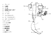
(イ)本発明の喉頭鏡システムは、歯列から喉頭蓋に至る気道に沿った湾曲のブレード(1)を有する。ブレード(1)は金属または合成樹脂で作成され、先端部(2)は、喉頭蓋(21)を挙上するに必要最小な面積と丸みを持たせ喉頭および喉頭蓋を保護する。舌圧板(3)は扁桃腺を避ける逃げ(4)を有し、先端から徐々に広くなり十分に舌を上方手前圧排できる。従来のブレード(17)は舌を左側に圧排した後、上方足側に押しやっていたが(25)、本発明のブレード(1)は舌を上方頭側に圧排する(24)ために最適な形とした。ブレード(1)とハンドル(5)の接続は、汎用性を考え従来と同じ機構で行う。ブレードの先端部(2)から約1〜2cmにカメラを装着保持するホルダー(7)とブレードの接続部(18)から1〜2cmにケーブルを保持するホルダー(8)をもうける。
(ロ)カメラはカラーCCDカメラ(9)とし、先端にLEDの照明(11)を設けることが望ましい。前記照明は一体でも別体でもよい。カメラは防水処理されたケーブル(6)によりハンドル内装置とつながる。
(ハ)ハンドル(5)は内部に映像処理装置、送信装置、カメラやLED照明や各装置に電力を供給する電源を納める。又ハンドル内部または外部にアンテナ(10)をそなえ、システムの送受信機は複数チャンネルを有し混信を避ける。
(ニ)モニター(12)は薄型カラーモニターで内部受信機および電源を持つ。なお電力は内蔵電源からでも外部からコード等を通して供給されてもよい。
(ホ)記録装置(13)は電源および受信機を内蔵し、信号を受信すると自動的に録画が始まり信号が切れると停止する。また受信した信号を携帯電話(14)に送ることもできる。なお記録装置は手動で操作してもよい。
本発明は以上のような内容で構成され、これを用いて気管挿管するときには、準備として舌圧板(3)に潤滑剤を塗り、各機器の電源スイッチを入れておく。気管内チューブはスパイラルチューブを用い、スタイレットは気管内チューブが喉頭鏡ブレード(1)と同じカーブになるように曲げておき、よく潤滑してチューブに挿入しておく。モニター(12)を一台患者の胸部付近にアームを介して固定し、施行医が咽喉頭内の操作を観察できるようにする。又指導医および介助看護師も各自が携帯するモニター(12)で施行医の操作を見られるため、施行医に適切な指導および介助ができる。麻酔導入時にマスク換気を十分に行うのは従来と同じである。頚部の固定等でマスク換気の困難が予想されるときには経鼻エアウエーを挿入するとよい。喉頭展開が可能な状態となれば、左手で患者頭部を保持し右手で十分に開口する。右手はそのまま開口位をとりながら、左手で本喉頭鏡を持つ。正中から口腔へブレード(1)を挿入する。ブレード先端(2)が視野から外れたところでモニターに視線を移し、手首を用いて気道の線に沿ってブレードを進める。このとき舌が押し込まれないようにブレードの舌圧板(3)で舌を上方手前に圧排(24)しつつゆっくりと進める。これで喉頭前腔を広くすることができる。喉頭蓋(21)と声門(20)の一部が見えたら軽く舌を手前に圧した後、喉頭蓋(21)をブレード先端(2)で愛護的に挙上する。このとき喉頭を圧迫するような動きをしてはならない。声門(20)を確認したら右手を口から外し気管内チューブを受け取る。右口角からチューブを入れ、先端がモニター映像に現れたらそのまま気管に挿入する。チューブが声門を約1cm超えたところでスタイレットをチュープから5cm抜きチューブに自由度を持たせる。スタイレットを抜きつつカフが完全に声帯を超えるまで挿入し、正しい位置でチューブを保持してスタイレットを抜き去りバイトブロック設置、チューブを固定する。最終的に人工換気がきちんとなされるか確認する。
本発明喉頭鏡で従来の挿管手技のように喉頭鏡のブレード(1)を上方足側に進めると、頚部の強い屈曲を招いたり扁桃喉頭を傷つけ、又舌を奥に押しやる事になりかねない。又従来の喉頭鏡ブレード(17)では手首を使う操作は御法度であったが、本喉頭鏡では気道をイメージしながら手首を柔らかく使ってブレード(1)を進めるとよい。Embodiments of the present invention will be described below.
(A) The laryngoscope system of the present invention has a curved blade (1) along the airway from the dentition to the epiglottis. The blade (1) is made of metal or synthetic resin, and the tip (2) has a minimum area and roundness necessary for raising the epiglottis (21) to protect the larynx and epiglottis. The tongue pressure plate (3) has a relief (4) that avoids the tonsils, gradually widens from the tip, and can sufficiently remove the tongue from the front side. The conventional blade (17) has pushed the tongue to the left side and then pushed it to the upper foot side (25), but the blade (1) of the present invention is optimal for pushing the tongue to the upper head side (24). Shaped. The blade (1) and the handle (5) are connected by the same mechanism as before in consideration of versatility. A holder (7) for mounting and holding the camera at about 1 to 2 cm from the tip (2) of the blade and a holder (8) for holding the cable from 1 to 2 cm from the connecting portion (18) of the blade are provided.
(B) The camera is a color CCD camera (9), and it is desirable to provide an LED illumination (11) at the tip. The illumination may be integral or separate. The camera is connected to the in-handle device by a waterproof cable (6).
(C) The handle (5) accommodates a video processing device, a transmission device, a camera, LED lighting, and a power source for supplying power to each device. An antenna (10) is provided inside or outside the handle, and the system transceiver has multiple channels to avoid interference.
(D) The monitor (12) is a thin color monitor having an internal receiver and a power source. The power may be supplied from a built-in power source or from the outside through a cord or the like.
(E) The recording device (13) has a built-in power supply and receiver, and automatically starts recording when a signal is received and stops when the signal is cut off. The received signal can also be sent to the mobile phone (14). The recording device may be operated manually.
The present invention is configured as described above, and when performing tracheal intubation using this, as a preparation, a lubricant is applied to the tongue pressure plate (3), and the power switch of each device is turned on. A spiral tube is used as the endotracheal tube, and the stylet is bent so that the endotracheal tube has the same curve as the laryngoscope blade (1), and is lubricated and inserted into the tube. A monitor (12) is fixed near the chest of the patient via an arm so that the practitioner can observe the operation in the larynx. In addition, since the supervising doctor and the assisting nurse can see the operation of the practicing doctor on the monitor (12) carried by each, the supervising doctor and the assisting nurse can appropriately give guidance and assistance. It is the same as before that mask ventilation is adequately performed when anesthesia is introduced. A nasal airway should be inserted when mask ventilation is expected due to cervical fixation. When the larynx can be deployed, the patient's head is held with the left hand and the right hand opens sufficiently. Hold the laryngoscope with your left hand while keeping your right hand open. Insert the blade (1) from the midline into the oral cavity. When the blade tip (2) deviates from the field of view, the line of sight is transferred to the monitor, and the blade is advanced along the airway line using the wrist. At this time, the tongue is pushed forward slowly with the tongue pressure plate (3) of the blade so as not to be pushed in (24). This can widen the prelaryngeal cavity. When the epiglottis (21) and part of the glottis (20) can be seen, the tongue is lightly pressed forward, and then the epiglottis (21) is lifted up with the blade tip (2). At this time, it should not move to press the larynx. After checking the glottis (20), remove the right hand from the mouth and receive the endotracheal tube. Insert the tube from the right mouth corner, and when the tip appears in the monitor image, insert it into the trachea as it is. When the tube exceeds the glottis by approximately 1 cm, the stylet is removed from the tube by 5 cm, and the tube has a degree of freedom. Insert the stylet until the cuff completely exceeds the vocal cord, hold the tube in the correct position, remove the stylet, install the bite block, and secure the tube. Finally, check whether artificial ventilation is properly performed.
When the laryngoscope blade (1) is advanced to the upper foot side as in the conventional intubation procedure with the present laryngoscope, it may cause strong neck flexion, damage the tonsils and larynx, and push the tongue back. . In the conventional laryngoscope blade (17), the operation of using the wrist is legal, but in the present laryngoscope, the blade (1) may be advanced using the wrist softly while imagining the airway.
本発明の第2の実施の形態を説明する。当該形態はハンドル(5)、ブレード(1)、LED照明付きカラーCCDカメラ(9)、ケーブル(6)から成る。前記最良の形態と異なる点は、小型薄型の直付モニター(16)が一軸継ぎ手(15)を介してハンドルに固定され、ハンドルから直接映像信号および電力が供給される。なお記録装置は設けても設けなくてもよい。この第2の形態が気管挿管で必要な最小ユニットの喉頭鏡システムである。 A second embodiment of the present invention will be described. The form comprises a handle (5), a blade (1), a color CCD camera (9) with LED illumination, and a cable (6). The difference from the best mode is that a small and thin direct monitor (16) is fixed to the handle via a uniaxial joint (15), and a video signal and power are directly supplied from the handle. Note that a recording device may or may not be provided. This second form is a minimum unit laryngoscope system necessary for tracheal intubation.
1 ブレード
2 先端部
3 舌圧板
4 扁桃腺を避ける逃げ
5 ハンドル
6 ケーブル
7 カメラのホルダー
8 ケーブルのホルダー
9 カラーCCDカメラ
10 アンテナ
11 LED照明
12 モニター
13 記録装置
14 携帯電話
15 一軸継手
16 直付モニター
17 従来のブレード
18 安静時頚椎
19 強度後屈頚椎
20 声門
21 喉頭蓋
22 カメラの視野
23 従来の医師の視野
24 本発明での医師の操作力
25 従来の医師の操作力DESCRIPTION OF
Claims (3)
Priority Applications (1)
| Application Number | Priority Date | Filing Date | Title |
|---|---|---|---|
| JP2007183712A JP2008307350A (en) | 2007-06-16 | 2007-06-16 | Laryngoscope system |
Applications Claiming Priority (1)
| Application Number | Priority Date | Filing Date | Title |
|---|---|---|---|
| JP2007183712A JP2008307350A (en) | 2007-06-16 | 2007-06-16 | Laryngoscope system |
Publications (1)
| Publication Number | Publication Date |
|---|---|
| JP2008307350A true JP2008307350A (en) | 2008-12-25 |
Family
ID=40235490
Family Applications (1)
| Application Number | Title | Priority Date | Filing Date |
|---|---|---|---|
| JP2007183712A Pending JP2008307350A (en) | 2007-06-16 | 2007-06-16 | Laryngoscope system |
Country Status (1)
| Country | Link |
|---|---|
| JP (1) | JP2008307350A (en) |
Cited By (2)
| Publication number | Priority date | Publication date | Assignee | Title |
|---|---|---|---|---|
| JP2012532689A (en) * | 2009-07-10 | 2012-12-20 | アクシス サージカル テクノロジーズ,インク. | Hand-held minimum-sized diagnostic device with integrated distal end visualization |
| ES2524654A1 (en) * | 2014-01-13 | 2014-12-10 | Bernat CARNER BONET | Video-laryngoscope blade with connection to smartphones (Machine-translation by Google Translate, not legally binding) |
Citations (2)
| Publication number | Priority date | Publication date | Assignee | Title |
|---|---|---|---|---|
| WO2003068056A1 (en) * | 2002-02-15 | 2003-08-21 | Yoshinori Iwase | Laryngoscope |
| JP2006326111A (en) * | 2005-05-27 | 2006-12-07 | Daiken Iki Kk | Laryngoscope |
-
2007
- 2007-06-16 JP JP2007183712A patent/JP2008307350A/en active Pending
Patent Citations (2)
| Publication number | Priority date | Publication date | Assignee | Title |
|---|---|---|---|---|
| WO2003068056A1 (en) * | 2002-02-15 | 2003-08-21 | Yoshinori Iwase | Laryngoscope |
| JP2006326111A (en) * | 2005-05-27 | 2006-12-07 | Daiken Iki Kk | Laryngoscope |
Cited By (3)
| Publication number | Priority date | Publication date | Assignee | Title |
|---|---|---|---|---|
| JP2012532689A (en) * | 2009-07-10 | 2012-12-20 | アクシス サージカル テクノロジーズ,インク. | Hand-held minimum-sized diagnostic device with integrated distal end visualization |
| ES2524654A1 (en) * | 2014-01-13 | 2014-12-10 | Bernat CARNER BONET | Video-laryngoscope blade with connection to smartphones (Machine-translation by Google Translate, not legally binding) |
| WO2015104444A1 (en) * | 2014-01-13 | 2015-07-16 | Bernat Carner Bonet | Video laryngoscope blade having smartphone connection |
Similar Documents
| Publication | Publication Date | Title |
|---|---|---|
| EP1738789B1 (en) | Endotracheal video device | |
| US8479739B2 (en) | System and method for managing difficult airways | |
| JP4717843B2 (en) | Wireless optical endoscopic device | |
| EP1847214B1 (en) | Ultra wide band wireless optical endoscopic device | |
| US20050279355A1 (en) | Superglottic and peri-laryngeal apparatus having video components for structural visualization and for placement of supraglottic, intraglottic, tracheal and esophageal conduits | |
| US7946981B1 (en) | Two-piece video laryngoscope | |
| US6929600B2 (en) | Apparatus for intubation | |
| US8382665B1 (en) | Endotracheal tube placement system and method | |
| US20030181789A1 (en) | Laryngoscope with image sensor | |
| US9622651B2 (en) | Wireless laryngoscope simulator with onboard event recording adapted for laryngoscopy training | |
| JP2009524482A (en) | Device for introducing an airway tube into a trachea having visualization ability and method of using the same | |
| US9662068B2 (en) | Medical device for conducting a medical examination and/or intervention | |
| US20240366895A1 (en) | System and method for video assisted percutaneous needle cricothyrotomy and tracheostomy | |
| Hagberg | Special devices and techniques | |
| CN105169540A (en) | Brightness-adjustable double positioning video light stick for tracheal cannula | |
| JP2019509115A (en) | Disposable intubation device | |
| JP2008307350A (en) | Laryngoscope system | |
| JP3108837U (en) | Small camera radio system for tracheal intubation | |
| JPH11113843A (en) | Stylet having observing optical system for endotracheal tube inserting pipe | |
| AU2013239345A1 (en) | Endotracheal tube introducer | |
| JP2015016177A (en) | Medical instrument | |
| Van Zundert et al. | Video-assisted laryngoscopy: a useful adjunct in endotracheal intubation | |
| CN218853276U (en) | Double-mirror display device for difficult trachea cannula | |
| KR20170121388A (en) | Video laryngoscope holder | |
| KR101706066B1 (en) | A camera device for a detachable use laryngoscope |
Legal Events
| Date | Code | Title | Description |
|---|---|---|---|
| A621 | Written request for application examination |
Free format text: JAPANESE INTERMEDIATE CODE: A621 Effective date: 20100302 |
|
| A521 | Written amendment |
Free format text: JAPANESE INTERMEDIATE CODE: A523 Effective date: 20100416 |
|
| A521 | Written amendment |
Free format text: JAPANESE INTERMEDIATE CODE: A523 Effective date: 20100602 |
|
| A977 | Report on retrieval |
Free format text: JAPANESE INTERMEDIATE CODE: A971007 Effective date: 20111215 |
|
| A131 | Notification of reasons for refusal |
Free format text: JAPANESE INTERMEDIATE CODE: A131 Effective date: 20111220 |
|
| A02 | Decision of refusal |
Free format text: JAPANESE INTERMEDIATE CODE: A02 Effective date: 20120417 |


