-
The present invention relates to methods of cancer prognosis and kits for cancer prognosis as well as the use of specific genes in cancer prognosis.
-
The first task of a clinician in assessing a patient suspected of having cancer because of the presence of a suspicious lesion (e.g. a node in the breast), is to investigate to what entity the lesion belongs (cyst, tumor, inflammation, etc.) and to what dignity the lesion belongs (malignant, benign). Traditionally, this is done clinically using imaging methods including ultrasound, (digital) mammography, MRI, etc. and histo-pathologically after tissue sampling or tissue removal.
-
Once a lesion has been diagnosed as cancer, further classification is important with respect to prognosis (patient outcome) and selection of treatment (therapy prediction). Currently classification is achieved based largely on histo-pathological tumor features (grade, tumor size, nodal involvement), as well as patient age. For example, in breast cancer, the St. Gallen and NIH consensus criteria are currently the most widely used classification systems. These guidelines classify the majority of primary breast cancers into intermediate or high-risk subgroups. This leads to the recommended administration of adjuvant systemic therapy for nearly 90% of all newly diagnosed breast cancer patients. However, these classification systems do not assess individual biological changes on a molecular level in a systematic way.
-
Gene expression profiling using microarrays can be used to assess such changes by measuring hundreds of genes simultaneously. However this method has the major disadvantage that the cost, complexity and interpretation of DNA micro-arrays make them currently unsuitable for routine testing used in standard clinical settings.
-
Other workers have focused on individual genes as biomarkers involved in cancer, for example (SEQ ID NO. 37, 38) ERBB2 (also known as HER2: Human Epidermal Growth Factor Receptor 2 and neu: neuro/glioblastoma derived oncogene homolog (avian)). (SEQ ID NO. 37, 38) ERBB2 has been mechanistically linked with a variety of malignant processes including lymph node and distant metastasis. Amplification and/or overexpression of (SEQ ID NO. 37, 38) ERBB2 is observed in 15 to 30% of all newly diagnosed breast cancers, as well as in 15 to 30% of ovarian carcinomas. It has also been found to be overexpressed in lung and prostate cancer. It is associated with poor prognosis and serves as a predictor of clinical responsiveness to the anti-(SEQ ID NO. 37, 38) ERBB2 therapeutics such as trastuzumab (Herceptin®) as well as chemo- and endocrine therapy.
-
In the most recent (9th) St. Gallen consensus panel determination, all (SEQ ID NO. 37, 38) ERBB2-positive breast cancer cases are to be considered high-risk (Goldhirsch et al, Annals of Oncology, 2005, doi:10.1093/annonc/mdi326).
-
This is unsatisfactory, since (SEQ ID NO. 37, 38) ERBB2-positive breast cancers display varying amounts of clinical aggressiveness i.e. not all (SEQ ID NO. 37, 38) ERBB2-positive patients develop metastasis, and thus not all are at high risk. Thus some patients are unnecessarily subjected to aggressive chemotherapy and unnecessary suffering as a consequence.
-
In addition not all (SEQ ID NO. 37, 38) ERBB2-positive patients respond to anti-(SEQ ID NO. 37, 38) ERBB2 therapy. In fact only up to 30% of (SEQ ID NO. 37, 38) ERBB2-positive patients will respond to trastuzumab therapy. This implies that other factors may also be influential in determining responsiveness to trastuzumab therapy.
-
Therefore, additional biomarkers are needed to improve classification of (SEQ ID NO. 37, 38) ERBB2-positive patients with respect to prognosis and therapeutic responsiveness in order to optimize the clinical management of these patients. Similarly additional biomarkers are needed to improve classification of (SEQ ID NO. 37, 38) ERBB2-negative patients with respect to prognosis and therapeutic responsiveness.
-
Thus the present invention provides a method used for assessing the probability of survival of a cancer patient by assaying one or more ex-vivo samples from that patient, wherein the (SEQ ID NO. 37, 38) ERBB2 status of that patient is determined to be positive or negative; characterized in that
- a) in (SEQ ID NO. 37, 38) ERBB2-positive patients, at least one of a first set of cancer related genes is analysed in the sample or in one of the samples from said patient
and - b) in (SEQ ID NO. 37, 38) ERBB2-negative patients, at least one of a second set of cancer related genes is analysed in the sample or in one of the samples from said patient.
-
Preferably the probability of survival of the cancer patient is assessed in terms of metastasis free survival (MFS).
-
Alternatively, the probability of survival of the cancer patient is assessed in terms of relapse free survival (RFS).
-
The term cancer related genes relates to genes particularly the following (SEQ ID NO. 73, 74) PLAU, (SEQ ID NO. 77, 78) PLAUR, (SEQ ID NO. 65, 66) MMP11, (SEQ ID NO. 49, 50) MMP1, (SEQ ID NO. 57, 58) MMP3, (SEQ ID NO. 93, 94) TIMP3, (SEQ ID NO. 41, 42) ERBB3, (SEQ ID NO. 33, 34) ECGF1, (SEQ ID NO. 81, 82) SERPINE1, (SEQ ID NO. 53, 54) MMP2, (SEQ ID NO. 9, 10) BIRC5, (SEQ ID NO. 29, 30) E2F1, (SEQ ID NO. 105, 106) TOP2A, (SEQ ID NO. 101, 102) TK1, (SEQ ID NO. 109, 110) TYMS, (SEQ ID NO. 113, 114) VEGF, (SEQ ID NO. 85, 86) SRD5A1, (SEQ ID NO. 13, 14) CCND1, (SEQ ID NO. 25, 26) CTSD, (SEQ ID NO. 5, 6) BAX, (SEQ ID NO. 21, 22) CTSB, (SEQ ID NO. 17, 18) CTNNB1, (SEQ ID NO. 1, 2) ADM, (SEQ ID NO. 61, 62) MMP9, (SEQ ID NO. 89, 90) STK11 and (SEQ ID NO. 97, 98) TIMP4.
-
In a preferred embodiment said first set of genes comprises (SEQ ID NO. 73, 74) PLAU, (SEQ ID NO. 77, 78) PLAUR, (SEQ ID NO. 65, 66) MMP11, (SEQ ID NO. 49, 50) MMP1, (SEQ ID NO. 57, 58) MMP3, (SEQ ID NO. 93, 94) TIMP3, (SEQ ID NO. 41, 42) ERBB3, (SEQ ID NO. 33, 34) ECGF1, (SEQ ID NO. 81, 82) SERPINE1, and (SEQ ID NO. 53, 54) MMP2.
-
In another preferred embodiment of the invention, said second set of genes comprises (SEQ ID NO. 9, 10) BIRC5, (SEQ ID NO. 29, 30) E2F1, (SEQ ID NO. 105, 106) TOP2A, (SEQ ID NO. 101, 102) TK1, (SEQ ID NO. 109, 110) TYMS, (SEQ ID NO. 113, 114) VEGF, (SEQ ID NO. 85, 86) SRD5A1, (SEQ ID NO. 13, 14) CCND1, (SEQ ID NO. 25, 26) CTSD, (SEQ ID NO. 5, 6) BAX, (SEQ ID NO. 21, 22) CTSB, (SEQ ID NO. 17, 18) CTNNB1, (SEQ ID NO. 1, 2) ADM, (SEQ ID NO. 61, 62) MMP9, (SEQ ID NO. 89, 90) STK11 and (SEQ ID NO. 97, 98) TIMP4.
-
Preferably, in (SEQ ID NO. 37, 38) ERBB2-positive patients the combination of genes of (SEQ ID NO. 73, 74) PLAU, (SEQ ID NO. 77, 78) PLAUR, (SEQ ID NO. 65, 66) MMP11 and (SEQ ID NO. 49, 50) MMP1 is analysed.
-
In one embodiment of the invention, in (SEQ ID NO. 37, 38) ERBB2-positive patients the combination of genes of (SEQ ID NO. 73, 74) PLAU, (SEQ ID NO. 77, 78) PLAUR and (SEQ ID NO. 65, 66) MMP11 is analysed.
-
In another embodiment of the invention, in (SEQ ID NO. 37, 38) ERBB2-positive patients the combination of genes of (SEQ ID NO. 77, 78) PLAUR, (SEQ ID NO. 65, 66) MMP11 and (SEQ ID NO. 49, 50) MMP1 is analysed.
-
In yet another embodiment of the invention, in (SEQ ID NO. 37, 38) ERBB2-positive patients in addition to the aforementioned combinations of genes, the gene (SEQ ID NO. 57, 58) MMP3 is analysed, namely the combination of (SEQ ID NO. 73, 74) PLAU, (SEQ ID NO. 77, 78) PLAUR, (SEQ ID NO. 65, 66) MMP11, (SEQ ID NO. 49, 50) MMP1 and (SEQ ID NO. 57, 58) MMP3 or the combination (SEQ ID NO. 73, 74) PLAU, (SEQ ID NO. 77, 78) PLAUR, (SEQ ID NO. 65, 66) MMP11 and (SEQ ID NO. 57, 58) MMP3 or the combination (SEQ ID NO. 77, 78) PLAUR, (SEQ ID NO. 65, 66) MMP11, (SEQ ID NO. 49, 50) MMP1 and (SEQ ID NO. 57, 58) MMP3.
-
In a preferred embodiment of the invention, in (SEQ ID NO. 37, 38) ERBB2-positive patients in addition to the aforementioned combinations, the gene (SEQ ID NO. 93, 94) TIMP3 is analysed, such as for example the combination of (SEQ ID NO. 73, 74) PLAU, (SEQ ID NO. 77, 78) PLAUR, (SEQ ID NO. 65, 66) MMP11, (SEQ ID NO. 49, 50) MMP1, (SEQ ID NO. 57, 58) MMP3 and (SEQ ID NO. 93, 94) TIMP3.
-
In yet a further embodiment of the invention, in (SEQ ID NO. 37, 38) ERBB2-positive patients in addition to the aforementioned combinations the gene (SEQ ID NO. 53, 54) MMP2 is analysed, such as for example the combination of (SEQ ID NO. 73, 74) PLAU, (SEQ ID NO. 77, 78) PLAUR, (SEQ ID NO. 65, 66) MMP11, (SEQ ID NO. 49, 50) MMP1, (SEQ ID NO. 57, 58) MMP3, (SEQ ID NO. 93, 94) TIMP3 and (SEQ ID NO. 53, 54) MMP2.
-
In a still yet further embodiment of the invention, in (SEQ ID NO. 37, 38) ERBB2-positive patients in addition to the aforementioned combinations the gene (SEQ ID NO. 81, 82) SERPINE1 is analysed, such as for example the combination of (SEQ ID NO. 73, 74) PLAU, (SEQ ID NO. 77, 78) PLAUR, (SEQ ID NO. 65, 66) MMP11, (SEQ ID NO. 49, 50) MMP1, (SEQ ID NO. 57, 58) MMP3, (SEQ ID NO. 93, 94) TIMP3, (SEQ ID NO. 53, 54) MMP2 and (SEQ ID NO. 81, 82) SERPINE1.
-
Preferably in (SEQ ID NO. 37, 38) ERBB2-negative patients, the combination of genes of (SEQ ID NO. 9, 10) BIRC5, (SEQ ID NO. 29, 30) E2F1, (SEQ ID NO. 105, 106) TOP2A and (SEQ ID NO. 101, 102) TK1 is analysed.
-
In a preferred embodiment of the invention, in (SEQ ID NO. 37, 38) ERBB2-negative patients the combination of genes of (SEQ ID NO. 9, 10) BIRC5, (SEQ ID NO. 29, 30) E2F1 and (SEQ ID NO. 105, 106) TOP2A is analysed.
-
In another embodiment of the invention, in (SEQ ID NO. 37, 38) ERBB2-negative patients the combination of genes of (SEQ ID NO. 9, 10) BIRC5, (SEQ ID NO. 29, 30) E2F1 and (SEQ ID NO. 101, 102) TK1 is analysed.
-
In yet another embodiment of the invention, in (SEQ ID NO. 37, 38) ERBB2-negative patients the combination of genes of (SEQ ID NO. 9, 10) BIRC5, (SEQ ID NO. 29, 30) E2F1 and (SEQ ID NO. 109, 110) TYMS is analysed.
-
In still yet another embodiment of the invention, in (SEQ ID NO. 37, 38) ERBB2-negative patients the combination of genes of (SEQ ID NO. 9, 10) BIRC5, (SEQ ID NO. 29, 30) E2F1, (SEQ ID NO. 101, 102) TK1 and (SEQ ID NO. 109, 110) TYMS is analysed.
-
In a further embodiment of the invention, in (SEQ ID NO. 37, 38) ERBB2-negative patients in addition to the aforementioned combinations, the gene (SEQ ID NO. 21, 22) CTSB is analysed, such as for example the combination of (SEQ ID NO. 9, 10) BIRC5, (SEQ ID NO. 29, 30) E2F1, (SEQ ID NO. 105, 106) TOP2A, (SEQ ID NO. 101, 102) TK1 and (SEQ ID NO. 21, 22) CTSB.
-
In a yet further embodiment of the invention, in (SEQ ID NO. 37, 38) ERBB2-negative patients in addition to the aforementioned combinations, the gene (SEQ ID NO. 25, 26) CTSD is analysed, such as for example the combination of (SEQ ID NO. 9, 10) BIRC5, (SEQ ID NO. 29, 30) E2F1, (SEQ ID NO. 105, 106) TOP2A, (SEQ ID NO. 101, 102) TK1, (SEQ ID NO. 21, 22) CTSB and (SEQ ID NO. 25, 26) CTSD.
-
Suitable methods known in the art may be used to measure the expression levels of the genes. In a preferred embodiment of the method of the invention the analysis of the genes involves measuring the expression levels of the genes at the mRNA level.
-
Preferably also the (SEQ ID NO. 37, 38) ERBB2 status is determined at the mRNA level by qRT-PCR by measuring the cycle threshold value for (SEQ ID NO. 37, 38) ERBB2 and for 18S rRNA, subtracting the cycle threshold value for (SEQ ID NO. 37, 38) ERBB2 from the cycle threshold value for 18S rRNA. A value of ≥ -15.8 is determined to be positive (SEQ ID NO. 37, 38) ERBB2 status.
-
Most preferably in the method of the invention, in the first set of genes each gene is statistically weighted according to its association with survival in a population of cancer patients and within the second set of genes each gene is statistically weighted according to its association with survival in a population of cancer patients.
-
Preferably also the statistical weighting of the genes in the first set of genes and the statistical weighting of the genes in the second set of genes is expressed in terms of the Cox-coefficient. (Cox DR: Regression Models and Lifetables J.R. Soc. 34: 187-220, 1972) Another aspect of the invention is a kit for assessing the probability of survival of a patient of whom the (SEQ ID NO. 37, 38) ERBB2 status is known to be positive; comprising a set of oligonucleotide primers (SEQ ID NO. 75, 76, 79, 80, 67, 68, 51, 52), said primer set allowing PCR to be performed on mRNA transcribed from the following genes: (SEQ ID NO. 73, 74) PLAU, (SEQ ID NO. 77, 78) PLAUR, (SEQ ID NO. 65, 66) MMP11 and (SEQ ID NO. 49, 50) MMP1.
-
A further aspect of the invention is a kit for assessing the probability of survival of a patient of whom the (SEQ ID NO. 37, 38) ERBB2 status is known to be negative; comprising a set of oligonucleotide primers (SEQ ID NO. 11, 12, 31, 32, 107, 108, 103, 104), said primer set allowing PCR to be performed on mRNA transcribed from the following genes: (SEQ ID NO. 9, 10) BIRC5, (SEQ ID NO. 29, 30) E2F1, (SEQ ID NO. 105, 106) TOP2A and (SEQ ID NO. 101, 102) TK1.
-
Preferably, there is provided a kit comprising a set of oligonucleotide primers (SEQ ID NO. 75, 76, 79, 80, 67, 68, 51, 52, 11, 12, 31, 32, 107, 108, 103, 104), said primer set allowing PCR to be performed on mRNA transcribed from the following genes: (SEQ ID NO. 73, 74) PLAU, (SEQ ID NO. 77, 78) PLAUR, (SEQ ID NO. 65, 66) MMP11, (SEQ ID NO. 49, 50) MMP1, (SEQ ID NO. 9, 10) BIRC5, (SEQ ID NO. 29, 30) E2F1, (SEQ ID NO. 105, 106) TOP2A and (SEQ ID NO. 101, 102) TK1.
-
More preferably there is provided a kit with all of the primers in Table 2 (SEQ ID NO. 3, 7, 11, 15, 19, 23, 27, 31, 35, 39, 43, 47, 51, 55, 59, 63, 67, 71, 75, 79, 83, 87, 91, 95, 99, 103, 107, 111, 115, 117, 119, 121, 123, 125, 127, 129, 131, 133, 135, 137, 139, 141, 143, 145, 147, 149, 151, 153, 155, 157, 159, 161, 163, 165, 167, 169, 171, 173, 175, 177, 179, 181, 183, 185, 187, 189, 191, 193, 195, 197, 199, 201, 203, 205, 207), and table 3 (SEQ ID NO. 4, 8, 12, 16, 20, 24, 28, 32, 36, 40, 44, 48, 52, 56, 60, 64, 68, 72, 76, 80, 84, 88, 92, 96, 100, 104, 108, 112, 116, 118, 120, 122, 124, 126, 128, 130, 132, 134, 136, 138, 140, 142, 144, 146, 148, 150, 152, 154, 156, 158, 160, 162, 164, 166, 168, 170, 172, 174, 176, 178, 180, 182, 184, 186, 188, 190, 192, 194, 196, 198, 200, 202, 204, 206, 208).
-
The aforementioned kits may also contain suitable reagents (other than primers) to perform PCR.
-
Another aspect of the invention is use of any of the genes or any combination of the genes disclosed herein in cancer prognosis.
-
Preferably the invention relates to the use of the genes (SEQ ID NO. 73, 74) PLAU, (SEQ ID NO. 77, 78) PLAUR, (SEQ ID NO. 65, 66) MMP11 and (SEQ ID NO. 49, 50) MMP1 in cancer prognosis.
-
A further aspect of the invention is use of the genes (SEQ ID NO. 9, 10) BIRC5, (SEQ ID NO. 29, 30) E2F1, (SEQ ID NO. 105, 106) TOP2A and (SEQ ID NO. 101, 102) TK1 in cancer prognosis.
Brief Description of the Figures
-
For a more detailed description of the terms used in the figure legends see the detailed description section and the examples.
- Figure 1: Distribution of (SEQ ID NO. 37, 38) ERBB2 mRNA expression levels in breast cancer biopsies from 575 patients. X: axis: (SEQ ID NO. 37, 38) ERBB2 expression level (delta cT), Y: axis: density (probability distribution)
- Figure 2: ROC (Receiver operator characteristics) analysis of (SEQ ID NO. 37, 38) ERBB2 mRNA expression levels measured by PCR (n=100) (SEQ ID NO. 37, 38) ERBB2 amplification measured by FISH was used as reference. X: axis 1-specificity, Y: axis: sensitivity
- Figure 3: Ranked (SEQ ID NO. 37, 38) ERBB2 mRNA expression levels (n=100). Black circles: FISH positive, empty circles: FISH negative samples, Broken line: cut off value for (SEQ ID NO. 37, 38) ERBB2 status by PCR (-15.8) X:axis: samples ranked according to (SEQ ID NO. 37, 38) ERBB2 expression level (ordered samples) Y:axis: (SEQ ID NO. 37, 38) ERBB2 expression (delta cT)
- Figure 4: Distribution of mRNA expression levels in 317 breast cancer biopsies. X-axis: (SEQ ID NO. 45, 46) ESR1 expression (delta cT). Y-axis: density (probability distribution)
- Figure 5: ROC (Receiver operator characteristics) analysis of ER mRNA expression levels measured by PCR (n=317). (SEQ ID NO. 45, 46) ESR1 protein cut off of 20 fmol/mg protein measured by ELISA was used as reference. X: axis 1-specificity, Y: axis: sensitivity
- Figure 6: Correlation between (SEQ ID NO. 45, 46) ESR1 levels detected by PCR and ELISA. Solid line represents the regression line. Horizontal broken line shows the cut off value (20 fmol/mg protein) and vertical broken line shows the corresponding PCR cut off value (delta cT value of -19. X-axis: (SEQ ID NO. 45, 46) ESR1 expression measured by PCR (delta cT). Y-axis: (SEQ ID NO. 45, 46) ESR1 Expression levels measured by ELISA (log transformed)
- Figure 7: Prognosis graph for (SEQ ID NO. 37, 38) ERBB2-positive patients determined by analyzing the gene combination (SEQ ID NO. 73, 74) PLAU, (SEQ ID NO. 77, 78) PLAUR, (SEQ ID NO. 49, 50) MMP1 and (SEQ ID NO. 65, 66) MMP11. Shown is the probability of 5-years metastasis-free survival (Y-axis) as a function of different cut-offs of the prognostic score in percentiles (X-axis).
- Figure 8: Prognosis graph for (SEQ ID NO. 37, 38) ERBB2-positive patients determined by analyzing the gene combination (SEQ ID NO. 73, 74) PLAU, (SEQ ID NO. 77, 78) PLAUR, (SEQ ID NO. 49, 50) MMP1 and (SEQ ID NO. 65, 66) MMP11. Shown is the probability of 5-years relapse-free survival (Y-axis) as a function of different cut-offs of the prognostic score in percentiles (X-axis).
- Figure 9: Prognosis graph for (SEQ ID NO. 37, 38) ERBB2-positive patients determined by analyzing the gene combination (SEQ ID NO. 73, 74) PLAU, (SEQ ID NO. 77, 78) PLAUR, and (SEQ ID NO. 65, 66) MMP11. Shown is the probability of 5-years metastasis-free survival (Y-axis) as a function of different cut-offs of the prognostic score in percentiles (X-axis).
- Figure 10: Prognosis graph for (SEQ ID NO. 37, 38) ERBB2-positive patients determined by analyzing the gene combination (SEQ ID NO. 73, 74) PLAU, (SEQ ID NO. 77, 78) PLAUR, and (SEQ ID NO. 65, 66) MMP11. Shown is the probability of 5-years relapse-free survival (Y-axis) as a function of different cut-offs of the prognostic score in percentiles (X-axis).
- Figure 11: Prognosis graph for (SEQ ID NO. 37, 38) ERBB2-positive patients determined by analyzing the gene combination (SEQ ID NO. 73, 74) PLAU, (SEQ ID NO. 77, 78) PLAUR, (SEQ ID NO. 65, 66) MMP11, (SEQ ID NO. 49, 50) MMP1, (SEQ ID NO. 81, 82) SERPINE1 and (SEQ ID NO. 57, 58) MMP3. Shown is the probability of 5-years metastasis-free survival (Y-axis) as a function of different cut-offs of the prognostic score in percentiles (X-axis).
- Figure 12: Prognosis graph for (SEQ ID NO. 37, 38) ERBB2-positive patients determined by analyzing the gene combination (SEQ ID NO. 73, 74) PLAU, (SEQ ID NO. 77, 78) PLAUR, (SEQ ID NO. 65, 66) MMP11, (SEQ ID NO. 49, 50) MMP1, (SEQ ID NO. 81, 82) SERPINE1 and (SEQ ID NO. 57, 58) MMP3. Shown is the probability of 5-years relapse-free survival (Y-axis) as a function of different cut-offs of the prognostic score in percentiles (X-axis).
- Figure 13: Prognosis graph for (SEQ ID NO. 37, 38) ERBB2-positive patients determined by analyzing the gene (SEQ ID NO. 73, 74) PLAU. Shown is the probability of 5-years metastasis-free survival (Y-axis) as a function of different cut-offs of the prognostic score in percentiles (X-axis).
- Figure 14: Prognosis graph for (SEQ ID NO. 37, 38) ERBB-positive patients determined by analyzing the gene (SEQ ID NO. 73, 74) PLAU. Shown is the probability of 5-years relapse-free survival (Y-axis) as a function of different cut-offs of the prognostic score in percentiles (X-axis).
- Figure 15: Prognosis graph for (SEQ ID NO. 37, 38) ERBB2-positive patients determined by analyzing the gene (SEQ ID NO. 77, 78) PLAUR. Shown is the probability of 5-years metastasis-free survival (Y-axis) as a function of different cut-offs of the prognostic score in percentiles (X-axis).
- Figure 16: Prognosis graph for (SEQ ID NO. 37, 38) ERBB2-positive patients determined by analyzing the gene (SEQ ID NO. 77, 78) PLAUR. Shown is the probability of 5-years relapse-free survival (Y-axis) as a function of different cut-offs of the prognostic score in percentiles (X-axis).
- Figure 17: Prognosis graph for (SEQ ID NO. 37, 38) ERBB2-negative patients determined by analyzing the gene combination (SEQ ID NO. 9, 10) BIRC5, (SEQ ID NO. 29, 30) E2F1, (SEQ ID NO. 105, 106) TOP2A and (SEQ ID NO. 101, 102) TK1. Shown is the probability of 5-years metastasis-free survival (Y-axis) as a function of different cut-offs of the prognostic score in percentiles (X-axis).
- Figure 18: Prognosis graph for (SEQ ID NO. 37, 38) ERBB2-negative patients determined by analyzing the gene combination (SEQ ID NO. 9, 10) BIRC5, (SEQ ID NO. 29, 30) E2F1, (SEQ ID NO. 105, 106) TOP2A and (SEQ ID NO. 101, 102) TK1. Shown is the probability of 5-years relapse-free survival (Y-axis) as a function of different cut-offs of the prognostic score in percentiles (X-axis).
- Figure 19: Prognosis graph for (SEQ ID NO. 37, 38) ERBB2-negative patients determined by analyzing the gene combination (SEQ ID NO. 9, 10) BIRC5, (SEQ ID NO. 29, 30) E2F1, and (SEQ ID NO. 105, 106) TOP2A. Shown is the probability of 5-years metastasis-free survival (Y-axis) as a function of different cut-offs of the prognostic score in percentiles (X-axis).
- Figure 20: Prognosis graph for (SEQ ID NO. 37, 38) ERBB2-negative patients determined by analyzing the gene combination (SEQ ID NO. 9, 10) BIRC5, (SEQ ID NO. 29, 30) E2F1, and (SEQ ID NO. 105, 106) TOP2A. Shown is the probability of 5-years relapse-free survival (Y-axis) as a function of different cut-offs of the prognostic score in percentiles (X-axis).
- Figure 21: Prognosis graph for (SEQ ID NO. 37, 38) ERBB2-negative patients determined by analyzing the gene combination (SEQ ID NO. 9, 10) BIRC5, (SEQ ID NO. 29, 30) E2F1, (SEQ ID NO. 105, 106) TOP2A, (SEQ ID NO. 21, 22) CTSB and (SEQ ID NO. 25, 26) CTSD. Shown is the probability of 5-years metastasis-free survival (Y-axis) as a function of different cut-offs of the prognostic score in percentiles (X-axis).
- Figure 22: Prognosis graph for (SEQ ID NO. 37, 38) ERBB2-negative patients determined by analyzing the gene combination (SEQ ID NO. 9, 10) BIRC5, (SEQ ID NO. 29, 30) E2F1, (SEQ ID NO. 105, 106) TOP2A, (SEQ ID NO. 21, 22) CTSB and (SEQ ID NO. 25, 26) CTSD. Shown is the probability of 5-years relapse-free survival (Y-axis) as a function of different cut-offs of the prognostic score in percentiles (X-axis).
- Figure 23: Prognosis graph for (SEQ ID NO. 37, 38) ERBB2-negative patients determined by analyzing the gene (SEQ ID NO. 9, 10) BIRC5. Shown is the probability of 5-years metastasis-free survival (Y-axis) as a function of different cut-offs of the prognostic score in percentiles (X-axis).
- Figure 24: Prognosis graph for (SEQ ID NO. 37, 38) ERBB2-negative patients determined by analyzing the gene (SEQ ID NO. 9, 10) BIRC5. Shown is the probability of 5-years relapse-free survival (Y-axis) as a function of different cut-offs of the prognostic score in percentiles (X-axis).
- Figure 25: Prognosis graph for (SEQ ID NO. 37, 38) ERBB2-negative patients determined by analyzing the gene (SEQ ID NO. 29, 30) E2F1. Shown is the probability of 5-years metastasis-free survival (Y-axis) as a function of different cut-offs of the prognostic score in percentiles (X-axis).
- Figure 26: Prognosis graph for (SEQ ID NO. 37, 38) ERBB2-negative patients determined by analyzing the gene (SEQ ID NO. 29, 30) E2F1. Shown is the probability of 5-years relapse-free survival (Y-axis) as a function of different cut-offs of the prognostic score in percentiles (X-axis).
Detailed description
-
This invention provides a method of assessing the probability of survival of a cancer patient, which may be assessed in terms of metastasis free survival, relapse free survival or overall survival.
-
The term metastasis-free survival (MFS) refers to distant metastasis-free survival MFS (DMFS) i.e. it refers to living without the occurrence of metastasis at a site distant from the primary tumour.
-
The term relapse-free survival (RFS) refers to living without the recurrence of cancer at any site, except as second primary cancer.
-
The term overall survival as used herein refers to disease-specific overall survival, i.e. it refers to living rather than dying from a specific disease.
-
The survival prediction is made based on the analysis of one or more ex-vivo samples from a patient. Ex-vivo samples include tissue samples, stool samples, cell aspirates, ductal lavages, serum samples, urine and blood samples.
-
The (SEQ ID NO. 37, 38) ERBB2 status of the patient is determined to be positive or negative. The following methods may be used to determine the (SEQ ID NO. 37, 38) ERBB2 status and are routinely used in the field. They are intended to be illustrative of the techniques available, however measurement of the (SEQ ID NO. 37, 38) ERBB2 status is not intended to be restricted to the assays described below. The methods may also be used in combination to determine the (SEQ ID NO. 37, 38) ERBB2 status.
-
The (SEQ ID NO. 37, 38) ERBB2 status may be determined at the protein level for example by IHC (immunohistochemistry), ELISA (enzyme linked immunosorption assay) or western blot. IHC based testing is possible through use of the HercepTest (DAKO) kit, which has received US FDA approval for clinical use. The kit contains standardized reagents and control slides. A scoring system is employed in which a score ranging in whole numbers from 0 to 3 is given which reflects the staining intensity and pattern in 10% or more of tumor cells. A score of 3+ is considered as positive. Scores of 2+ require additional confirmation by another method such as FISH (see below) to determine positivity. (Hanna, W. Testing for HER2 Status., Oncology 2001; 61 (suppl.2) 22-30; Leo. A et al., Current Status of HER2 Testing; Oncology 2002, 63 (suppl. 1)25-32)).
-
The (SEQ ID NO. 37, 38) ERBB2 status may be determined at the DNA level for example by FISH (fluorescence in situ hybridization), CISH (chromogenic in situ hybridization), PCR (polymerase chain reaction) or southern blot. With FISH the number of (SEQ ID NO. 37, 38) ERBB2 genes in the cell nucleus can be counted to measure (SEQ ID NO. 37, 38) ERBB2 amplification. Some FISH assays measure (SEQ ID NO. 37, 38) ERBB2 amplification as an absolute number of gene copies, while others involve the ratio between (SEQ ID NO. 37, 38) ERBB2 gene copies and chromosome 17 copies (the (SEQ ID NO. 37, 38) ERBB2 gene resides on chromosome 17). Standardized kits for use for FISH are currently available, which have been approved for use by the FDA. The Inform (SEQ ID NO. 37, 38) ERBB2 kit (ONCOR/Ventana) is an example of a FISH assay which measures the absolute number of (SEQ ID NO. 37, 38) ERBB2 gene copies. The PathVysion® Kit (Vysis) on the other hand uses two DNA probes, one which specifically recognizes the (SEQ ID NO. 37, 38) ERBB2 gene and the other the chromosome 17 centromere. (SEQ ID NO. 37, 38) ERBB2 and centromere signals are measured in 60 nuclei and then the (SEQ ID NO. 37, 38) ERBB2 gene:chromosome 17 centromere ratio is calculated. A ratio of ≥ 2 indicates (SEQ ID NO. 37, 38) ERBB2 positivity. (Hanna, W. Testing for HER2 Status., Oncology 2001; 61 (suppl.2) 22-30; Leo. A et al., Current Status of HER2 Testing; Oncology 2002, 63 (suppl. 1)25-32)).
-
(SEQ ID NO. 37, 38) ERBB2 status may also determined using a gene expression based signature based on the expression levels of other marker genes.
-
Importantly also, the (SEQ ID NO. 37, 38) ERBB2 status may be determined at the RNA level using PCR, microarray analysis or northern blot.
PCR includes competitive PCR and quantitative real-time PCR (qRT-PCR). QRT-PCR measurement of RNA expression is a highly sensitive, reproducible, quantitative and cost effective method. A description of the use of qRT-PCR to determine (SEQ ID NO. 37, 38) ERBB2 status is to be found in Examples 1, 2 and 3.
-
In addition to (SEQ ID NO. 37, 38) ERBB2 status additional genes are analysed in accordance with this invention. If (SEQ ID NO. 37, 38) ERBB2 status is determined to be positive, then at least one of a first set of cancer related genes is analysed in the or one of the samples from the patient and in (SEQ ID NO. 37, 38) ERBB2-negative patients, at least one of a second set of cancer related genes is analysed in the or one of the samples from the patient.
-
Genes analysed according to the invention are related to full-length nucleic acid sequences that code for the production of a protein or peptide. A skilled person in the art will recognize that portions of these sequences or ESTs can be used to assess gene expression for the corresponding gene. A skilled person in the art will also recognize that complements of the sequences disclosed therein can be measured according to the invention.
-
It is possible to measure the expression at the protein level by well known methods in the art such as IHC. It is also possible to measure gene amplification at the DNA level by well known methods in the art such as FISH.
-
Preferably the genes are analysed by measuring the expression levels of those genes at the mRNA level. Suitable methods for such measurement are PCR, microarray analysis or northern blot. PCR includes competitive PCR and quantitative real-time PCR (qRT-PCR). Most preferably the method of the invention employs the use of qrt-PCR to measure the mRNA expression levels of the genes in the first set and the second set.
-
Most preferably the analysis of the genes involves measuring expression levels of the genes and then a determination of the probability of survival. The (SEQ ID NO. 37, 38) ERBB2 status may be determined before, after or simultaneously as the measurement of the genes in either the first set and/or the second set of cancer related genes.
-
In a preferred method of the invention, after the expression levels of the genes are measured from a specific patient, the analysis of those genes further involves the calculation of a prognostic score and then comparison of that score value with a patient population data set. This then allows a determination of what percentile in the population that patient to be analysed falls within. From this a prediction regarding the survival probability for that patient can be given. The probability of survival of the cancer patient can be expressed in terms of MFS, RFS or OS by using patient population data concerning MFS, RFS and OS respectively.
-
The prognostic score (risk index) is based on univariate Cox regression models. The score is calculated as follows:
Prognostic Score:
where β
i is the Cox-coefficient (weighting factor) for gene i,
xi,j the expression level of gene i in sample j, and n the number of genes in the score (n≥1).
-
The cox-coefficient is determined for each gene based on analysis of samples from cancer patient populations.
-
The invention is further illustrated by the following nonlimiting examples.
Example 1: Tissue processing, RNA extraction and cDNA preparation
-
A patient population of 575 patients diagnosed with primary breast cancer were studied. Breast tissue samples, which included core needle and excisional biopsies, were taken from the patient and immediately stored in either ammonium sulfate, liquid nitrogen, or dry ice to preserve tissue quality and prevent (RNA) degradation. Samples were either used immediately or stored at -80°C until further processing (i.e. gene expression profiling using qrt-PCR).
-
The following steps were performed to process the sample:
- a) 5 to 10 µm thick frozen sections were prepared from each biopsy using a cryostat (cut at -25 to -20°C). Subsequently, tissues were stained using Hematoxylin & Eosin (staining kit from Richard & Allan International) according to the manufacturers instructions. For each sample, amount of tumors cells, stromal component, fatty tissue, leukocyte infiltration and necrosis was quantified.
- b) Ten consecutive sections of 5 µm which were directly put into a lysis buffer (Qiazol, RNeasy Kit, Qiagen) and subjected to nucleic acid extraction.
- c) After the ten sections taken for nucleic acid extraction, another consecutive 5µm section was taken for a second control at the end of the tissue opposite to the end from which the sections in step a) were taken. Histological control of this tissue was performed to determine the tumor cells content, stromal component and lymphocytic infiltration.
-
Total RNA was extracted from the sections taken in step b) using the Lipid Tissues RNeasy Kit (Qiagen) according to the manufacturer's recommendations.
-
The amount of RNA and RNA quality was assessed with a Bioanalyzer 2100 (Agilent Technologies) and RNA 6000 Nano LabChip-Kit (Agilent Technologies) following the manufacturer's recommendations.
-
The degree of rRNA degradation in the sample was used as an estimate of overall RNA quality. The 28S and 18S rRNA species were electrophoretically separated in the Bioananalyzer 2100, and the sharpness of the two RNA bands on the gel was assessed. Furthermore the ratio of 28S to 18S rRNA provided an estimate of the RNA quality. Since the amplicons generated in the following PCR reactions were short, even samples showing partial rRNA degradation were used. Samples having a 28S/18S rRNA ratio 0.5 or more were used in the following examples.
Reverse transcription
-
100ng to 1µg total RNA of the target sample was reverse transcribed to cDNA in a final volume of 20µl using a MMLV reverse transcription kit (Invitrogen Corp., Carlsbad, Calif) according to the manufacturer's recommendations. After reverse transcription, the 20 µl of cDNA was diluted by adding 180µl of molecular biology grade water (Eppendorf).
Example 2: qRT-PCR
-
A total of 75 genes were analysed, selected because of their involvement in cancer.
Table 1: genes studied
-
Gene Name, Gene Symbol and Genbank accession numbers
| Gene Name | Official Symbol | Reference Sequence |
| Cathepsin B | CTSB | NM_001908 |
| Cathepsin D | CTSD | NM_001909 |
| Plasminogen activator, urokinase | PLAU | NM_002658 |
| Plasminogen activator, urokinase receptor | PLAUR | NM_002659 |
| Metalloprotease 1 | MMP1 | NM_002421 |
| Metalloprotease 2 | MMP2 | NM_004530 |
| Metalloprotease 3 | MMP3 | NM_002422 |
| Metalloprotease 7 | MMP7 | NM_002423 |
| Metalloprotease 9 | MMP9 | NM_004994 |
| Metalloprotease 11 | MMP11 | NM_005940 |
| Tissue inhibitor of Metalloproteases 1 Metalloproteases 1 | TIMP1 | NM_003254 |
| Tissue inhibitor of Metalloproteases 2 | TIMP2 | NM_003255 |
| Tissue inhibitor of Metalloproteases 3 | TIMP3 | NM_000362 |
| Tissue inhibitor of Metalloproteases 4 | TIMP4 | NM_003256 |
| Plasminogen Activator Inhibitor-1 | SERPINE1 | NM_000602 |
| Thymidilate synthase | TYMS | NM_001071 |
| Thymidine kinase 1 | TK1 | NM_003258 |
| Topoisomerase II alpha | TOP2A | NM_001067 |
| Thymidine Phosphorylase | ECGF1 | NM_001953 |
| Dihydropyrimidine deshydrogenase | DPYD | NM_000110 |
| Multidrug restistance 1 | ABCB1 | NM_000927 |
| Transforming Growth factor, alpha | TGFA | NM_003236 |
| Insulin Growth Factor 1 | IGF1 | NM_000618 |
| Insulin Growth Factor 2 | IGF2 | NM_000612 |
| Transforming Growth Factor, beta 1 | TGFB1 | NM_000660 |
| Amphiregulin | AREG | NM_001657 |
| Epidermal Growth Factor | EGF | NM_001963 |
| Vascular Endothelial Growth Factor A | VEGF | NM_003376 |
| Vascular Endothelial Growth Factor B | VEGFB | NM_003377 |
| Vascular Endothelial Growth Factor C | VEGFC | NM_005429 |
| Vascular Endothelial Growth Factor D | FIGF | NM_004469 |
| Epidermal Growth Factor receptor 1 | EGFR | NM_005228 |
| Epidermal Growth Factor receptor 2 | ERBB2 | NM_004448 |
| Epidermal Growth Factor receptor 3 | ERBB3 | NM_001982 |
| Epidermal Growth Factor receptor 4 | ERBB4 | NM_005235 |
| Insulin Growth Factor 1 receptor | IGF1R | NM_000875 |
| Insulin Growth Factor 2 receptor | IGF2R | NM_000876 |
| Vascular Endothelial Growth Factor receptor 1/Flt1 | FLT1 | NM_002019 |
| Vascular Endothelial Growth Factor receptor 2/KDR | KDR | NM_002253 |
| Vascular Endothelial Growth Factor receptor 3/Flt4 | FLT4 | NM_002020 |
| Fibroblast growth factor receptor 1 | FGFR1 | NM_000604 |
| Estrogen receptor alpha | ESR1 | NM_000125 |
| Progesterone receptor | PGR | NM_000926 |
| Androgen receptor | AR | NM_000044 |
| Hydroxy (17-beta) Steroid Deshydrogenase 1 | HSD17B1 | NM_000413 |
| Aromatase | CYP19A1 | NM_000103 |
| Steroid 5-alpha-reductase | SRD5A1 | NM_001047 |
| Cyclooxygenase 2b | PTGS2 | NM_000963 |
| Peptidylglycine alpha-amidating Monooxygenase | PAM | NM_000919 |
| BCL2-associated protein X | BAX | NM_138761 |
| B-cell CLL/lymphoma 2 | BCL2 | NM_000633 |
| Survivin | BIRC5 | NM_001168 |
| Keratin 19 | KRT19 | NM_002276 |
| Keratin 7 | KRT7 | NM_005556 |
| Glutathione S-transferase pi | GSTP1 | NM_000852 |
| Cyclin D1 | CCND1 | NM_053056 |
| P21/Cip | CDKN1A | NM_078467 |
| P27/Kip | CDKN1B | NM_004064 |
| MDM2 | MDM2 | NM_002392 |
| Retinoblastoma 1 | RB1 | NM_000321 |
| Adrenomedullin | ADM | NM_001124 |
| Transcription factor E2F1 | E2F1 | NM_005225 |
| Hypoxia inducible factor 1, alpha | HIF1A | NM_001530 |
| Transcription Factor 4 | TCF4 | NM_003199 |
| Catentin, beta 1 | CTNNB1 | NM_001904 |
| Amplified in Breast cancer1 (AlB1) | NCOA3 | NM_006534 |
| NCOR1 | NCOR1 | NM_006311 |
| P53 | TP53 | NM_000546 |
| c-Myc | MYC | NM_002467 |
| v-ETS erythroblastosis virus E26 oncogene homolog 1 | ETS1 | NM_005238 |
| Secreted frizzled-related protein 1 | SFRP1 | NM_003012 |
| Mammaglobin 1 | SCGB2A2 | NM_002411 |
| Mammaglobin 2 | SCGB2A1 | NM_002407 |
| 18S ribosomal RNA pseudogene | LOC359724 | NC_000024 |
| LKB1 | STK11 | NM_000455 |
Primer selection:
-
Primers were designed using the Primer Express
™ v2.0 software (Applied Biosystems, Forster City, CA). All primer sets were selected to anneal at the same conditions to the target sequence (AT=60°C) and to be cDNA specific. Primers were further designed to be compatible with (gene-specific) fluorescent detection probes which were designed in parallel. In addition, primers were tested on the LightCycler Probe design software to validate their design. To check primer specificity all primer sequences were also blasted against the dbEST and nr databases.
Table 2: Forward primers | Official Symbol | Forward Primer | Seq. ID |
| CTSB | GGAGAATGGCACACCCTACTG | 23 |
| CTSD | TGATTCAGGGCGAGTACATGAT | 27 |
| PLAU | GGAAAACCTCATCCTACACAAGGA | 75 |
| PLAUR | CCGAGGTTGTGTGTGGGTTAG | 79 |
| MMP1 | ACACCTCTGACATTCACCAAGGT | 51 |
| MMP2 | CCTGAGCTCCCGGAAAAGA | 55 |
| MMP3 | AAAGGATACAACAGGGACCAATTTA | 59 |
| MMP7 | GGATGGTAGCAGTCTAGGGATTAACTT | 117 |
| MMP9 | CAGTACCGAGAGAAAGCCTATTTCTG | 63 |
| MMP11 | AAGACGGACCTCACCTACAGGAT | 67 |
| TIMP1 | CCAGCGCCCAGAGAGACA | 119 |
| TIMP2 | CACCCAGAAGAAGAGCCTGAA | 121 |
| TIMP3 | CTGCTGACAGGTCGCGTCTA | 95 |
| TIMP4 | CACCCTCAGCAGCACATCTG | 99 |
| SERPINE1 | GGCTGACTTCACGAGTCTTTCAG | 83 |
| TYMS | GAGTGATTGACACCATCAAAACCA | 111 |
| TK1 | ATTCTCGGGCCGATGTTCT | 103 |
| TOP2A | GATTCATTGAAGACGCTTCGTTATG | 107 |
| ECGF1 | CCTGCGGACGGAATCCTATA | 35 |
| DPYD | AAAGTGGTCTTCAGTTTCTCCATAGTG | 123 |
| ABCB1 | CCTAATGCCGAACACAT | 125 |
| TGFA | CCTGGCTGTCCTTATCATCACA | 127 |
| IGF1 | TGTATTGCGCACCCCTCAA | 129 |
| IGF2 | CCGTGCTTCCGGACAACTT | 131 |
| TGFB1 | AAATTGAGGGCTTTCGCCTTA | 133 |
| AREG | CGGCTCAGGCCATTATGC | 135 |
| EGF | TGCAGCTTCAGGACCACAAC | 137 |
| VEGF | GGGCAGAATCATCACGAAGTG | 115 |
| VEGFB | GACGATGGCCTGGAGTGTGT | 139 |
| VEGFC | TACCTCAGCAAGACGTTATTTGAAA | 141 |
| FIGF | GAGGAAAATCCACTTGCTGGAA | 143 |
| EGFR | GGACTATGTCCGGGAACACAA | 145 |
| ERBB2 | CTGAACTGGTGTATGCAGATTGC | 39 |
| ERBB3 | AATAAAAGGGCTATGAGGCGATACT | 43 |
| ERBB4 | GTCCAGATAGCTAAGGGAATGATGTAC | 147 |
| IGF1R | GCATACCTCAACGCCAATAAGTTC | 149 |
| IGF2R | GCAGAAGCTGGGTGTCATAGGT | 151 |
| FLT1 | GCTAAAAATCTTGACCCACATTGG | 153 |
| KDR | CTGAAACGGCGCTTGGA | 155 |
| FLT4 | GGAAAGAATAAGACTGTGAGCAAGCT | 157 |
| FGFR1 | GAGGCTACAAGGTCCGTTATGC | 159 |
| ESR1 | CTTGCTCTTGGACAGGAACCA | 47 |
| PGR | TGTCGAGCTCACAGCGTTTC | 71 |
| AR | CCGCTGAAGGGAAACAGAAG | 161 |
| HSD17B1 | TGCCTTTCAATGACGTTTATTGC | 163 |
| CYP19A1 | TGCTCCTCACTGGCCTTTTT | 165 |
| SRD5A1 | CCACTGTTGGCATGTACAATGG | 87 |
| PTGS2 | GAATCATTCACCAGGCAAATTG | 167 |
| PAM | ACCATTTAGGTAAGGTAGTAAGTGGATACAG | 169 |
| BAX | TGGAGCTGCAGAGGATGATTG | 7 |
| BCL2 | CCTGTGGATGACTGAGTACCTGAA | 171 |
| BIRC5 | GACGACCCCATAGAGGAACATAAA | 11 |
| KRT19 | TGCGGGACAAGATTCTTGGT | 173 |
| KRT7 | ATGCTGCCTACATGAGCAAGGT | 175 |
| GSTP1 | GAGACCCTGCTGTCCCAGAAC | 177 |
| CCND1 | CTGGAGGTCTGCGAGGAACA |
| | 15 |
| CDKN1A | GCAGACCAGCATGACAGATTTC | 179 |
| CDKN1B | CCTGCAACCGACGATTCTTC | 181 |
| MDM2 | AAAGAGCACAGGAAAATATATACCATGA | 183 |
| RB1 | CGGATTCCTGGAGGGAACA | 185 |
| ADM | CCGTCGCCCTGATGTACCT | | 3 |
| E2F1 | GAGCAGATGGTTATGGTGATCAAAG | 31 |
| HIF1A | TGCCCCAGATTCAGGATCAG | 187 |
| TCF4 | CTCCAGGTTTGCCATCTTCAGT | 189 |
| CTNNB1 | ATAAAGGCTACTGTTGGATTGATTCG | 19 |
| NCOA3 | CAGCCCCAGCAGGGTTTT | 191 |
| NCOR1 | TGAAACACCTAGCGATGCTATTGA | 193 |
| TP53 | GATGGAGAATATTTCACCCTTCAGAT | 195 |
| MYC | CAGCTGCTTAGACGCTGGATTT | 197 |
| ETS1 | TATACCTCGGATTACTTCATTAGCTATGGTA | 199 |
| SFRP1 | ACGTCTGCATCGCCATGAC | 201 |
| SCGB2A2 | TTCTTAACCAAACGGATGAAACTCT | 203 |
| SCGB2A1 | GGGAAATTCAAGCAGTGTTTCC | 205 |
| LOC359724 | CTACCACATCCAAGGAAGGCA | 207 |
| STK11 | GGGAGGCCAACGTGAAGA | 91 |
Table 3: Reverse primers | Official Symbol | Reverse Primer | Seq. ID |
| CTSB | CTGAGTATTTTAAAGAAGCCATTGTCA |
| | 24 |
| CTSD | GGGACAGCTTGTAGCCTTTGC | 28 |
| PLAU | ACGGATCTTCAGCAAGGCAAT | 76 |
| PLAUR | GGCTTCGGGAATAGGTGACA |
| | 80 |
| MMP1 | CCGATGATCTCCCCTGACAA | 52 |
| MMP2 | ATTCATTCCCTGCAAAGAACACA | 56 |
| MMP3 | CAGTGTTGGCTGAGTGAAAGAGA | | 60 |
| MMP7 | GGAATGTCCCATACCCAAAGAA | 118 |
| MMP9 | TAGGTCACGTAGCCCACTTGGT | 64 |
| MMP11 | GGCGTCACATCGCTCCAT | 68 |
| TIMP1 | AGCAACAACAGGATGCCAGAA | 120 |
| TIMP2 | GGCAGCGCGTGATCTTG | 122 |
| TIMP3 | TTACAACCCAGGTGATACCGATAGT | 96 |
| TIMP4 | TGGCCGGAACTACCTTCTCA | | 100 |
| SERPINE1 | CGTTCACCTCGATCTTCACTTT | 84 |
| TYMS | GCGCCATCAGAGGAAGATCTC | 112 |
| TK1 | GCACTTGTACTGAGCAATCTGGAA | 104 |
| TOP2A | GAAGAGAGGGCCAGTTGTGATG | 108 |
| ECGF1 | TTACTGAGAATGGAGGCTGTGATG | 36 |
| DPYD | CTTCGATCACAGTGAAATCCAGAT | 124 |
| ABCB1 | TCCAGGCTCAGTCCCTGAAG | 126 |
| TGFA | GGGCGCTGGGCTTCTC | 128 |
| IGF1 | CCTCTACTTGCGTTCTTCAAATGTAC | 130 |
| IGF2 | GTGGACTGCTTCCAGGTGTCA | 132 |
| TGFB1 | CCGGTAGTGAACCCGTTGAT | 134 |
| AREG | GGTCCCCAGAAAATGGTTCAC | 136 |
| EGF | TTCCATAGTCTGTTCCATCAAAATG | 138 |
| VEGF | GGTCTCGATTGGATGGCAGTA | 116 |
| VEGFB | GGTACCGGATCATGAGGATCTG | 140 |
| VEGFC | AAGTGTGATTGGCAAAACTGATTGT | 142 |
| FIGF | ATCATGTGTGGCCCACAGAGA | 144 |
| EGFR | CCAAGTAGTTCATGCCCTTTGC | 146 |
| ERBB2 | TTCCGAGCGGCCAAGTC |
| | 40 |
| ERBB3 | AGCTTCCTTAGCTCTGTCTCTTTGA | 44 |
| ERBB4 | CTAGCCCAAAATCTGTGATTTTCAC | 148 |
| IGF1R | CGTCATACCAAAATCTCCGATTTT | 150 |
| IGF2R | CACGGAGGATGCGGTCTTATT | 152 |
| FLT1 | AGTCACGTTTGCTCTTGAGGTAGTT | 154 |
| KDR | CAGATCTTCAGGAGCTTCCTCTTC | 156 |
| FLT4 | CATAGAAGTAGATGAGCCGCTCATC | 158 |
| FGFR1 | CTGCCGTACTCATTCTCCACAA | 160 |
| ESR1 | CAAACTCCTCTCCCTGCAGATT | 48 |
| PGR | TACAGATGAAGTTGTTTGACAAGATCA | 72 |
| AR | TCATAACATTTCCGAAGACGACAA | 162 |
| HSD17B1 | GGCTCAAGTGGACCCCAAA | 164 |
| CYP19A1 | ATGCAGTAGCCAGGAC | 166 |
| SRD5A1 | GTTAACCACAAGCCAAAACCTATTAGA | 88 |
| PTGS2 | TCTGTACTGCGGGTGGAACA | 168 |
| PAM | GCAGCCAGTAGGTCACCAAAA | 170 |
| BAX | GCTGCCACTCGGAAAAAGAC | | 8 |
| BCL2 | CAGAGACAGCCAGGAGAAATCA | 172 |
| BIRC5 | CACCAAGGGTTAATTCTTCAAACTG |
| | 12 |
| KRT19 | GCAGCCAGACGGGCATT | 174 |
| KRT7 | GCTCTGTCAACTCCGTCTCATTG | 176 |
| GSTP1 | AGGTTGTAGTCAGCGAAGGAGATCT | 178 |
| CCND1 | TGCAGGCGGCTCTTTTTC |
| | 16 |
| CDKN1A | GCGGATTAGGGCTTCCTCTT | 180 |
| CDKN1B | GGGCGTCTGCTCCACAGA | 182 |
| MDM2 | GGTGACACCTGTTCTCACTCACA | 184 |
| RB1 | CAATTGATACTAAGATTCTTGATCTTGGA | 186 |
| ADM | CCCACTTATTCCACTTCTTTCGAA |
| | 4 |
| E2F1 | GGAGATCTGAAAGTTCTCCGAAGA | 32 |
| HIF1A | TGGGACTATTAGGCTCAGGTGAAC | 188 |
| TCF4 | GCCTGGCGAGTCCCTATTG | 190 |
| CTNNB1 | CTCACGCAAAGGTGCATGATT | | 20 |
| NCOA3 | CACCCTCTGTTGTCGGAAGTG | 192 |
| NCOR1 | GGTAGGATCATTTTCCGCTTGA | 194 |
| TP53 | CCTCATTCAGCTCTCGGAACA | 196 |
| MYC | TTCCTGTTGGTGAAGCTAACGTT | 198 |
| ETS1 | CCGAGCTGATGGGATGGA | 200 |
| SFRP1 | ATGGCCTCAGATTTCAACTCGTT | 202 |
| SCGB2A2 | CCAAAGGTCTTGCAGAAAGTTAAAA | 204 |
| SCGB2A1 | CCAGTCACATAGAACTCGAAAAACTTTGGAC TGATG | 206 |
| LOC359724 | TTTTTCGTCACTACCTCCCCG | 208 |
| STK11 | CATCACCATATACATTTTCTGCTTCTC | 92 |
-
The primers were designed to yield small amplicon sizes, ranging from 65 to 146 nucleotides, the mean being around 90 nucleotides. This was because short amplicon sizes are relatively insensitive to RNA degradation compared to longer ones and are expected to have better sensitivity.
-
Primers were then manufactured by GeneScan Europe (Freiburg, Germany). Primer sets were tested in qRT-PCR reactions with universal human reference RNA (Stratagene, La Jolla, CA) and/or samples that express the target gene. After qrt-PCR all amplicons were submitted to a temperature ramping to be analyzed for their melting points. In addition, amplicons were each individually sequenced. Finally, amplicon sizes were checked using the Bioanalyzer 2100 DNA 500 Lab Chip Kit (Agilent).
PCR plate preparation
-
For each gene a mixture was prepared containing 1 µM each of reverse and forward primers in molecular biology grade water. 5 µl of each primer mixture were distributed per well on a standard 96 well PCR plate (Applied Biosystems). The PCR plate was closed using adhesive covers (Applied Biosystems) shortly centrifuged and then stored at -20°C until use.
Preparation of PCR mixtures
-
200µl cDNA target sample prepared as described in Example 1, was diluted in a mixture containing 395 µl molecular biology grade water (Eppendorf) and 988 µl of 2X SYBR® GREEN PCR Master Mix (Applied Biosystems), which contained enzyme, dye (SYBR GREEN), dNTP's, MgCl. This is referred to later as the PCR reaction mixture.
-
Also an inter-assay PCR control was prepared. 1µg of pre-aliquoted universal human reference RNA (Stratagene, La Jolla, CA)) was reverse transcribed using a MMLV reverse transcription kit (Invitrogen Corp., Carlsbad, Calif) according to the manufacturer's recommendations in a final volume of 20µl. After reverse transcription 180µl of molecular biology grade water was added and aliquoted in tubes with 15µl each. Subsequently, 1 aliquot (15µl) was diluted into a mixture containing 7.5 µl molecular biology grade water (Eppendorf/) and 37.5 µl of 2X SYBR® GREEN PCR Master Mix (Applied Biosystems).
Mixture plating:
-
A PCR plate prepared as described above was defrosted 5 minute before use. 20 µl of the PCR reaction mixture containing target sample were distributed per well into wells of the PCR plate. 20 µl of interassay PCR control was distributed into 2 wells which did not contain target sample PCR reaction mixture. The PCR plate was closed using adhesive covers, vortexed and quickly centrifuged.
-
Performing a PCR run using ABI Prism 7000 Taqman instrument: The PCR plate was inserted into an ABI Prism 7000 Taqman instrument (Applied Biosystems) and a PCR run was performed according to the instructions of the manufacturer. PCR cycling was carried out as follow: 1) plateau at 95°C for 10 minutes 2) 40 cycles with 20 seconds at 95°C and for 1 minute at 60°C, followed by dissociation protocols (melting point analysis).
-
At the end of the run, the relative gene quantities were obtained after normalization to the 18S rRNA according to the following formula
-
The cT (cycle threshold) value specifies after how many PCR cycles a gene is detected at a specified threshold baseline (fluorescence level). Background fluorescence (noise) calculation was performed between cycle 6 to 15 for all genes except 18SrRNA. A threshold baseline was fixed at 0.25 for all genes and cT values calculated. The delta cT value corresponds to a normalised cT value for individual genes by subtracting the cT value of a gene of interest from the cT-value of the reference gene (18S rRNA).
Example 3: Quantitative real-time PCR assay to determine (SEQ ID NO. 37, 38) ERBB2 status
-
After evaluation of 575 breast cancer biopsies using PCR (methodology and conditions are as described in Example 1 and 2), (SEQ ID NO. 37, 38) ERBB2 RNA expression levels revealed a clear bi-modal distribution (Figure 1).
-
Results suggest that FISH is more accurate than IHC in predicting patient outcome and response to trastuzumab (Herceptin®) (Duffy MJ. Predictive markers in breast and other cancers: a review. Clin Chem 2005;51(3):494-503.). Therefore, (SEQ ID NO. 37, 38) ERBB2 DNA amplification measured by FISH (n=100 cases) was used to define a cutoff value for (SEQ ID NO. 37, 38) ERBB2 mRNA status measured by PCR. Importantly, all 100 biopsies analyzed by PCR were obtained from a single pathological institute (Institute of Pathology, University of Basel) which also performed all FISH analysis, and which is a reference center for FISH analysis. Receiver operator characteristics (ROC) analysis ( Zweig M et al., Receiver operating characteristic (ROC) plots: A fundamental evaluation tool in clinical medicine, Clin Chem. 1993 Apr 39(4)561-77) was applied to assess sensitivity and specificity of the PCR method compared to FISH. There was very good concordance between the two methods (Figure 2).
-
The sensitivity of a test is the probability that the test is positive when the true test result is positive.
where TP and FN are the number of true positive and false negative results, respectively.
-
The specificity of a test is the probability that the test will be negative when the true test result is negative.
where TN and FP and the number of true negative and false positive results, respectively.
-
Subsequently, a cutoff value for (SEQ ID NO. 37, 38) ERBB2 mRNA status by PCR was calculated setting the sensitivity to be 95% and using (SEQ ID NO. 37, 38) ERBB2 amplification by FISH as the reference. The resulting specificity and accuracy were 94% each. This corresponded to a delta-CT value for (SEQ ID NO. 37, 38) ERBB2 mRNA expression level of -15.8 (cT[18S]-cT[(SEQ ID NO. 37, 38) ERBB2]) (Figure 3) or a "raw" cT value of 22.3 (cT[(SEQ ID NO. 37, 38) ERBB2]) per 12.5ng total RNA. Thus, a patient would be considered as (SEQ ID NO. 37, 38) ERBB2-if:
delta-cT (cT[18S]-cT[ERBB2]) > -15.8 (cycles)
or cT[ERBB2] < 22.3 cT/12.5 ng total RNA
Example 4: Quantitative real-time PCR assay to determine (SEQ ID NO. 45, 46) ESR1 Status
-
Estrogen receptor (SEQ ID NO. 45, 46) ESR1, also commonly referred to as ER) status was also determined based on mRNA expression levels. Methodology and conditions used to extract RNA and perform PCR are described in detail in Example 1 and 2.
-
(SEQ ID NO. 45, 46) ESR1 mRNA expression levels measured by PCR showed a bi-modal distribution (Figure 4). (SEQ ID NO. 45, 46) ESR1 mRNA expression levels by PCR were compared to (SEQ ID NO. 45, 46) ESR1 protein expression levels detected by ELISA in 317 primary breast cancer biopsies. ROC analysis showed high concordance between the two methods (Figure 5). Cutoff value for (SEQ ID NO. 45, 46) ESR1 mRNA expression levels was determined using regression analysis and set to be equivalent to a protein cutoff (ELISA) of 20 fmol/mg protein (Figure 6). This corresponds to a delta-CT value of -19 (cT[18S]-cT[(SEQ ID NO. 45, 46) ESR1]) or a "raw" cT value of 25.5 (cT[(SEQ ID NO. 45, 46) ESR1]) per 12.5 ng total RNA. The sensitivity was 94%, specificity 82% and accuracy 91%. In summary, a patient would be considered as (SEQ ID NO. 45, 46) ESR1 positive if:
delta-cT(cT[18S]-cT[ESR1]) > -19 (cycles)
or cT[ESR1] < 25.5 cT/12.5 ng total RNA
-
Therapy decisions can be made based on the method according to the invention in combination with (SEQ ID NO. 45, 46) ESR1 status determination.
Example 5: Identification and Ranking of Prognostic Genes
-
317 breast cancer biopsy samples were analysed. All patients underwent primary surgery between 1992 and 1996. Median age at diagnosis was 60 years (range 27 to 88). 90 (30%) patients relapsed and 57 (18%) patients developed distant metastasis. Median relapse-free survival and median metastasis-free survival time was 44 months each. 143 (53%) patients were node-positive, 231 (73%) (SEQ ID NO. 45, 46) ESR1-positive (>20 fmol/mg protein), and 82 (26%) (SEQ ID NO. 37, 38) ERBB2-positive (> -15.8 cycles by PCR). Systemic adjuvant hormone therapy was administrated to 135 (43%), chemotherapy to 72 (22%) and combination therapy to 50 (16%) patients. None of the patients received neoadjuvant therapy or (SEQ ID NO. 37, 38) ERBB2-targeted therapy.
-
The samples were prepared and the gene expression levels measured using the methodology and conditions as described in Examples 1 and 2. The genes studied were those listed in Table 1. The (SEQ ID NO. 37, 38) ERBB2 status and (SEQ ID NO. 45, 46) ESR1 status were determined according to the cut off values determined in Examples 3 and 4 ie
(SEQ ID NO. 37, 38) ERBB2 positive if:
delta-cT (cT[18S]-cT[(ERBB2]) > -15.8 (cycles)
or cT[ERBB2] < 22.3 cT/12.5 ng total RNA
(SEQ ID NO. 45, 46) ESR1 positive if:
delta-cT (cT[18S]-cT[ESR1]) > -19 (cycles)
or cT[ESR1] < 25.5 cT/12.5 ng total RNA
-
The prognostic value of genes was assessed by univariate Cox regression against relapse-free (RFS), distant metastasis-free (MFS) and overall survival (OS). A description of Cox-regression is to be found in the publication Cox DR: Regression Models and Lifetables J.R. Soc. 34: 187-220, 1972.
-
Analysis was performed in (SEQ ID NO. 37, 38) ERBB2-positive and (SEQ ID NO. 37, 38) ERBB2-negative patients separately, and with and without stratification by (SEQ ID NO. 45, 46) ESR1 status and treatment group. Genes were ranked according to their p-value in each group ((SEQ ID NO. 37, 38) ERBB2 positive and (SEQ ID NO. 37, 38) ERBB2 negative). The results revealed that the prognostic value of the 75 genes surprisingly differed substantially according to (SEQ ID NO. 37, 38) ERBB2 status. Those with a p-value less than 0.1 are shown for MFS in Tables 4 and 5.
-
The tables 4 and 5 also include the Cox-coefficient, hazard ratio (HR) and level of significance (p-value) for each gene. Cox-coefficients were used as weights in the prognostic score (see below).
Table 4. Univariate Cox, MFS, (SEQ ID NO. 37, 38) ERBB2-positive samples. | | Cox | Hazard | |
| Gene | Coefficient | Ratio | p-Value |
| (SEQ ID NO. 73, 74) | | | |
| PLAU | 0.519404159 | 1.681025727 | 0.010035908 |
| (SEQ ID NO. 77, 78) | | | |
| PLAUR | 0.513453392 | 1.671052039 | 0.021442621 |
| (SEQ ID NO. 65, 66) | | | |
| MMP11 | 0.329722687 | 1.390582448 | 0.022349904 |
| (SEQ ID NO. 49, 50) | | | |
| MMP1 | 0.178773462 | 1.195749831 | 0.02493824 |
| (SEQ ID NO. 57, 58) | | | |
| MMP3 | 0.286152341 | 1.331295251 | 0.036556477 |
| (SEQ ID NO. 93, 94) | | | |
| TIMP3 | 0.314571596 | 1.369672412 | 0.05542383 |
| (SEQ ID NO. 41, 42) | | | |
| ERBB3 | -0.33623338 | 0.714456344 | 0.056972358 |
| (SEQ ID NO. 33, 34) | | | |
| ECGF1 | 0.422523747 | 1.525807452 | 0.076088667 |
| (SEQ ID NO. 81, 82) | | | |
| SERPINE1 | 0.275693224 | 1.317443642 | 0.076627596 |
| (SEQ ID NO. 53, 54) | | | |
| MMP2 | 0.285592031 | 1.330549522 | 0.098816859 |
Table 5. Univariate Cox, MFS, (SEQ ID NO. 37, 38) ERBB2-negative samples. | | Cox | Hazard | |
| Gene | Coefficient | Ratio | p-Value |
| (SEQ ID NO. 9, 10) | | | |
| BIRC5 | 0.315945286 | 1.37155521 | 0.000431973 |
| (SEQ ID NO. 29, 30) | 0.375411367 | 1.455590073 | 0.000811719 |
| E2F1 | | | |
| (SEQ ID NO. 105, 106) | | | |
| TOP2A | 0.269379935 | 1.309152439 | 0.00318208 |
| (SEQ ID NO. 101, 102) | | | |
| TK1 | 0.3453796 | 1.412526013 | 0.003898848 |
| (SEQ ID NO. 109, 110) | | | |
| TYMS | 0.313585409 | 1.368322325 | 0.006069729 |
| (SEQ ID NO. 113, 114) | | | |
| VEGF | 0.270465209 | 1.310574 | 0.013399219 |
| (SEQ ID NO. 85, 86) | | | |
| SRD5A1 | 0.202704459 | 1.224710463 | 0.018686172 |
| (SEQ ID NO. 13, 14) | | | |
| CCND1 | 0.250139642 | 1.284204733 | 0.023188033 |
| (SEQ ID NO. 25, 26) | | | |
| CTSD | 0.347293242 | 1.41523167 | 0.032034051 |
| (SEQ ID NO. 5, 6) BAX | 0.513971316 | 1.671917742 | 0.038030789 |
| (SEQ ID NO. 21, 22) | | | |
| CTSB | 0.267234911 | 1.306347286 | 0.042570705 |
| (SEQ ID NO. 17, 18) | | | |
| CTNNB1 | 0.373064118 | 1.452177447 | 0.053195743 |
| (SEQ ID NO. 1, 2) ADM | 0.177200297 | 1.193870198 | 0.054674004 |
| (SEQ ID NO. 61, 62) | | | |
| MMP9 | 0.153711516 | 1.166154422 | 0.054743765 |
| (SEQ ID NO. 89, 90) | | | |
| STK11 | 0.26430051 | 1.302519558 | 0.070879025 |
| (SEQ ID NO. 97, 98) | | | |
| TIMP4 | -0.19485774 | 0.822951723 | 0.078932524 |
-
Therefore this analysis revealed that a specific subset of genes is important in the prognosis of (SEQ ID NO. 37, 38) ERBB2-positive patients (Table 4), and another second subset is important in the prognosis of (SEQ ID NO. 37, 38) ERBB2-negative patients (Table 5).
Table 6. Genes important in prognosis of cancer | Official Symbol | Name | Ref. Seq. | cDNA SEQ ID NO | DNA SEQ ID NO |
| ERBB2 | Epidermal Growth Factor receptor 2 | NM_004448 | 37 | 38 |
| PLAU | Plasminogen activator, urokinase | NM_002658 | 73 | 74 |
| PLAUR | Plasminogen activator, urokinase receptor | NM_002659 | 77 | 78 |
| MMP11 | Metalloprotease 11 | NM_005940 | 65 | 66 |
| MMP1 | Metalloprotease 1 | NM_002421 | 49 | 50 |
| MMP3 | Metalloprotease 3 | NM_002422 | 57 | 58 |
| TIMP3 | Tissue inhibitor of Metalloproteases 3 | NM_000362 | 93 | 94 |
| ERBB3 | Epidermal Growth Factor receptor 3 | NM_001982 | 41 | 42 |
| ECGF1 | Thymidine Phosphorylase | NM_001953 | 33 | 34 |
| SERPINE1 | Plasminogen Activator Inhibitor 1 | NM_000602 | 81 | 82 |
| MMP2 | Metalloprotease 2 | NM_004530 | 53 | 58 |
| BIRC5 | Survivin | NM_001168 | | 9 | 10 |
| E2F1 | Transcription factor E2F1 | NM_005225 | 29 | 30 |
| TOP2A | Topoisomerase II alpha | NM_001067 | 105 | 106 |
| TK1 | Thymidine kinase | 1 | NM_003258 | 101 | 102 |
| TYMS | Thymidilate synthase | NM_001071 | 109 | 110 |
| VEGF | Vascular Endothelial Growth Factor A | NM_003376 | 113 | 114 |
| SRDSA1 | Steroid 5-alpha-reductase | NM_001047 | | 85 | 86 |
| CCND1 | Cyclin D1 | NM_053056 | 13 | 14 |
| ( CTSD | Cathepsin D | NM_001909 | | 25 | 26 |
| BAX | BCL2-associated protein X | NM_138761 | | 5 | 6 |
| CTSB | Cathepsin B | NM_001908 | 21 | 22 |
| CTNNB1 | Catenin, beta 1 | NM_001904 | 17 | 18 |
| ADM | Adrenomedullin | NM_001124 | | 1 | 2 |
| MMP9 | Metalloprotease | 9 | NM_004994 | 63 | 64 |
| STK11 | LKB1 | NM_000455 | 89 | 90 |
| TIMP4 | Tissue inhibitor of Metalloproteases 4 | NM_003256 | 97 | 98 |
| ERBB2 | Epidermal Growth Factor receptor 2 | NM_004448 | 37 | 38 |
| ESR1 | Estrogen receptor alpha | NM_000125 | 45 | 46 |
-
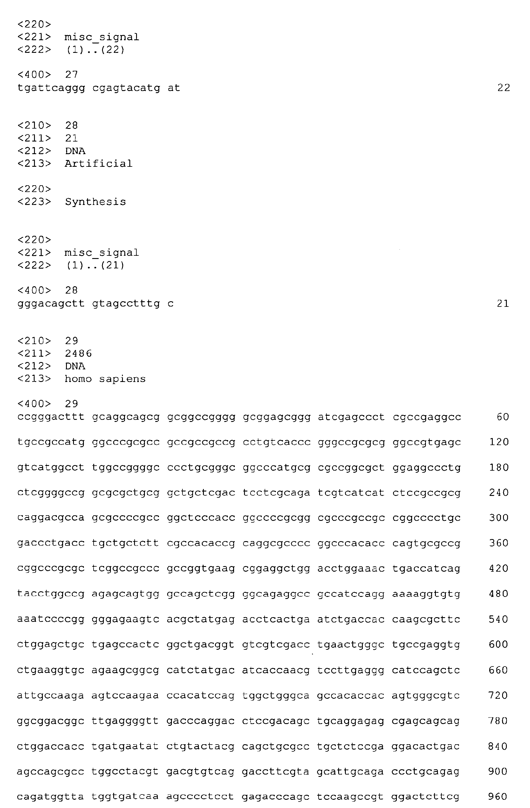
Listed above are the sequence ID numbers for each gene in table 6. Accompanying this application is the DNA sequence for each gene and a corresponding mRNA sequence for each gene, taken from the GenBank database. The GenBank accession numbers listed above in Table 6 (with the prefix NM_) are for the corresponding mRNA sequence. As the mRNA sequence is listed in the GenBank database as the corresponding cDNA sequence, this has been indicated in the accompanying sequence listings as DNA and referred to with the sequence ID name gene X cDNA. However it is intended with this to disclose the mRNA sequence. These mRNA sequences were used to design the corresponding primers listed in Tables 2 and 3. The corresponding primers in Tables 2 and 3 are also listed in the accompanying sequence listings. Thus, for example, for the gene ADM, the DNA sequence is SEQ ID NO. 1, the cDNA (corresponding to mRNA) sequence is SEQ ID NO 2, the forward primer is SEQ ID NO. 3 and the reverse primer is SEQ ID NO. 4. The information recorded in computer readable form is identical to the written sequence listing.
Example 6: Evaluation and Performance Assessment of the Prognostic score
-
317 breast cancer samples were used as described in example 5 and gene expression levels were measured using the methodology and conditions as described in Examples 1 and 2. The genes studied were those listed in Table 1. The (SEQ ID NO. 37, 38) ERBB2 status was determined according to the cut off value determined in Examples 3.
-
To assess the performance of the prognostic score as described in formula I and the ability to predict new data, a cross-validation procedure was used where a rule was created on one set of data (the training set) and then was applied to an independent set of data (the validation set). The test set emulates the set of future samples for which class labels or the outcome variable is to be predicted. Consequently, the test samples are not used for the development of the prediction model (R Simon, British Journal of Cancer 2003 (89), p1599).
-
Therefore, samples were randomly rearranged and split into a training set (two thirds of the samples) and a test set (remaining third). Univariate Cox-coefficients were assessed for each gene using only the training set samples. Prognostic scores were calculated for both the training set and test set samples using the coefficients established in the training set.
-
In order to estimate the outcome as a function of the prognostic score (e.g. the 5-year survival or hazard ratio for a given prognostic score), a cutoff-value (in percentiles) was determined for the distribution of prognostic scores in training set and applied to dichotomize the test set samples according to their prognostic scores. Subsequently, the probability of 5-year survival (RFS, MFS or OS) was determined for each of the dichotomized groups in the test set (e.g. the samples above the cutoff value and the samples below the the training set cutoff value). The entire procedure was repeated 1000 times for each individual cutoff starting typically with the 10th percentile up to the 90th percentile of the distribution of prognostic scores determined in the training set. 5-year survival probabilities were summarized in graphs and plotted as a function of the cutoff (in percentiles). Survival probabilities were calculated according to the Kaplan-Meier method (M Tableman et al , Survival Analysis Using S, 2004, Chapman & Hall/CRC). Coefficients (and hazard ratios) were assessed by univariate Cox regression analysis. The results of this procedure are shown in figures 7 to 26 and summarized in tables 7 and 8.
-
Each percentile cutoff has two corresponding data points: the clear circle represents the probability of 5-year survival (e.g. being metastasis-free or relapse-free after 5 years) given the PS of the test set (test sample) is lower than the x-th percentile (below cutoff) of the PS in the training set. Accordingly, the black, filled-in circles correspond to the probability of 5-year survival (e.g. MFS, or RFS) given the PS of the test set (test sample) is higher than the x-th percentile (above cutoff) of the PS in the training set. Each data point itself summarises the result of 1000 random training/test sets for each cutoff. These graphs are the basis for tables 7 and 8, and to estimate the outcome of a new sample as described in example 7.
-
Analogous procedure was used to assess hazard ratios as a function of the prognostic score and to estimate the performance depending on the number of genes used in the prognostic score, thereby using a specific cutoff value for the prognostic score (e.g. the 30
th and 70
th percentile) and increasing the number of genes in the score. It was found that the performance (hazard ratio) increases with the number of genes used in the prognostic score and that at least three genes, more preferably four or more genes should be used.
Table 7. Expected prognosis (5-year MFS and RFS) for different genes and gene combinations (prognostic scores) depending on different cutoffs, in (SEQ ID NO. 37, 38) ERBB2-positives. | Gene/ Combination | Cutoff MFS (Percentil e) | Prognosis 5year-MFS | Cutoff RFS (Percentil e) | Prognosis 5year-RFS |
| (SEQ ID NO. | <40 | 80-80% | <50 | 80-90% |
| 73, 74) PLAU | >50-80 | 50-60% | >50 | 50-60% |
| | >80 | <50 | | |
| (SEQ ID NO. | <15 | >90% | <15 | >90% |
| 77, 78) PLAUR | <15-30 | 80-90% | <15-30 | 80-90% |
| | <30-50 | 70-80% | <30-50 | 60-80% |
| | >50-75 | 50-60% | >50 | <50% |
| | >75 | <50% | >65 | <40% |
| (SEQ ID NO. | <15 | >90% | <15 | >90% |
| 65, 66) MMP11 | <15-30 | 80-90% | <15-50 | 70-80% |
| | >50-75 | 50-60% | >50 | <50% |
| | >75 | <50% | >60 | <40% |
| (SEQ ID NO. | <15 | >90% | <15 | >90% |
| 73, 74) PLAU, | <15-40 | 80-90% | <15-25 | 80-90% |
| (SEQ ID NO. | >50-70 | 50-60% | <25-50 | 60-80% |
| 77, 78) PLAUR, | >70 | <50% | >50 | <50% |
| (SEQ ID NO. | | | >70 | <40% |
| 65, 66) MMP11 | | | | |
| (SEQ ID NO. | <15 | >90% | <15 | >90% |
| 77, 78) PLAUR, | <15-30 | 80-90% | <15-25 | 80-90% |
| (SEQ ID NO. | <30-50 | 70-80% | <25-50 | 60-80% |
| 65, 66) MMP11, | >50-70 | 50-60% | >50 | <50% |
| (SEQ ID NO. | >70 | <50% | >70 | <40% |
| 49, 50) MMP1 | | | | |
| (SEQ ID NO. | <15 | >90% | <15 | >90% |
| 73, 74) PLAU, | <15-30 | 80-90% | <15-25 | 80-90% |
| (SEQ ID NO. | <30-50 | 70-80% | <25-50 | 60-80% |
| 77, 78) PLAUR, | >50-70 | 50-60% | >50 | <50% |
| (SEQ ID NO. | >70 | <50% | >65 | <40% |
| 65, 66) MMP11, | | | | |
| (SEQ ID NO. | | | | |
| 49, 50) MMP1 | | | | |
| (SEQ ID NO. | <20 | >90% | <15 | >90% |
| 73, 74) PLAU, | <20-30 | 80-90% | <15-25 | 80-90% |
| (SEQ ID NO. | <30-50 | 70-80% | <25-50 | 60-80% |
| 77, 78) PLAUR, | >50-70 | 50-60% | >50 | <50% |
| (SEQ ID NO. | >70 | <50% | >65 | <40% |
| 65, 66) MMP11, | | | | |
| (SEQ ID NO. | | | | |
| 49, 50) MMP1, | | | | |
| (SEQ ID NO. | | | | |
| 57, 58) MMP3, | | | | |
| (SEQ ID NO. | | | | |
| 81, 82) | | | | |
| SERPINE1 | | | | |
Table 8. Expected prognosis (5-year MFS and RFS) for different genes and gene combinations (prognostic scores) depending on different cutoffs, in (SEQ ID NO. 37, 38) ERBB2-negatives. | (SEQ ID NO. | <20 | >90% | <20 | >90% |
| 29, 30) E2F1 | <20-70 | 80-90% | <20-70 | 70-80% |
| | >15 | 60-80% | >15 | 60-80% |
| (SEQ ID NO. 9, | <50 | 80-90% | <50 | >90% |
| 10) BIRC5 | >50-85 | 60-70% | >50-85 | 70-80% |
| | >85 | <60% | >85 | 60-80% |
| (SEQ ID NO. | <50 | 80-90% | <15 | 80-90% |
| 105, 106) | >50-75 | 70-80% | <15-60 | 60-70% |
| TOP2A | >75 | <70% | >15 | <60% |
| (SEQ ID NO. | <20 | >90% | <15 | 80-90% |
| 29, 30) E2F1, | <20-50 | 80-90% | <15-25 | 70-80% |
| (SEQ ID NO. 9, | >50 | 60-70% | <25-50 | 60-70 |
| 10) BIRC5, | | | >50 | |
| (SEQ ID NO. | | | | |
| 105, 106) | | | | |
| TOP2A | | | | |
| (SEQ ID NO. | <25 | >90% | <15 | >90% |
| 29, 30) E2F1, | <25-50 | 80-90% | <15-30 | 80-90% |
| (SEQ ID NO. 9, | >50 | 60-70% | <30-50 | 70-80% |
| 10) BIRC5, | | | >50 | 60-70% |
| (SEQ ID NO. | | | | |
| 101, 102) TK1 | | | | |
| (SEQ ID NO. | <20 | >90% | <15 | >90% |
| 29, 30) E2F1, | <20-50 | 80-90% | <15-25 | 80-90% |
| (SEQ ID NO. 9, | >50 | 60-70% | <25-50 | 70-80% |
| 10) BIRC5, | | | >50 | 60-70% |
| (SEQ ID NO. | | | | |
| 109, 110) TYMS | | | | |
| (SEQ ID NO. | <20 | >90% | <15 | >90% |
| 29, 30) E2F1, | <20-50 | 80-90% | <15-30 | 80-90% |
| (SEQ ID NO. 9, | >50 | 60-70% | <30-50 | 70-80% |
| 10) BIRC5, | | | >50 | 60-70% |
| (SEQ ID NO. | | | | |
| 105, 106) | | | | |
| TOP2A, (SEQ ID | | | | |
| NO. 101, 102) | | | | |
| TK1 | | | | |
| (SEQ ID NO. | <20 | >90% | <15 | >90% |
| 29, 30) E2F1, | <20-50 | 80-90% | <15-30 | 80-90% |
| (SEQ ID NO. 9, | >50 | 60-70% | <30-50 | 70-80% |
| 10) BIRC5, | | | >50 | 60-70% |
| (SEQ ID NO. | | | | |
| 105, 106) | | | | |
| TOP2A, (SEQ ID | | | | |
| NO. 101, 102) | | | | |
| TK1, (SEQ ID | | | | |
| NO. 21, 22) | | | | |
| CTSB, (SEQ ID | | | | |
| NO. 25, 26) | | | | |
| CTSD | | | | |
Example 7: Analysis and Classification of a New Sample
-
The following example illustrates use of the method of the invention. A core biopsy is taken from patient X diagnosed with breast cancer. This core biopsy is analyzed using the tissue preparation, RNA extraction and qRT-PCR methods as described in examples 1 and 2. (SEQ ID NO. 37, 38) ERBB2 status is determined as described example 3. The delta CT value for (SEQ ID NO. 37, 38) ERBB2 is -14.5, and thus the sample is classified (SEQ ID NO. 37, 38) ERBB2 positive.
-
Subsequently, a prognostic score is calculated using a gene combination of 4 genes ((SEQ ID NO. 73, 74) PLAU, (SEQ ID NO. 77, 78) PLAUR, (SEQ ID NO. 65, 66) MMP11, (SEQ ID NO. 49, 50) MMP1) by applying the prognostic score formula I and the weights (coefficients) from table 4. The established score of patient X is -29. Compared with the distribution of prognostic scores in a reference population (e.g. 317 breast cancer samples as described in example 5) determined with the same 4 genes and the formula I, the score of patient X corresponds to the 18th percentile. According to figure 7 and table 7 patient X has an expected 5-year MFS of 80 to 90%. Figure 7 and table 7 were established using the methods described in example 6.
-
Although patient X is (SEQ ID NO. 37, 38) ERBB2-positive, according to his low prognostic score (18
th percentile) patient X is expected to have a good prognosis (estimated 5-year MFS of 80 to 90%). This is in contrast to the latest St. Gallen consensus meeting recommendations which classifies all (SEQ ID NO. 37, 38) ERBB2-positive patients in the highest risk category.


