Molecule of the Month: Ebola Virus Proteins
Structures of ebola virus proteins are giving new hope for fighting this deadly virus

Getting Into Cells
The Matrix

Nucleocapsid
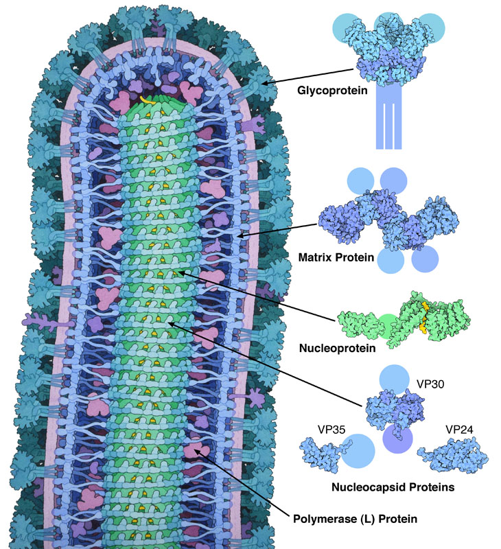
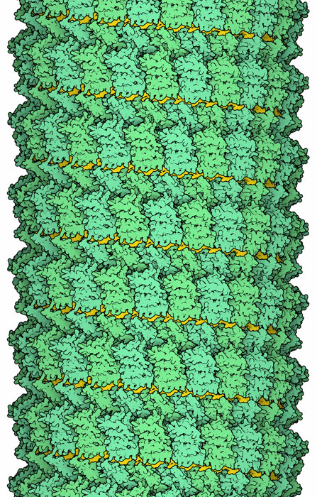
As with the other ebola proteins, the nucleocapsid proteins contain several flexibly-connected domains, so researchers have studied them in parts. The RNA-binding portion of nucleoprotein has been studied by cryoelectron microscopy (PDB entry 5z9w), and x-ray crystallography has been used to study other parts of the protein (PDB entry 4qb0). Several other nucleocapsid proteins, which assist with formation of the structure, have also been studied (PDB entries 3vne, 3fke and 2i8b.)

Moonlighting Proteins
Exploring the Structure
Ebola Glycoprotein and Antibodies (PDB entry 3csy)

Researchers are looking hard for ways to fight infection by ebola, both with drugs and with vaccines. The glycoprotein is the major target for vaccines, since it is on the surface of the virus and is accessible to antibodies. The structure shown here, PDB entry 3csy , includes neutralizing antibodies (in red and orange) from a person who survived infection by the virus. The antibodies bind to the underside of the glycoprotein, to a portion of the protein that is not usually masked by carbohydrates and that is essential for the process of fusion. Hopefully, vaccines will be able to elicit these types of antibodies in patients, protecting them from infection. To explore this structure in more detail, click on the image for an interactive JSmol.
Topics for Further Discussion
- Entry 3csy is thought to be the ebola glycoprotein before it binds to a cell surface. You can look at entry 2ebo to see a portion of the glycoprotein after it fuses with the cell.
Related PDB-101 Resources
- Browse Viruses
- Browse Immune System
References
- 5z9w: Y. Sugita, H. Matsunami, Y. Kawaoka, T. Noda & M. Wolf (2018) Cryo-EM structure of the Ebola virus nucleoprotein-RNA complex at 3.6 angstrom resolution. Nature 563, 137-140.
- 4qb0: P. J. Dziubanska, U. Derewenda, J. F. Ellena, D. A. Engle & Z. S. Derewenda (2014) The structure of the C-terminal domain of Zaire ebolavirus nucleoprotein. Acta Crystallographica Section D 70, 2420-2429.
- T. F. Booth, M. J. Rabb & D. R. Beniac (2013) How do filovirus filaments bend without breaking? Trends in Microbiology 21, 583-593.
- 4ldb, 4ldd: Z. A. Bornholdt, T. Noda, D. M. Abelson, P. Halfmann, M. R. Wood, Y. Kawaoka & E. O. Saphire (2013) Structural rearrangement of ebola virus VP40 begets multiple functions in the virus life cycle. Cell 154, 763-774.
- 3csy: J. E. Lee, M. L. Fusco, A. J. Hessell, W. B. Oswald, D. R. Burton & E. O Saphire (2008) Structure of the ebola virus glycoprotein bound to an antibody from a human survivor. Nature 454, 177-182.
- 3vne: A. P. P. Zhang, Z. A. Bornholdt, T. Liu, D. M. Abelson, D. E. Lee, S. Li, V. L. Woods & E. O. Saphire (2012) The ebola virus interferon antagonist VP24 directly binds STAT1 and has a novel, pyramidal fold. PLoS Pathogens 8: e1002550.
- 3fke: D. W. Leung, N. D. Ginder, D. B. Fulton, J. Nix, C. F. Basler, R. B. Honzatko & G. K. Amarasinghe (2009) Structure of the ebola VP35 interferon inhibitory domain. Proceedings of the National Academy of Science USA 106, 411-416.
- 2i8b: B. Hartlieb, T. Muziol, W. Weissenhorn & S. Becker (2007) Crystal structure of the C-terminal domain of ebola virus VP30 reveals a role in transcription and nucleocapsid association. Proceedings of the National Academy of Science USA 104, 624-629.
- 1h2c: F. X. Gomis-Ruth, A. Dessen, J. Timmins, A. Bracher, L. Kolesnikowa, S. Becker, H. D. Klenk & W. Weissenhorn (2003) The matrix protein VP40 from ebola virus octamerizes into pore-like structures with specific RNA binding properties. Structure 11, 423-433.
October 2014, David Goodsell
https://doi.org/10.2210/rcsb_pdb/mom_2014_10


