Read Link | Google CoLab Demo | Docker | Report Bug | Request Feature
Reporting biomarkers assessed by routine immunohistochemical (IHC) staining of tissue is broadly used in diagnostic pathology laboratories for patient care. To date, clinical reporting is predominantly qualitative or semi-quantitative. By creating a multitask deep learning framework referred to as DeepLIIF, we present a single-step solution to stain deconvolution/separation, cell segmentation, and quantitative single-cell IHC scoring. Leveraging a unique de novo dataset of co-registered IHC and multiplex immunofluorescence (mpIF) staining of the same slides, we segment and translate low-cost and prevalent IHC slides to more expensive-yet-informative mpIF images, while simultaneously providing the essential ground truth for the superimposed brightfield IHC channels. Moreover, a new nuclear-envelop stain, LAP2beta, with high (>95%) cell coverage is introduced to improve cell delineation/segmentation and protein expression quantification on IHC slides. By simultaneously translating input IHC images to clean/separated mpIF channels and performing cell segmentation/classification, we show that our model trained on clean IHC Ki67 data can generalize to more noisy and artifact-ridden images as well as other nuclear and non-nuclear markers such as CD3, CD8, BCL2, BCL6, MYC, MUM1, CD10, and TP53. We thoroughly evaluate our method on publicly available benchmark datasets as well as against pathologists' semi-quantitative scoring.
© This code is made available for non-commercial academic purposes.
 Figure1. Overview of DeepLIIF pipeline and sample input IHCs (different brown/DAB markers -- BCL2, BCL6, CD10, CD3/CD8, Ki67) with corresponding DeepLIIF-generated hematoxylin/mpIF modalities and classified (positive (red) and negative (blue) cell) segmentation masks. (a) Overview of DeepLIIF. Given an IHC input, our multitask deep learning framework simultaneously infers corresponding Hematoxylin channel, mpIF DAPI, mpIF protein expression (Ki67, CD3, CD8, etc.), and the positive/negative protein cell segmentation, baking explainability and interpretability into the model itself rather than relying on coarse activation/attention maps. In the segmentation mask, the red cells denote cells with positive protein expression (brown/DAB cells in the input IHC), whereas blue cells represent negative cells (blue cells in the input IHC). (b) Example DeepLIIF-generated hematoxylin/mpIF modalities and segmentation masks for different IHC markers. DeepLIIF, trained on clean IHC Ki67 nuclear marker images, can generalize to noisier as well as other IHC nuclear/cytoplasmic marker images.
NVIDIA GPU (Tested on NVIDIA QUADRO RTX 6000)
CUDA CuDNN (CPU mode and CUDA without CuDNN may work with minimal modification)
Python 3
Pytorch>=0.4.0
torchvision>=0.2.1
dominate>=2.3.1
visdom>=0.1.8.3
All image pairs must be 512x512 and paired together in 3072x512 images (6 images of size 512x512 stitched together horizontally). For testing purpose, for any unavailable image in the pairing set, just put the original IHC image or a blank image in the final paired image in place of the missing image in the pair (example images are provided in the Dataset folder). For testing, you can skip stitching the images and only put the original IHC image in the test folder, but make sure to set the --dataset_mode to 'single'. Data needs to be arranged in the following order:
XXX_Dataset
    ├── test
    ├── val
    └── train
We have provided two simple functions in the DeepLIIF/PrepareDataset.py file for preparing data for testing and training purposes.
- To prepare data for training, you need to have the paired data including IHC, Hematoxylin Channel, mpIF DAPI, mpIF Lap2, mpIF marker, and segmentation mask in the input directory. The script gets the address of directory containing paired data and the address of the dataset directory. It, first, creates the train and validation directories inside the given dataset directory. Then it reads all images in the folder and saves the pairs in the train or validation directory, based on the given validation_ratio.
python PrepareDataForTraining.py --input_dir /path/to/input/images
                                 --output_dir /path/to/dataset/directory
                                 --validation_ratio The ratio of the number of the images in the validation set to the total number of images.
- To prepare data for testing, you only need to have IHC images in the input directory. The function gets the address of directory containing the IHC data and the address of the dataset directory. It, first, creates the test directory inside the given dataset directory. Then it reads the IHC images in the folder and saves a pair in the test directory.
python PrepareDataForTesting.py --input_dir /path/to/input/images
                                --output_dir /path/to/dataset/directory
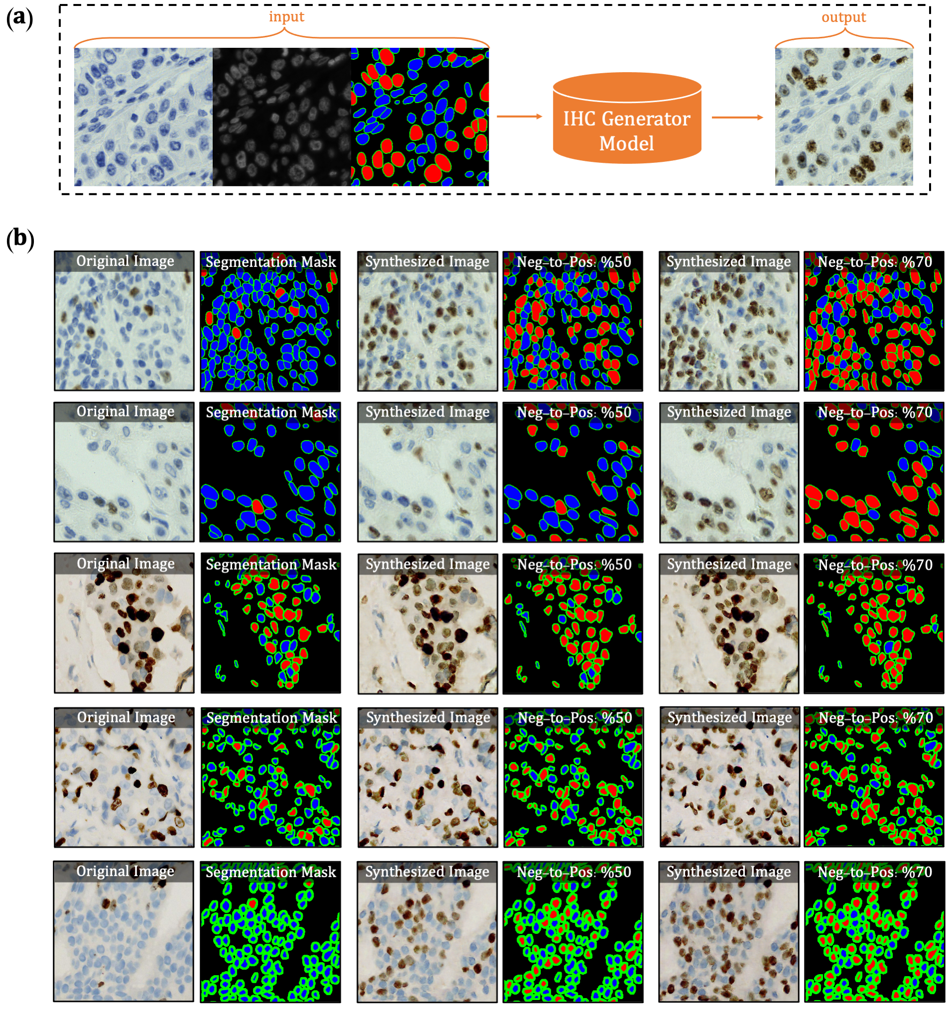
The first version of DeepLIIF model suffered from its inability to separate IHC positive cells in some large clusters, resulting from the absence of clustered positive cells in our training data. To infuse more information about the clustered positive cells into our model, we present a novel approach for the synthetic generation of IHC images using co-registered data. We design a GAN-based model that receives the Hematoxylin channel, the mpIF DAPI image, and the segmentation mask and generates the corresponding IHC image. The model converts the Hematoxylin channel to gray-scale to infer more helpful information such as the texture and discard unnecessary information such as color. The Hematoxylin image guides the network to synthesize the background of the IHC image by preserving the shape and texture of the cells and artifacts in the background. The DAPI image assists the network in identifying the location, shape, and texture of the cells to better isolate the cells from the background. The segmentation mask helps the network specify the color of cells based on the type of the cell (positive cell: a brown hue, negative: a blue hue).
In the next step, we generate synthetic IHC images with more clustered positive cells. To do so, we change the segmentation mask by choosing a percentage of random negative cells in the segmentation mask (called as Neg-to-Pos) and converting them into positive cells. Some samples of the synthesized IHC images along with the original IHC image are shown in Figure 2.
 Figure2. Overview of synthetic IHC image generation. (a) A training sample of the IHC-generator model. (b) Some samples of synthesized IHC images using the trained IHC-Generator model. The Neg-to-Pos shows the percentage of the negative cells in the segmentation mask converted to positive cells.
We created a new dataset using the original IHC images and synthetic IHC images. We synthesize each image in the dataset two times by setting the Neg-to-Pos parameter to %50 and %70. We re-trained our network with the new dataset. You can find the new trained model here.
To train a model:
python train.py --dataroot /path/to/input/images
                --name Model_Name
                --model DeepLIIF
- To view training losses and results, open the URL https://localhost:8097. For cloud servers replace localhost with your IP.
- To epoch-wise intermediate training results, DeepLIIF/checkpoints/Model_Name/web/index.html
- Trained models will be by default save in DeepLIIF/checkpoints/Model_Name.
- Training datasets can be downloaded here.
To test the model:
python test.py --dataroot /path/to/input/images
               --name Model_Name
               --model DeepLIIF
- The test results will be by default saved to DeepLIIF/results/Model_Name/test_latest/images.
- The latest version of the pretrained models can be downloaded here.
- Place the pretrained model in DeepLIIF/checkpoints/DeepLIIF_Latest_Model and set the Model_Name as DeepLIIF_Latest_Model.
- To test the model on large tissues, we have provided two scripts for pre-processing (breaking tissue into smaller tiles) and post-processing (stitching the tiles to create the corresponding inferred images to the original tissue). A brief tutorial on how to use these scripts is given.
- Testing datasets can be downloaded here.
This script is used for testing purposes. You can use this script to infer modalities and classified segmentation mask for large images. If the output_dir not given the script, it automatically saves the inferred modalities in the input_dir next to the original images.
python inference.py  --input_dir /path/to/preprocessed/images
                     --output_dir /path/to/output/images
                     --tile_size size_of_each_cropped_tile
                     --overlap_size overlap_size_between_crops
You can use the docker file to create the docker image for running the model. First, you need to install the Docker Engine. After installing the Docker, you need to follow these steps:
- Download the pretrained model and place them in DeepLIIF/checkpoints/DeepLIIF_Latest_Model.
- Change XXX of the WORKDIR line in the DockerFile to the directory containing the DeepLIIF project.
- To create a docker image from the docker file:
docker build -t deepliif_image .
The image is then used as a base. You can copy and use it to run an application. The application needs an isolated environment in which to run, referred to as a container.
- To create and run a container:
docker run --gpus all --name deepliif_container -it deepliif_image
When you run a container from the image, a conda environment is activated in the specified WORKDIR. You can easily run the preprocessing.py, postprocessing.py, train.py and test.py in the activated environment and copy the results from the docker container to the host.
- A quick sample of testing the pre-trained model on the sample images:
python preprocessing.py --input_dir Sample_Large_Tissues/ --output_dir Sample_Large_Tissues_Tiled/test/
python test.py --dataroot Sample_Large_Tissues_Tiled/ --name DeepLIIF_Latest_Model --model DeepLIIF
python postprocessing.py --output_dir Sample_Large_Tissues/ --input_dir results/DeepLIIF_Latest_Model/test_latest/images/ --input_orig_dir Sample_Large_Tissues/
If you don't have access to GPU or appropriate hardware, we have also created Google CoLab project for your convenience. Please follow the steps in the provided notebook to install the requirements and run the training and testing scripts. All the libraries and pretrained models have already been set up there. The user can directly run DeepLIIF on their images using the instructions given in the Google CoLab project.
To register the denovo stained mpIF and IHC images, you can use the registration framework in the 'Registration' directory. Please refer to the README file provided in the same directory for more details.
To train DeepLIIF, we used a dataset of lung and bladder tissues containing IHC, hematoxylin, mpIF DAPI, mpIF Lap2, and mpIF Ki67 of the same tissue scanned using ZEISS Axioscan. These images were scaled and co-registered with the fixed IHC images using affine transformations, resulting in 1667 co-registered sets of IHC and corresponding multiplex images of size 512x512. We randomly selected 709 sets for training, 358 sets for validation, and 600 sets for testing the model. We also randomly selected and segmented 41 images of size 640x640 from recently released BCDataset which contains Ki67 stained sections of breast carcinoma with Ki67+ and Ki67- cell centroid annotations (for cell detection rather than cell instance segmentation task). We split these tiles into 164 images of size 512x512; the test set varies widely in the density of tumor cells and the Ki67 index. You can find this dataset here.
We are also creating a self-configurable version of DeepLIIF which will take as input any co-registered H&E/IHC and multiplex images and produce the optimal output. If you are generating or have generated H&E/IHC and multiplex staining for the same slide (denovo staining) and would like to contribute that data for DeepLIIF, we can perform co-registration, whole-cell multiplex segmentation, train the DeepLIIF model and release back to the community with full credit to the contributors.
You can find more options in:
- DeepLIIF/options/base_option.py for basic options for training and testing purposes.
- DeepLIIF/options/train_options.py for advanced training options.
- DeepLIIF/options/test_options.py for advanced testing options.
- DeepLIIF/options/processing_options.py for advanced pre/post-processing options.
- DeepLIIF/options/Registration_App.py for registering pathology crops and slides.
Please report all issues on the public forum.
© Nadeem Lab - DeepLIIF code is distributed under Apache 2.0 with Commons Clause license, and is available for non-commercial academic purposes.
- This code is inspired by CycleGAN and pix2pix in PyTorch.
If you find our work useful in your research or if you use parts of this code, please cite our paper:
@article{ghahremani2021deepliif,
  title={DeepLIIF: Deep Learning-Inferred Multiplex ImmunoFluorescence for IHC Image Quantification},
  author={Ghahremani, Parmida and Li, Yanyun and Kaufman, Arie and Vanguri, Rami and Greenwald, Noah and Angelo, Michael and Hollmann, Travis J and Nadeem, Saad},
  journal={bioRxiv},
  year={2021},
  publisher={Cold Spring Harbor Laboratory}
}
